
Abstract
A 9-year-old female llama (Lama glama) that served as a blood donor at The Ohio State University Veterinary Teaching Hospital developed multiple small, raised, firm, non-haired cutaneous masses on the right hip, left cheek, and right and left shoulders. Cytological evaluation of fine-needle aspirates from the cutaneous mass from the left shoulder and right hip comprised many well-differentiated, highly granulated mast cells with moderate numbers of eosinophils. Occasional mast cells exhibited erythrophagocytosis and contained a small amount of hemosiderin or several variably sized vacuoles. A cytologic diagnosis of mast cell tumor with evidence of prior hemorrhage was made, and the masses were surgically removed. Microscopically, each mass consisted of sheets of neoplastic round cells that formed nonencapsulated nodules in the dermis and infiltrated into the adjacent dermal collagen. Eosinophils were scattered among the mast cells at the periphery of the nodules. Neoplastic mast cells, but not eosinophils, exhibited positive membrane KIT expression and cytoplasmic vimentin staining. A final diagnosis of mast cell tumor was made based on cytology, histology, and immunohistochemistry.
Keywords
The most common malignant round cell tumor in llamas (Lama glama) and alpacas (Lama pacos) is lymphoma, with B-cell lymphoma more commonly reported than T-cell lymphoma. 13 About 25% of malignant round cell tumors in llamas and alpacas are primitive malignant round cell tumors, based on immunohistochemistry. 13 Mast cell tumors (MCTs) are classified as round cell tumors in domestic animals, but they have not been reported in llamas or alpacas. The current report describes the first case of mast cell neoplasia in a llama, which occurred as multiple cutaneous tumors.
A 9-year-old female llama boarding in The Ohio State University Veterinary Teaching Hospital (Columbus, Ohio) as a blood donor was evaluated as part of her routine health assessment. Four small, raised, firm, non-haired cutaneous masses were identified during physical examination. The masses were 4–10 mm in diameter and were located on the right hip, left and right shoulders, and left cheek. The llama was otherwise clinically healthy. Complete blood cell count and serum biochemical profiles were not performed; abdominal ultrasound was unremarkable.
Fine-needle aspirates from the left shoulder were performed for cytological evaluation. Samples were stained initially with Hema 3, a as shown in Figure 1A. The cellularity was high in a bloody background. The majority of the cells were large (20–25 μm in diameter) round cells with minimal anisocytosis and anisokaryosis. These cells had a moderate nuclear to cytoplasmic ratio, centrally placed round nuclei, and abundant clear cytoplasm that contained rare small, purple granules. To evaluate cell morphology, samples from another aspirate of the same mass were stained with Wright–Giemsa stain (Fig. 1B). A similar round cell population was presented, and numerous purple cytoplasmic granules often obscured the nucleus of the cells. Occasional cells exhibited erythrophagocytosis; rare cells contained a small amount of hemosiderin (Fig. 1B); a few cells also contained multiple variably sized vacuoles. These cells were interpreted as neoplastic mast cells. The cytological diagnosis was cutaneous MCT. There also were increased numbers of eosinophils, as is the case with MCTs from other species. The presence of hemosiderin was compatible with prior hemorrhage.
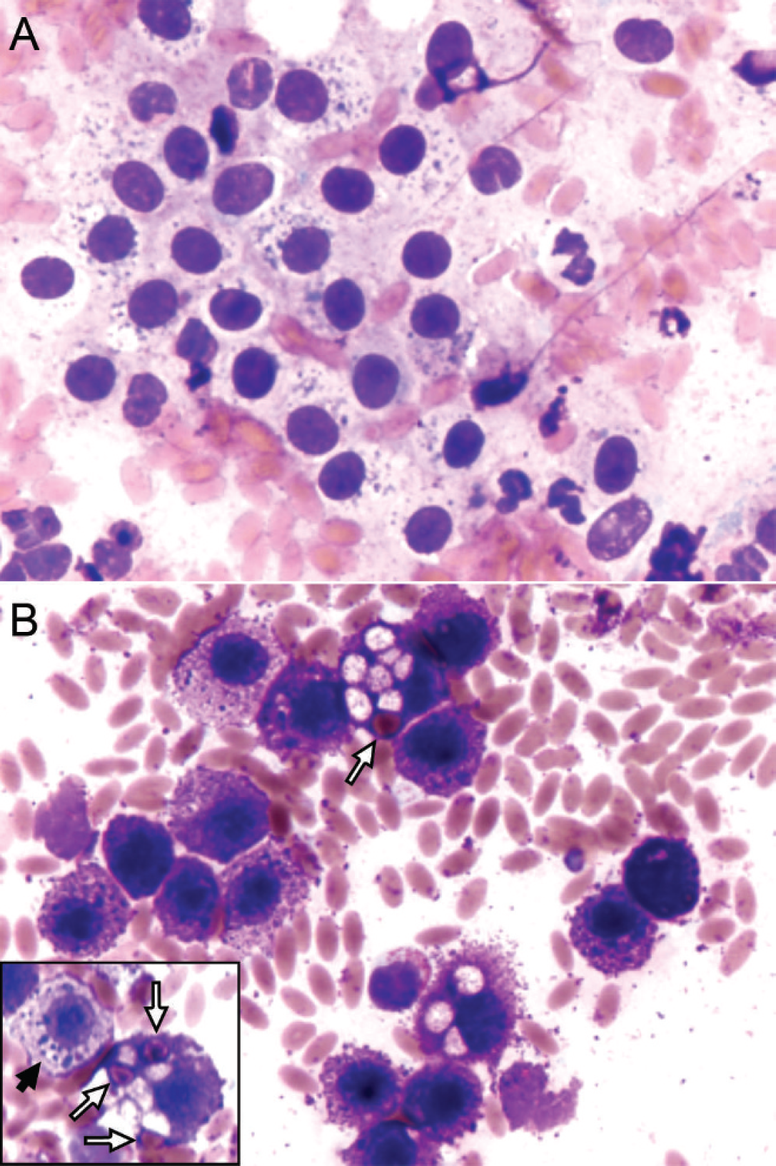
Fine-needle aspirate smear of a dermal mass from the left shoulder of a llama.
All 4 masses were surgically removed, fixed in formalin, and submitted for histologic evaluation. Masses from the right hip, cheek, and left and right shoulders formed large nonencapsulated nodules confined to the dermis. These nodules consisted of sheets of densely packed round cells that effaced portions of the dermis, infiltrated between bundles of dermal collagen and extended superficially to the dermal–epidermal junction (Fig. 2). The cells had distinct cell borders and abundant clear cytoplasm with faint basophilic granules measuring approximately 1 mm in diameter. Central nuclei were round to oval with clumped chromatin, and occasional nuclei had a single basophilic nucleolus. There were 2 mitotic figures per 10 high-power fields (40× objective). Small numbers of eosinophils were scattered among the mast cells at the deep edges of some sections. Sections of the masses were evaluated for the presence of KIT, vimentin, and cytokeratin and were stained with Giemsa and Toluidine blue using routine methods. Neoplastic mast cells were positive for KIT b expression in a membrane-associated pattern and for vimentin b expression in a cytoplasmic pattern (Fig. 3), whereas eosinophils were negative for KIT. Giemsa staining revealed numerous small cytoplasmic granules in nearly all of the neoplastic mast cells, whereas Toluidine blue staining yielded a basophilic granularity to the cytoplasm without distinct granule formation. The neo-plastic cells were negative for cytokeratin. b
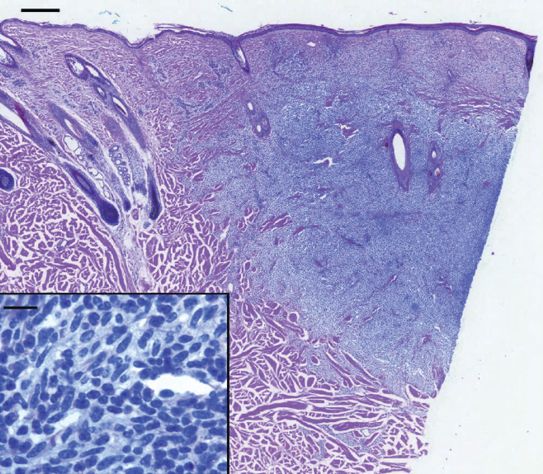
Histopathology of a dermal mass from the left shoulder of the llama. Bar = 0.35 mm. Eosinophils are present at the periphery of the mass. Inset: Bar = 15 μm.
Mast cell tumors are the most common skin tumors in dogs and also occur in humans, cats, horses, rodents, cows, goats, sheep, ferrets, and pigs. 3,5,7,16–19 In dogs, MCTs most commonly occur as cutaneous masses and tend to metastasize to local lymph nodes and internal organs, such as spleen and liver. 12 The low-grade, well-differentiated MCTs have a very low metastatic rate (10%), whereas medium- to high-grade, poorly differentiated MCTs have a much higher metastatic rate (55–96%). A histologic grading system has also been established for MCTs in dogs and may be associated with prognosis along with other proliferation markers, such as mitotic index, proliferating cell nuclear antigen, and argyrophil nucleolar organizer region. 20,23,24 Mast cell tumors in cats and human beings often occur as a systemic disease. The term systemic mastocytosis is used in human medicine to describe mastocytosis in blood, bone marrow, lymph nodes, and several internal organs. 1,22 In horses, cutaneous MCTs also are reported as one of the most common skin tumors, 26 and most are benign, with recurrence being unusual after complete excision. 14 Cutaneous and disseminated MCTs in cattle have been reported. Most are malignant, with tissue infiltration and necrosis, fibrosis, and mineralization within the tumor tissue. 6 Although multiple masses were identified in the llama in the present report, benign behavior was indicated based on the well-differentiated morphology of the mast cells, the noninvasive appearance (based on histological findings), and the absence of recurrence or progression after more than a year of follow-up. Cytology often is helpful in the diagnosis of MCTs because of the characteristic appearance of mast cells with routine staining. As is the case with findings from other species, Wright–Giemsa stain resulted in more intensely stained granules in the neoplastic mast cells than did the Diff-Quik or Hema 3 stain (Fig. 1A, 1B). The mechanism of the differences in staining is unclear. Several subtypes of mast cells have been identified in humans and mice based primarily on granule contents and biological function. In mice, mucosal mast cells contain only chymase, whereas connective tissue mast cells contain both chymase and tryptase. In humans, granules from “MCT” cells contain tryptase, whereas “MCTC” cells have both tryptase and chymase. Although mouse anti-human tryptase antibody has been used to detect mast cells in the small intestine from camels, 4 mast cell subtypes have not been adequately evaluated. Erythrophagocytosis and vacuolation in the mast cells noted in the current case have been reported 2,9,15 in both neoplastic and normal mast cells from several species, but the significance of this finding is unclear. According to the findings in the present case, it is also noteworthy that this antibody against KIT exhibits cross-reactivity with llama KIT and can be used to identify mast cells in this species.
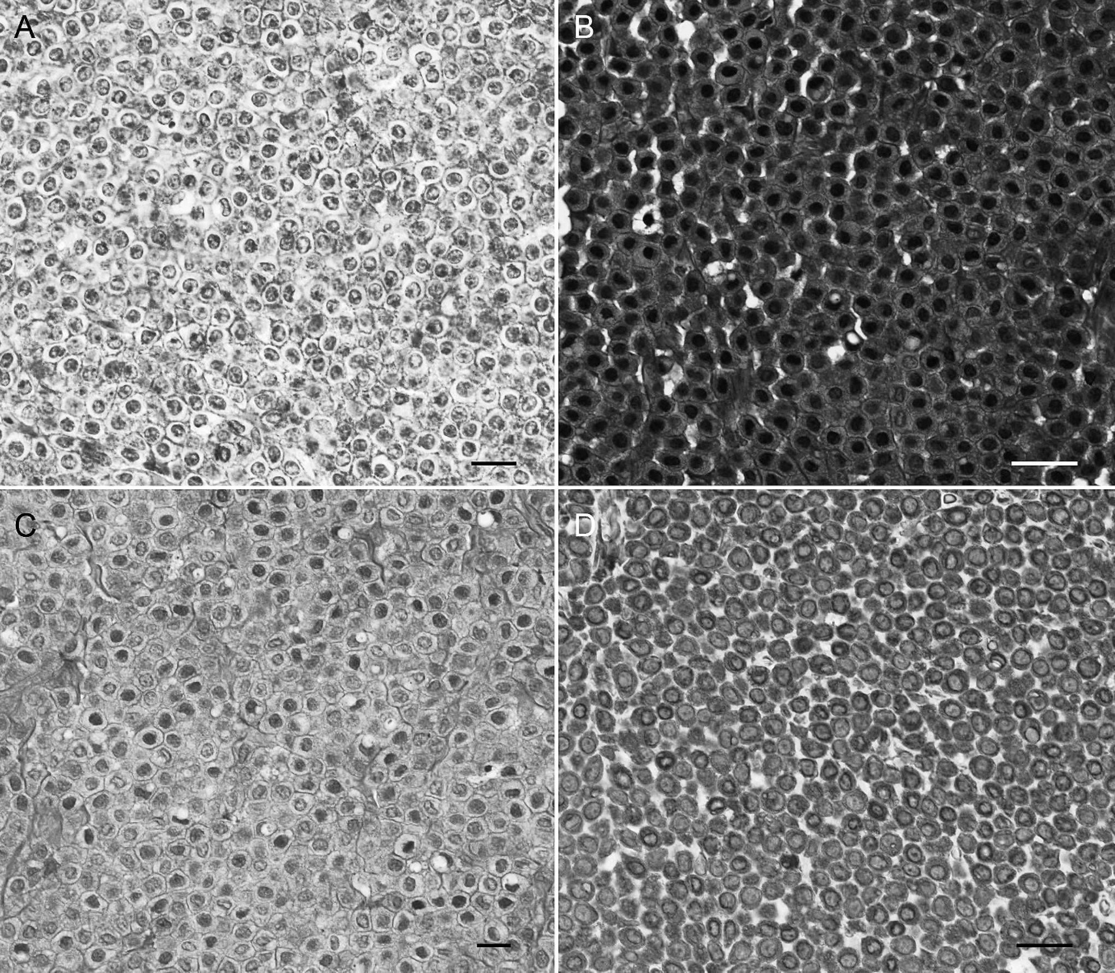
Immunohistochemistry of the dermal mass from the left shoulder from the llama:
KIT (cluster of differentiation 117) is an important oncogene in MCTs, and mutations of KIT have been described 8,11,25 in canine, human, and rodent MCTs. Over 80% of human patients with systemic mastocytosis carry the V816D KIT mutation in the kinase domain. 21 The internal random duplicate mutation in the juxtamembrane domain of KIT has been identified in up to 30% of high-grade canine MCTs, 11 while more recently a point mutation of the extracellular domain of KIT has been found in a small percentage of canine MCTs. 10 However, no further studies regarding KIT mutations have been performed in other domestic animals. This is the first report to demonstrate expression of KIT on llama neoplastic mast cells, which occurred in a membrane-associated pattern. KIT staining patterns have been linked to prognosis in dogs and horses. Cytoplasmic KIT staining has been associated with poor prognosis and recurrence in canine MCTs, 27 whereas a unique perinuclear KIT staining pattern has been proposed to be an aberrantly expressed marker of malignant mast cells in horses. 22 The clinical significance of a membrane-associated KIT expression pattern in neoplastic mast cells of llamas remains unclear. This is the first reported case of mast cell neoplasia in a llama supported by cytology, histopathology, and immunohistochemistry. After completely excising the tumors, no further treatments were attempted, and no recurrence has been noted to date (1.5 years).
Footnotes
a.
Fisher Diagnostics, Middletown, VA.
b.
Dako North America Inc., Carpinteria, CA.