
Abstract
A 4-year-old female Richardson's ground squirrel (Spermophilus richardsonii) presented with multicentric nodules arising from the skin of the middle of the tail and lumbosacral regions. Histologically, the nodules were composed of a proliferation of spindloid to pleomorphic cells that sometimes formed sheets and fascicular to storiform patterns. Diffuse infiltration of eosinophils was also noted. The results of immunohistochemistry indicated positive labeling for vimentin, mast cell tryptase, c-kit, and Ki-67. Toluidine blue stain revealed fine, metachromatic, cytoplasmic granules. The histologic diagnosis was mast cell tumor. The neoplasm recurred and metastasized to the right lumbar lymph node 1 month later.
Keywords
Mast cell tumor (MCT), or mastocytoma, has been reported in many mammalian species. This neoplasm can be focal or multicentric in the skin and may occasionally involve internal viscera such as the spleen, liver, and intestines. There is species variation in location and biologic behavior of MCT, but the similarities in histopathology outweigh the differences. 11 Mast cell tumor is the most common cutaneous tumor in dogs and the second most common in cats. 20 It also has been reported in horses, cattle, pigs, 11 and ferrets. 17
Richardson's ground squirrel (Spermophilus richardsonii) is a species of North American ground squirrel. This species has been used as an animal model of disease and most recently has been kept as a pet. Females live 4 years on average (maximum 6), while males usually live only 1 year (maximum 2–3). Although cancers are an uncommon cause of death in ground squirrel species, a number of cases of hepatocellular carcinoma have been reported in individuals infected with hepatitis virus. 4,10,12,19 Spontaneous adenocarcinoma in the buccal salivary gland of a Richardson's ground squirrel has also been described in a recent publication. 24
A 4-year-old female Richardson's ground squirrel developed multicentric primary masses arising from the skin of the mid tail and lumbosacral regions (Fig. 1). These masses appeared round, were red to white, and had a sunken appearance. The largest mass measured 3.5 cm in diameter, while the smaller mass was 2.0 cm in diameter. Neither ulceration nor hemorrhage was observed. The cut surfaces of the neoplastic masses were well delineated, solid, and gray to white. One month after surgical removal, the neoplasm of the tail recurred, and the right lumbar lymph node was enlarged. The recurrent neoplasms were firm and measured 1.5 cm in diameter.
The primary and recurrent cutaneous neoplasms and the right lumbar lymph node were excised or biopsied and evaluated histologically. Tissue samples were fixed in 10% neutral buffered formalin, processed routinely, embedded in paraffin, sectioned at 4 μm, and stained with hematoxylin and eosin, toluidine blue, and Masson trichrome. Paraffin sections also were prepared for immunohistochemical staining. The panel of antibodies used, primary antibody dilutions, and antigen retrieval methods are presented in Table 1. Primary mouse and rabbit immunoglobulin G primary antibodies were used at predetermined dilutions. The secondary biotinylated antibodies were goat anti-rabbit immunoglobulin G (IgG), goat anti-mouse IgG, a and rabbit anti-rat IgG. b Sites of binding of the biotinylated secondary antibodies were detected using streptavidin–biotin c reagent at a 1:300 dilution. The chromogen contained 0.05% 3,3′-diaminobenzidine (DAB) with 0.03% hydrogen peroxide in Tris-HCl buffer. The tissue sections subsequently were counterstained with methyl green, dehydrated, coverslipped, and examined microscopically. Samples of confirmed cutaneous MCTs from a dog and a ferret were used as positive controls. For negative controls, the primary antibodies were replaced with 8% skim milk in Tris-buffered saline solution.
Each neoplastic nodule had similar gross and histologic features. These neoplasms extended from the dermis into the subcutis and were not well encapsulated. Neoplastic cells were arranged in fascicles, in a storiform pattern, or in solid sheets and lacked excessive fibrous stroma (Fig. 2). Individual cells were pleomorphic and round to spindle. The nucleus varied in size, was round to elongate, and contained single to multiple nucleoli. The cytoplasm was scant to abundant, lightly eosinophilic, and had indistinct cell borders (Fig. 3). Infiltrates of eosinophils (Fig. 3) and neutrophils, focal necrosis, and hemorrhage were also present.
With toluidine blue staining, nonneoplastic mast cells in the dermis had intensely stained cytoplasmic granules. In contrast, most of the neoplastic cells in the dermis had faint to moderately staining cytoplasmic granules (Fig. 4). Masson's trichrome staining failed to show significant collagen production in the intercellular matrix. This observation did not support a diagnosis of primary fibrosarcoma.
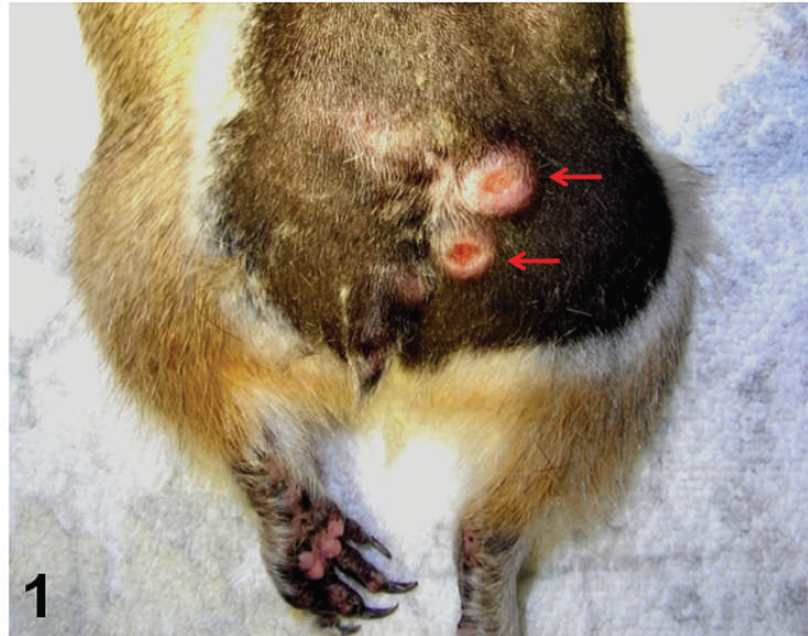
Two neoplastic nodules (arrows) are present at the lumbosacral region.

Neoplastic cells are arranged in fascicles, in storiform patterns, or in sheets. Hematoxylin and eosin. Bar = 20 μm.

Pleomorphic, round to spindle cells containing a variably sized nucleus are observed. Infiltration of eosinophils is prominent (arrows). Hematoxylin and eosin. Bar = 20 μm.

Toluidine blue (TB) staining reveals faintly to moderately stained, metachromatic, cytoplasmic granules in most of neoplastic cells. Inset: TB-positive nonneoplastic mast cells in the dermis with intensely stained cytoplasmic granules. Bar = 20 μm.

Most of the neoplastic cells are intensely immunopositive for mast cell tryptase. Inset: Tryptase-positive nonneoplastic mast cells in the dermis. Bar = 20 μm.

Most of the neoplastic cells are intensely immunopositive for c-kit. Inset: c-kit-positive nonneoplastic mast cell in the dermis. Bar = 20 μm.
Following immunohistochemical staining, most of the neoplastic cells were positive for mast cell tryptase (Fig. 5) and c-kit (Fig. 6). The reliability of the immunostaining technique was supported by intense immunoreactivity of nonneoplastic mast cells residing in the dermis of these tissue sections. Tryptase is a secretory granule-derived serine proteinase of mast cells that has recently been used as a marker for mast cell activation. 5 The c-kit protein is a tyrosine kinase receptor and a product of the c-kit protooncogene, 25 which is expressed in many cells and tissues, including glioblastoma cells, term placenta, brain, erythroid precursors, melanocytes, basophils, and mast cells. 2,13,25 Expression of the c-kit receptor in MCTs has been well established. 8,16,21 The combination of positive immuno-staining for mast cell tryptase and c-kit in conjunction with the presence of metachromatic cytoplasmic granules has been suggested to be a powerful tool for the diagnosis of gastrointestinal MCTs 14 ; the identification of highly atypical, hypogranulated mast cells (especially in mast cell leukemia); and the detection of small to minute mast cell infiltrates. 6 The neoplastic cells also stained strongly for vimentin. Moreover, many neoplastic mast cells were positive for Ki-67 using the anti-Ki-67 antibody MIB-1 (approximately 97 positive cells per high-power [400×] field in the primary neoplastic lesions). The neoplasm in the right lumbar lymph node also was confirmed as a metastatic MCT.
Antibodies used for the immunohistochemical evaluation of mast cell tumors in a Richardson's ground squirrel.*
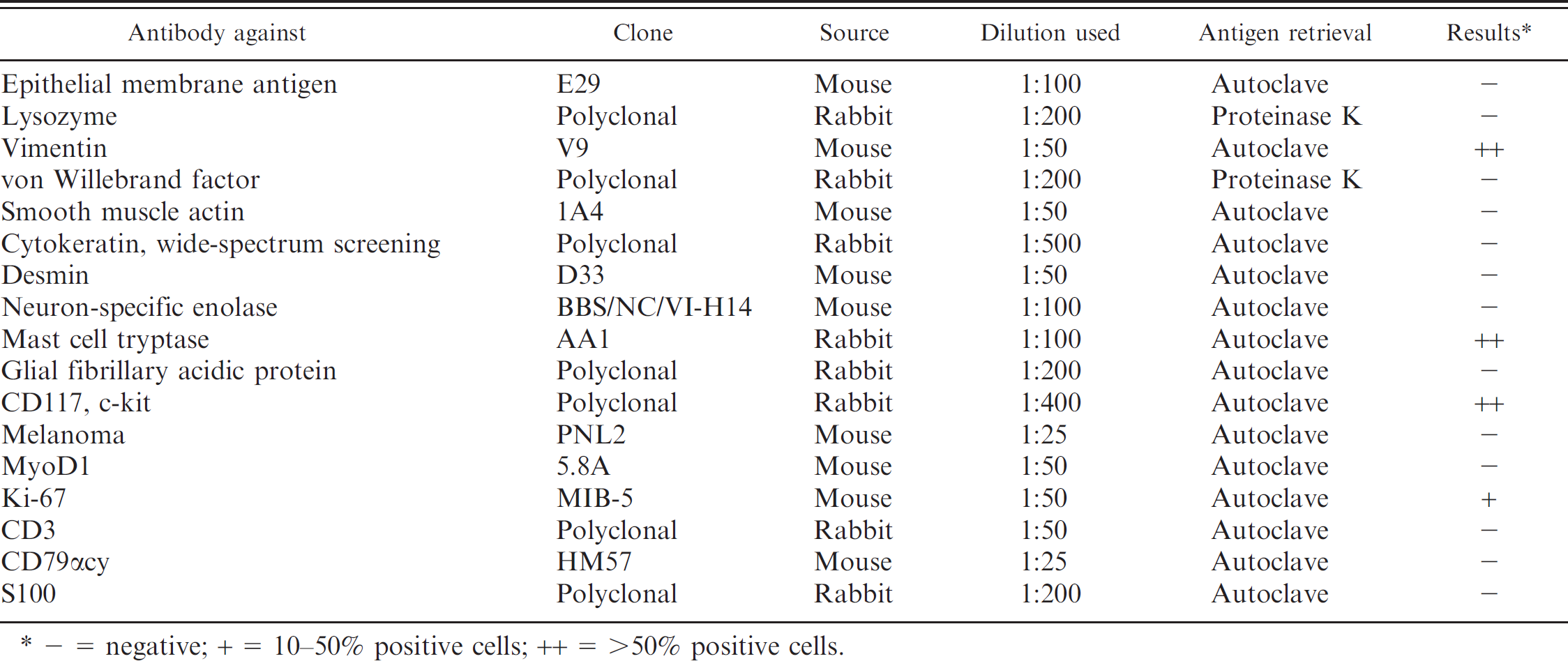
– = negative; + = 10–50% positive cells; ++ = >50% positive cells.
In the current case, it was difficult to make a definitive diagnosis of MCT by routine histology and common special stains neoplasms consisted of atypical spindle cells admixed with pleomorphic round cells that were arranged in fascicles, in storiform patterns, or in sheets. Thus, the histologic features of these neoplasms were similar to those of fibrosarcoma or leiomyosarcoma. Moreover, the histologic features of the present neoplasms were unlike those of MCTs in other rodent species (e.g., laboratory rats 1 and mice 3,7,9 ) or of poorly differentiated MCTs in canids (grade III) 18 and other animal species. 15,23 With regard to the spindle-shaped tumor cells, the following points should be considered: 1) normal dermal mast cells of Richardson's ground squirrels are pleomorphic and round to spindle, and 2) the present neoplasms may be poorly differentiated and the cells assume a more pleomorphic spindloid appearance. The results of immunohistochemical staining for tryptase and c-kit as well as positive toluidine blue staining support the histologic diagnosis of cutaneous MCT with metastasis to the right lumbar lymph node.
The biologic behavior of this MCT in Richardson's ground squirrel is similar to that of MCTs in other species, especially dogs. In the present case, recurrence of the MCT on the tail with metastasis to the right lumbar lymph node within 1 month probably correlates with the high proliferative activity of the tumor cells as revealed by marked Ki-67 labeling. In dogs, recurrence of MCTs after attempted surgical excision is fairly common in grade II and grade III neoplasms and metastases first involve the regional lymph nodes. 11,22 Nevertheless, generalizations concerning the biologic behavior of MCTs in Richardson's ground squirrel cannot be made until more animals with this neoplasm are reported.
Footnotes
a.
Kirkegaard & Perry Laboratories, Gaithersburg, MD.
b.
Vector Laboratories Inc., Burlingame, CA.
c.
Dako North America Inc., Carpinteria, CA.