
Abstract
Four adult mixed-breed beef cows from a cow–calf operation in West Virginia were referred to the Virginia-Maryland Regional College of Veterinary Medicine in March 2009 with weakness, ataxia, hind limb paresis progressing to lateral recumbency, and death within 2–3 days. Histologically, there was accumulation of light brown, granular pigment in neurons of the ventral gray horns of the spinal cord (more severe in thoracic and lumbar sections), brain stem, and pons, resulting in distortion and bulging of the cell body and displacement of the Nissl substance, suggestive of Phalaris sp. grass toxicosis. The most severely affected cow had accumulation of dark green–brown pigment in renal tubular epithelial cells. Reed canarygrass (Phalaris arundinacea) was identified in pastures, and the concentration of tryptamine alkaloids in new leaf blades was approximately 0.2% on a wet weight basis. These alkaloids are serotonergic receptor agonists, resulting in neurologic “staggers” in ruminants. Delayed onset times of up to 4–5 months have been reported in sheep after removal from Phalaris sp. pastures. Distribution of pigment in serotonergic tracts of the midbrain, brain stem, and spinal cord with Phalaris sp. toxicoses is distinct and differs from lipofuscin. Electron microscopy confirmed that the pigment was not lipofuscin. From these findings, a diagnosis of delayed P. arundinacea toxicosis was made. Over a 2-month period, 18 cows died with similar clinical signs.
Canarygrass (Phalaris spp.) toxicoses have been documented since 1942, primarily in Australia and New Zealand, 1,12,21 with recent poisonings reported in Argentina, South Africa, Norway, and the United States. 1,13,19,20 Most canarygrass toxicoses have been associated with bulbous canarygrass or hardinggrass (Phalaris aquatica, previously Phalaris tuberosa), 2,12,21 but timothy canarygrass (Phalaris angusta), 20 reed canarygrass (Phalaris arundinacea), 21 littleseed canarygrass or annual canarygrass (Phalaris minor),13 and Carolina canarygrass or May grass (Phalaris caroliniana) 19 have also been implicated in outbreaks. A single gene controls the production of tryptamine alkaloids, 23 and efforts to develop less toxic cultivars of canarygrass have been ongoing, with no absolute success. In the United States, outbreaks have been reported in feedlot lambs and sheep in California (P. aquatica), 10,15 as well as sheep and cows in Louisiana (P. caroliniana). 19 Sheep are considered to be the most susceptible species, but cattle 16 and horses 7 can also be affected.
Reed canarygrass is well adapted to the northern half of the United States 17 and has multiple desirable characteristics, including good crude protein content, high digestibility, drought and flood tolerance, high yield, and good cool season growth. 16,17 The toxicosis can manifest in 2 apparently unrelated forms. 4 The first is referred to as “sudden death,” 4,6,12 which may present as either a neurologic or a cardiac syndrome, 6,12 whereas the second form is referred to as “staggers” and affects the animal's locomotor abilities. Staggers occurs as either an acute and reversible or a chronic, irreversible, and typically lethal form. The latter, more common, form usually occurs as a large outbreak. Delayed or chronic Phalaris sp. toxicoses are associated with tryptamine alkaloids present in canarygrass, which structurally resemble serotonin. 1,2 These alkaloids act as serotonergic receptor agonists 5 within the central nervous system, and accumulation eventually results in neurologic staggers in ruminants. Delayed onset times of up to 4–5 months have been reported in sheep after removal from Phalaris sp. pastures. 3,10
Beef cattle producers, predominantly in Monroe County, West Virginia, reported multiple unexplained deaths in their cattle that all exhibited similar neurologic clinical signs. Affected cows were weak, ataxic, and staggering. They also had profound hind limb paresis that progressed rapidly from “dog sitting” to sternal recumbency, lateral recumbency, and spontaneous death over a course of 2–3 days. Extension of both the head and neck and the feet and leg was observed immediately preceding death. Cranial nerve deficits affecting prehension or tongue movement were not evident, as opposed to previous case reports in cattle. 1,14 Although many of these animals were somewhat emaciated, others were alert and able to eat. Producers in adjacent Greenbrier and Nicholas counties reported similar problems in their cattle; however, fewer animals were affected. A total of 18 adult mixed-breed beef cows were affected within a single cow–calf operation in Monroe County, from March to April 2009. Four of these cows were referred for necropsy examination to the Virginia–Maryland Regional College of Veterinary Medicine (Blacksburg, Virginia) from March 9 to 15, 2009. At the time of admission, the cows (age range: 4–12 years old) were in fair to poor body condition, 1 was pregnant, and all had a history of neurologic clinical signs that progressed rapidly over a 2–3-day period. Gross findings (ascites, pericardial effusion, lymphadenopathy, and colonic ulcers) were minor and not considered significant. The postmortem interval range was between 24 and 48 hr. One cow was pregnant with a bull calf of approximately 9 months gestation that measured 88 cm from crown to rump.
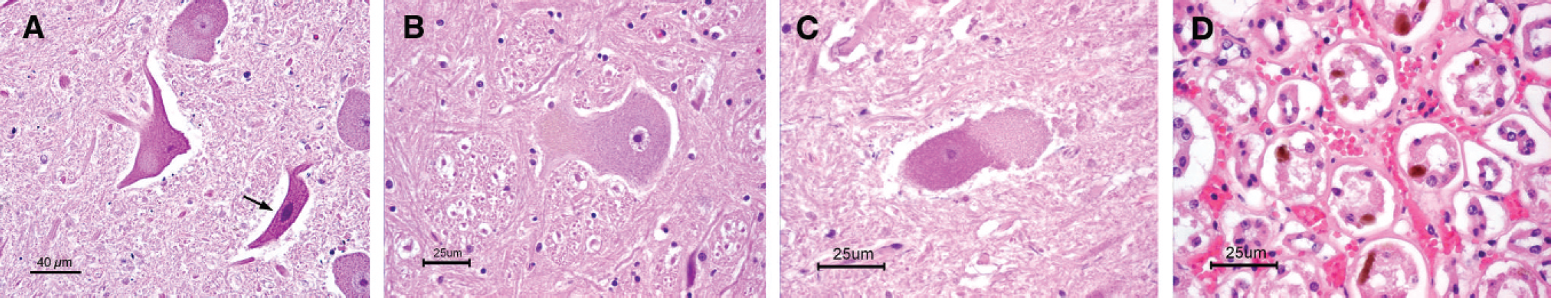
A, necrotic neurons with increased cytoplasmic eosinophilia, shrunken cell bodies, and condensed nuclei (arrow) were commonly observed. Some necrotic neurons contain moderate amounts of tan to brown, finely granular, intracytoplasmic pigment. The postmortem interval ranged from 24 to 48 hr. Hematoxylin and eosin. Bar = 40 μm.
The brain, cervical, thoracic, and lumbar spinal cord were examined. Histologic evaluation revealed lesions limited to spinal cord segments in the thoracic and lumbar spinal cord (Fig. 1A–C), pons, medulla, midbrain, and kidney (Fig. 1D). Neurons bilaterally within the ventral grey horns of the spinal cord were frequently necrotic with hypereosinophilic cytoplasm and condensed nuclei (Fig. 1A). Many neurons contained moderate to large amounts of intracytoplasmic pigment that was finely granular and tan to brown. This pigment often accumulated within the cytoplasm expanding axon hillocks (Fig. 1B) and distorted the overall shape, resulting in bulging surfaces (Fig. 1C). Nuclei affected in the pons, medulla, and midbrain included the red nucleus, olivary nuclei, nucleus ambiguus, and nucleus gracilis. Dark brown to green, intracytoplasmic pigment was also observed in the renal tubular epithelial cells at the corticomedullary junction and within the medulla (Fig. 1D). The bull calf fetus had similar pigment within its kidney. No other significant histologic lesions were observed.
Distribution of pigment in serotonergic tracts of the midbrain, brain stem, and spinal cord with Phalaris sp. toxicoses is rather distinct. 2,22 Similar lipofuscin pigment is present with normal aging in cattle and other species. 18 However, age-related lipofuscinosis has a different anatomical distribution than Phalaris sp. toxicosis and is not associated with clinical signs in animals. Pigment granules were positive for periodic acid–Schiff-like lipofuscin, so ultrastructural evaluation was performed. Transmission electron microscopy of the pigment granules within the spinal cord revealed electron-dense bodies contained within multilayered, concentrically arranged membranous lamellae (Fig. 2). These pigment granules are similar to findings observed in a Phalaris spp. outbreak in California sheep 10 and, thus, confirmed that the pigment granules were not lipofuscin. 8 This indole-like pigment is believed to be a postmetabolite breakdown of the tryptamine alkaloids. 2
A field investigation of the cow–calf operation in Monroe County was conducted on March 27, 2009. The herd consisted of 75 mixed-breed beef cows that were pregnant, housed on pasture, and fed grass hay during December 2008. Additionally, a similar group of 40 pregnant cows were recently purchased, kept adjacent to the other 75 cows, and fed grain silage. In preparation for February and March calving dates, all 115 cows that were previously housed on 2 different pastures were moved during the first 2 weeks of January to a pasture on the main farm. Cows were allowed to graze on a mixed grass and clover pasture that had only been harvested once during the 2008 growing season. Cattle were offered free-choice trace mineral salt. Water was available from a small stream in a gully area, as well as a pond. On February 1, 2009, all cattle were moved to a larger adjacent pasture that housed the barn and hay sheds. After the move, cattle were fed hay and haylage previously obtained from their current and initial pasture. The first affected animals showed clinical signs of ataxia and paresis in the first week of March.
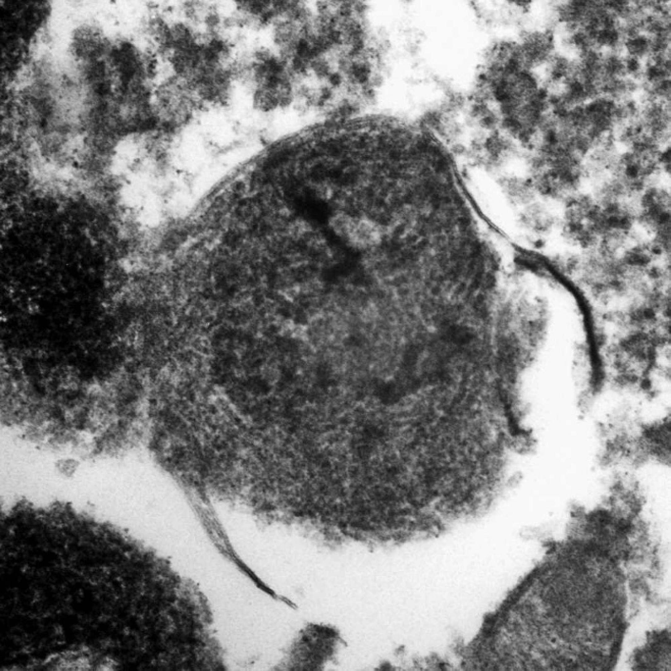
Pigment granule within the cytoplasm of a spinal cord neuron. An electron-dense body is surrounded by membranous lamellae that are multilayered and concentrically arranged, which is ultrastructurally different from lipofuscin. The tissues were initially fixed in 10% neutral buffered formalin at the time of necropsy. After histologic identification of pigment granules within neurons, tissues were placed in 2.5% glutaraldehyde in phosphate buffer and postfixed in osmium tetroxide. Transmission electron microscopy, 80,000×.
The initial pasture used in January was examined during the field investigation. The pasture grass was very short and consisted mainly of fescue, orchard grass, timothy, and red clover. The gully area that ran through the pasture had a small creek; multiple small trees; and tall, brown weed and grass remnants. Some grass stems in the gully from the previous growing season were approximately 1 m tall but lacked any dried leaf blades. However, new spring growth of grass near the base of these tall stems was approximately 15 cm high. Dry stems and new grass blades were identified as reed canarygrass (P. arundinacea) by county extension agronomists accompanying the field investigation. Representative samples of dried canarygrass stems, new spring canarygrass leaf blades, and hay and haylage from the initial pasture and main pasture were obtained.
Six samples were semiquantitatively analyzed for tryptamine alkaloids, as previously described. 11 Samples consisted of hay from 2007 and 2008 and of 2008 haylage from the initial pasture and from the main pasture, as well as new spring canarygrass leaf blades and old canarygrass dried stems remaining from January. Briefly, samples were finely chopped with a paper cutter; 5-g samples were weighed, macerated in chloroform, filtered, and extracted with dilute hydrochloric acid, followed by treatment with silicotungstic acid according to published procedures. 11 All samples except the new reed canarygrass leaf blades were perfectly clear solutions after addition of silicotungstic acid (i.e., less than 0.1% tryptamine alkaloids). The sample of new reed canarygrass blades became immediately turbid and developed precipitate within minutes after incubation with silicotungstic acid. According to the test scaling, the new leaf blades were ranked 3 or 4, which corresponds to a tryptamine alkaloid concentration of approximately 0.2% on a wet-weight basis (corresponding to approximately 1% on a dry-weight basis). Previous reports have demonstrated toxic total tryptamine levels to range from 0.1% to 0.3% dry matter. 12 This level is almost 3-fold higher than published reports stating the total tryptamine alkaloid concentration of toxic Phalaris spp. grasses to be as high as 0.3% dry matter. 11 Based on these results, known access to canarygrass, and histologic pigment accumulation, a diagnosis of Phalaris sp. grass toxicosis was made.
Tryptamine alkaloids identical to those in canarygrass species 16 are also reported in other plants. 22 A review of these plants exposed 2 other potential differential plants known to be in West Virginia. The most likely candidates were Desmodium spp. and Lespedeza bicolor shrub. Neither Desmodium spp. nor L. bicolor was recognized by West Virginia agronomists as present on the problem farm. Similar neuronal pigmentation and distribution of lesions have also been reported with a South African plant, Trachyandra spp. 13
Environmental conditions that promote high concentrations of tryptamine alkaloids in Phalaris spp. include drought, nitrogen fertilization, shady conditions, new growth or regrowth, top growth consumption, and leaf versus stem consumption. 16 The reason for the outbreak in the present report is likely multifactorial. The region has had a moderate drought for the last 2 summers, which could promote the expansion of the drought-resistant reed canarygrass and higher concentrations of tryptamine alkaloids in the grass. Exposure of cattle to a tall, ungrazed pasture in January is unusual and precluded the normal feeding of supplemental hay or haylage initially. After the tall pasture became sparse, cattle probably fed preferentially on reed canarygrass during the last 2 weeks in January. Finding bare stems of reed canarygrass without leaf blades led investigators to believe that the cattle had selectively peeled away the leaf material that was present in January.
Recommendations that were given to the producer included fencing off the gully area and applying herbicide to isolated patches of reed canarygrass outside of the fenced-off area. Increasing the cobalt concentration in the free-choice trace mineral mix to 250 ppm was also recommended. Cobalt boluses are used as preventative measures in Australia and New Zealand for Phalaris spp. staggers 9 and are believed to promote growth of rumen bacteria capable of destroying tryptamine alkaloids before the alkaloids are absorbed. 8 Cobalt supplementation does not have an effect on ongoing clinical problems. There is no treatment for animals affected with staggers. Other producers considering planting of reed canarygrass for its desirable characteristics should seek out a cultivar that does not produce tryptamine alkaloids. Multiple farms were affected in this outbreak, with the highest concentration occurring on a single farm in Monroe County, West Virginia. Over the course of the outbreak, 18 cows died. This is the fourth reported set of cases in the United States and to the authors' knowledge the first report of toxicity associated with reed canarygrass in the United States.