
Abstract
Mycoplasma haemolamae is a hemotropic mycoplasma that affects red blood cells of llamas (Lama glama) and alpacas (Lama pacos). It is variably associated with anemia, and most infections are subclinical. Development of a polymerase chain reaction assay has facilitated detection of this infection in llamas and alpacas in the United States and other countries. Whether the infection occurs in camelids in South America has previously been unknown. The current study documents a 15.8% infection rate among 76 Peruvian llamas, a 19.3% infection rate among Peruvian alpacas at one site, and a 9.26% infection rate in 108 Chilean alpacas from selected herds. All of the camelids tested appeared to be clinically healthy. No gender or species predilection was found. Only 1 positive camelid younger than 18 months was found. Infection is not associated with anemia, and the mean packed cell volume (PCV) in positive Peruvian camelids was slightly higher than the mean PCV in negative Peruvian camelids. In the Chilean alpacas, the positive alpacas had a slightly lower PCV than the negative alpacas, although the mean PCV was not in the anemic range in any of the groups.
Mycoplasma haemolamae (order Mycoplasmatales, family Mycoplasmataceae) is a hemotropic mycoplasma that attaches to the membrane of erythrocytes of llamas (Lama glama) and alpacas (Lama pacos). The organism was first described in llamas in 1990 and reported to be an Eperythrozoon-like organism based on light and scanning microscope morphology. 5,8 It was subsequently reclassified as a mycoplasma based on the 16S ribosomal RNA sequence that was most similar to Mycoplasma haemosuis (classified as Mycoplasma suis) and Mycoplasma wenyonii, mycoplasmas that infect the erythrocytes of swine and cattle, respectively. 7
Llamas and alpacas infected with M. haemolamae may have mild to severe anemia that variably appears to be regenerative. 5,8,9 Lethargy, depression, and fever are sometimes present in infected camelids. Deaths have occurred in heavily infected camelids. However, it appears that most infected camelids do not show any clinical or laboratory abnormalities associated with infection. In many cases, clinical disease is seen when animals are immune suppressed, stressed, or concurrently infected with other organisms, such as gastrointestinal parasites.
In 2001, a polymerase chain reaction (PCR)–based assay was developed to provide a more sensitive and specific diagnostic method than the blood smear examination used previously. The assay was used in experimental infections and showed that many camelids are subclinical chronic carriers. Some of the experimentally infected camelids were transiently anemic, while others were not, and they did not develop fever, depression, or hypoglycemia (Tornquist SJ, Boeder LJ, Parker JE, et al.: 2002, Use of a polymerase chain reaction assay to study the carrier state in infection with camelid Mycoplasma haemolama, formerly Eperythrozoon spp. infection camelids. Vet Clin Pathol 31:153–154. Abstract). 9
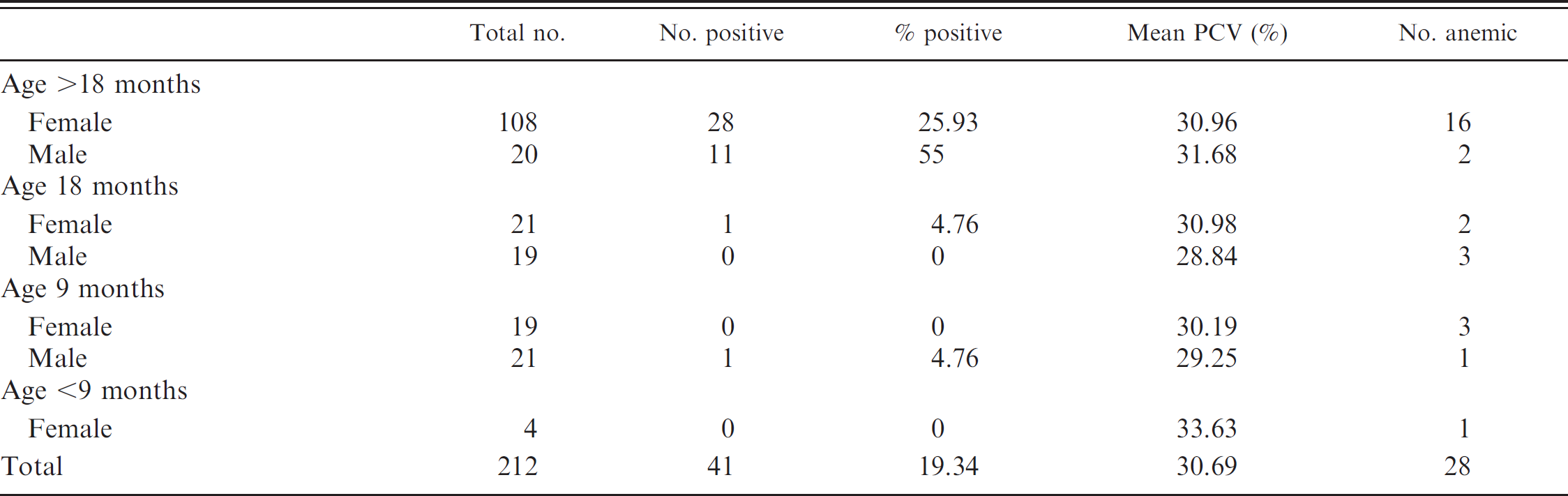
Peruvian alpacas, Mycoplasma haemolamae, and anemia status and packed cell volume (PCV).
The PCR assay had been used diagnostically in more than 6,000 blood samples from all over the United States as well as Australia, Canada, and the United Kingdom. There have been no reports documenting whether infection was present in llamas or alpacas in South America. The current study was undertaken to answer that question.
Samples were collected from study sites in Peru and Chile. The Peruvian samples were obtained from 212 alpacas and 76 llamas at the La Raya Research Station in Peru. The site houses approximately 3,000 alpacas and 1,000 llamas kept in herds of approximately 150 animals segregated by age, sex, and species. These are closed herds, with no animals being brought in from outside the station, which is situated at an altitude of 4,320 m. The alpacas in the study included 152 females and 60 males grouped by age: >9 months, 9 months, 18 months, and >18 months.
There were 57 females and 19 males among the llamas sampled, with similar age groups. None of the animals sampled showed any clinical evidence of disease.
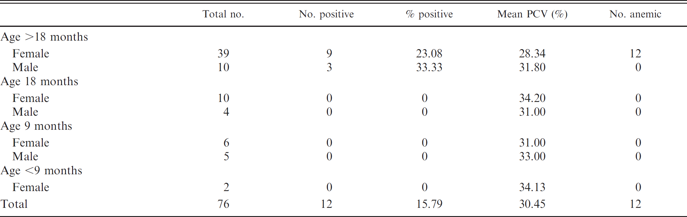
Peruvian llamas, Mycoplasma haemolamae, and anemia status and packed cell volume (PCV).
Samples from 107 Chilean alpacas were collected from 7 separate herds. Six of the sites were in altiplano at elevations of 3,400–4,400 m. One site was located at an elevation of 500 m. These alpacas were allowed to graze during the day and were housed in paddocks during the night. The alpacas included 48 males and 60 females with an age range of 1–11 years. All animals were clinically healthy. These were all closed herds.
Blood was collected by jugular venipuncture into tubes containing ethylenediamine tetra-acetic acid (EDTA). Blood was stored between 5 and 20°C until assays were performed. The PCR assay for M. haemolamae was performed as previously described (Tornquist SJ, et al.: 2002, Use of a polymerase chain reaction assay to study the carrier state in infection with camelid Mycoplasma haemolam). 9 Briefly, DNA was extracted from 200 μl of blood using a commercial extraction system. a To ensure there was no DNA contamination during the extraction process, a negative control consisting of all of the reagents without a DNA sample was included, as was a positive control that consisted of EDTA–anticoagulated blood from a naturally infected alpaca with large numbers of typical organisms seen on blood smears.
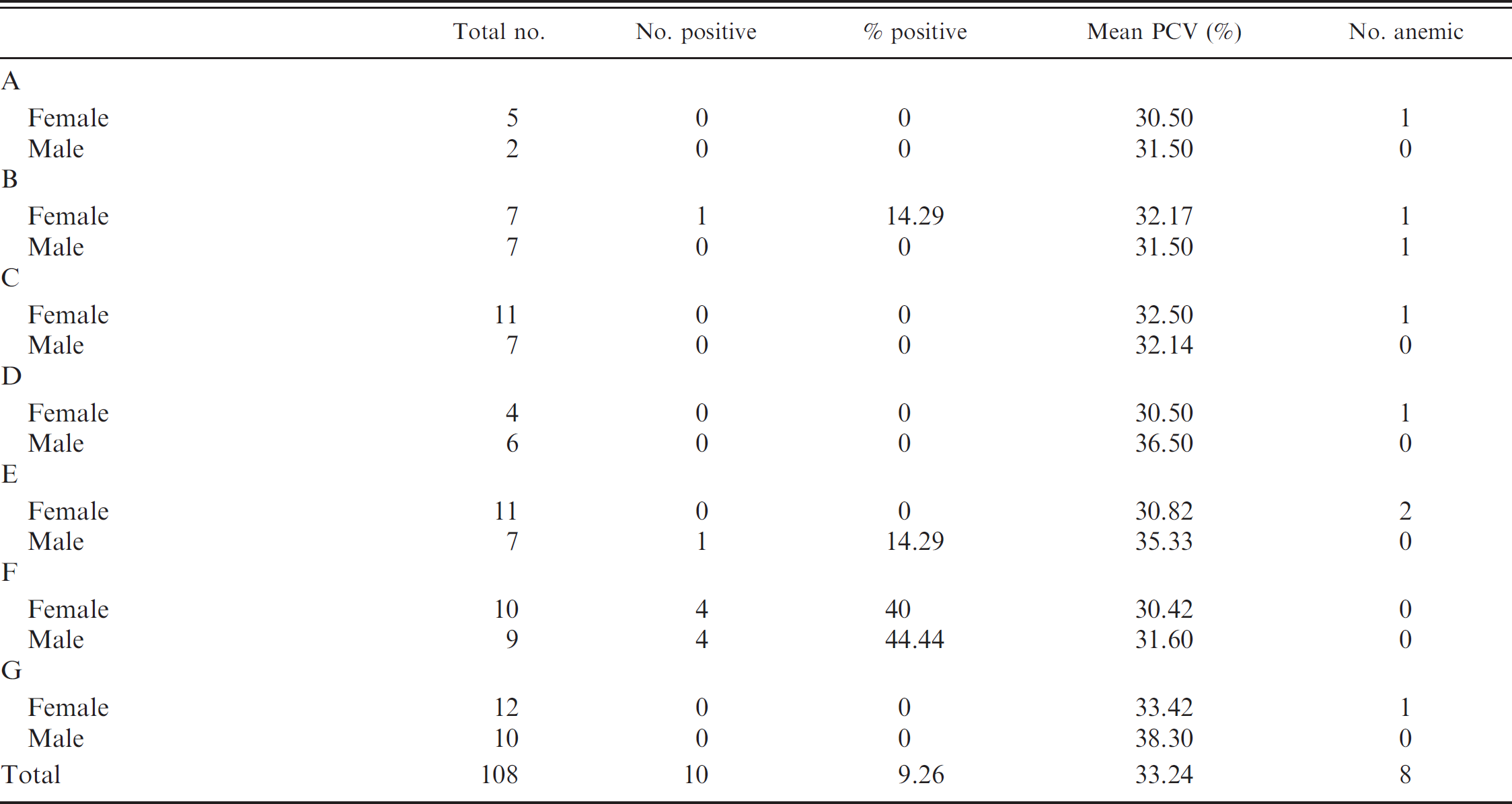
Chilean alpacas (all adults), Mycoplasma haemolamae, and anemia status and packed cell volume (PCV).
Amplification was accomplished using primers for a 318-bp sequence that is unique to the hypervariable region of the M. haemolamae 16S ribosomal RNA region as identified in GenBank. 7 A negative control consisting of a reaction mixture with no template DNA was included. Confirmed positive blood and distilled water were included in each assay as positive and negative controls, respectively.
Packed cell volumes (PCVs) were determined by centrifugation of blood in microhematocrit tubes. Anemia was defined as a PCV of less than 27% based on Oregon State University's Veterinary Diagnostic Laboratory (Corvallis, Oregon) hematology reference ranges. Comparisons of PCVs between infected and uninfected animals were made using Student's t-test, with P < 0.05 considered to be significant.
Out of a total of 288 Peruvian samples, there were 53 PCR-positive samples, for an overall 18.4% positive percentage. This included a 15.8% positive percentage for llamas and a 19.3% positive percentage for alpacas. There was no difference between llamas and alpacas in the percent of positive animals (P = 0.07; Tables 1, 2). With the exception of a single positive 9-month-old alpaca male, no infected alpacas or llamas younger than 18 months were found (Tables 1, 2). There was 1 positive 18-month-old alpaca female; all of the other positive animals were older than 18 months. Overall, there was no difference in the percentage of positives between females and males in the combined population of camelids (18.2% females and 18.9% males) or among llamas or alpacas. The mean PCV of infected camelids (31.75%) was slightly higher than that of uninfected camelids (30.44%; P = 0.03), although the difference is not clinically significant. Overall, of the negative group, 13.61% of animals were anemic; in the positive group, 8.87% of animals were anemic. There were 10 positive alpacas from the 108 Chilean samples, for an overall 9.26% positive percentage (Table 3). The positive animals included 5 females and 5 males, thus no sex difference was detected. Of the 6 separate groups sampled, there were 4 groups with no positive animals. The mean PCV of positive Chilean alpacas (30.3%) was significantly lower than the mean PCV of uninfected alpacas (33.24%). There were 7 anemic animals (7.14%) in the negative group and 1 anemic animal (10%) in the positive group.
The present study is the first to document M. haemolamae infections in llamas and alpacas in South America. Although the organism has been recognized for 20 years in the United States, it has not been known whether it is present in camelids in their regions of origin. The presence of infection in camelids in closed and isolated groups in the Peruvian Andes and the Chilean altiplano suggests that it is most likely present in the New World camelid population in general.
As with many infected camelids in the United States, the positive animals appear to have mostly subclinical infections. The positive animals did not show signs of clinical disease, and anemia was very rarely present. Although the mean PCV of positive Chilean alpacas was significantly lower than the mean PCV of negative alpacas, it was not low enough to be considered anemic, and the difference is of minimal clinical significance. In the Peruvian camelids, the mean PCV of negative camelids was actually lower than that of positive camelids; again, this difference is not great enough to be of any clinical significance, and other causes of anemia, including individual variation, nutrition, and other concurrent infections, may be contributing factors.
Although early reports of this infection suggested that younger camelids were more likely to be infected, 5,8 this has not been confirmed since the development of the PCR assay (Tornquist, unpublished observations), nor was it the case in the current study. Infection was very rarely seen in the Peruvian camelids younger than 18 months, and the average age of infected Chilean alpacas was 3.3 years. These findings suggest that there may be an increased risk with increased age.
The mode of transmission of M. haemolamae is not known, although insect vectors are suspected, as they are for other hemotropic mycoplasmas. 6 In the United States, more infections tend to be detected in the fall, which would be consistent with transmission by insect vectors (Tornquist, unpublished observations). In the present study, the Peruvian samples were all collected in October, and the Chilean samples were collected in November and December. At the Peruvian study site, insects are not seen at that altitude and time of year. At the Chilean sites, some of the herds were heavily infected with ticks (Amblyomma parvitarsum); however, there was no apparent correlation between the presence of ticks and the percent of infected alpacas.
In utero transmission of the infection has also been postulated and is supported by the finding of organism in crias younger than 24 hr (Tornquist, unpublished observations). 1,3 The fact that there is at most a 44% infection rate (and usually much lower) in a group of animals kept together in the current study suggests relatively low infectivity of the organisms, effective immune responses, and lack of direct animal-to-animal transmission.
Clinical signs and increased numbers of M. haemolamae are often associated with camelids that are stressed, are immune suppressed, or have concurrent infections (Torn-quist SJ, et al.: 2002, Use of a polymerase chain reaction assay to study the carrier state in infection with camelid Mycoplasma haemolam). 2,4,9 In the present study, the animals appeared healthy and in good bodily condition, but complete health status information was not collected. In summary, the current study documents the presence of M. haemolamae in selected llama and alpaca populations in 2 South American countries. It would be of interest to survey llamas and alpacas at other sites as well as test wild populations of guanacos (Lama guanicoe) and vicunas (Vicugna vicugna).
Acknowledgements
The authors thank the Department of Biomedical Sciences, College of Veterinary Medicine, Oregon State University, Corvallis, OR; Dr. Jorge Crossley, College of Veterinary Medicine, Universidad de Santo Tomas, Santiago, Chile, for funding; and Drs. Jorge Crossley, Alberto Raggi, Rodrigo Fuentes, Walter Bravo, and Johanna Rigas for help with sample collection.
Footnotes
a.
Generation™ Capture Column, Qiagen Inc., Valencia, CA.