
Abstract
A litter of 3-month-old Pharaoh Hound puppies presented to the referring veterinarian with severe generalized erythematous-crusted papules with pruritus, accompanied by exfoliation and erythema of footpads, inappetence, lethargy, and retarded growth. Three of 5 puppies (2 male and 1 female) were affected. Representative areas were biopsied from 1 affected male puppy and were routinely processed. Histologically, there was marked epidermal hyperplasia with a disorganized appearance of the epidermis and massive parakeratotic hyperkeratosis, compatible with zinc-responsive dermatosis. Low serum zinc concentrations were documented, and the affected animals partially responded to intravenous zinc supplementation but did not respond to oral supplementation. One male puppy died as a result of unrelated causes and was necropsied. The remaining 4 puppies were followed over 2 years. Growth was stunted, and enamel hypoplasia of permanent dentition developed compared with unaffected littermates. Intravenous zinc supplementation at 3-4 week intervals was required to prevent further skin lesion development. One dog died at 3 years of age of renal failure.
Skin diseases associated with zinc deficiency or failure to absorb or appropriately utilize zinc are uncommon in dogs. 4,10 Zinc responsive dermatosis is clinically subdivided into 2 groups. Syndrome I is seen almost exclusively in northern breed dogs and characterized as an inherited impairment in the absorption or metabolism of zinc. 6,10 Syndrome II is seen in rapidly growing large-breed puppies consuming a high phytate diet, zinc-deficient diet, or diet supplemented with components that may inhibit zinc absorption. Both of these syndromes are characterized clinically by mucocutaneous junction and pressure point erythema, scaling, crusting, alopecia, and lichenification.
Acrodermatitis is a disease specific to Bull Terriers 7,9 which is characterized by progressive dermatitis, stunting, abnormal behavior, diarrhea, and bronchopneumonia. Affected animals typically develop clinical signs as early as 8 weeks of age that include papular to pustular erythematous and exfoliative dermatitis of distal extremities, mucocutaneous junctions, muzzle, and ears. Footpad lesions are caused by abnormal cornification and are characterized by thickening, fissures, and exfoliation. Affected patients have variable plasma and tissue zinc concentrations and do not clinically respond to zinc supplementation. 5,7 Histologically, there is marked parakeratotic hyperkeratosis with moderate to severe epidermal hyperplasia, hypogranulosis, and dyskeratosis. Deeper keratinocytes may appear disorganized. Lymphoid organs are typically affected, with thymic atrophy, lymph node atrophy, and decreased white pulp in the spleen present. 5
An inherited zinc deficiency in humans, acrodermatitis enteropathica, shares clinical and pathologic features with acrodermatitis in Bull Terriers. Unlike Bull Terriers, human patients are often responsive to zinc supplementation. 2 An inherited zinc deficiency in cattle, known as lethal trait A46, shares similar clinical signs to acrodermatitis enteropathica and is responsive to zinc therapy. It has recently been proposed as an animal model for acrodermatitis enteropathica 11 based on the finding of a mutation in SLC39A4. The SLC39A4 gene encodes a zinc-specific transporter belonging to the zinc and/or iron-regulated transporter-like family, which is highly expressed in the duodenum and jejunum. 8 Other studies have identified a specific mutation proposed as the cause for acrodermatitis enteropathica. 1 A recent study demonstrated differential expression of 13 proteins by proteomic analysis between affected and unaffected Bull Terriers. 3 The proteins identified were involved in numerous cellular physiological functions, including chaperones, calcium binding, and energy metabolism, as well as being associated with the inflammatory response.
Three 11-week-old Pharaoh Hound littermates presented with a 50-day duration of pruritus rapidly progressing to erythematous-crusted papules affecting nearly every region of haired skin. Extremities and footpads were exfoliative, erythematous, and painful (Fig. 1). Constitutional signs included inappetence, lethargy, mental dullness, and inferior stature compared with the 2 unaffected littermates. Of the affected animals, the 2 males were severely affected, and the female was less affected. A complete blood count with differentials and standard serum chemistries were within normal limits for this age.
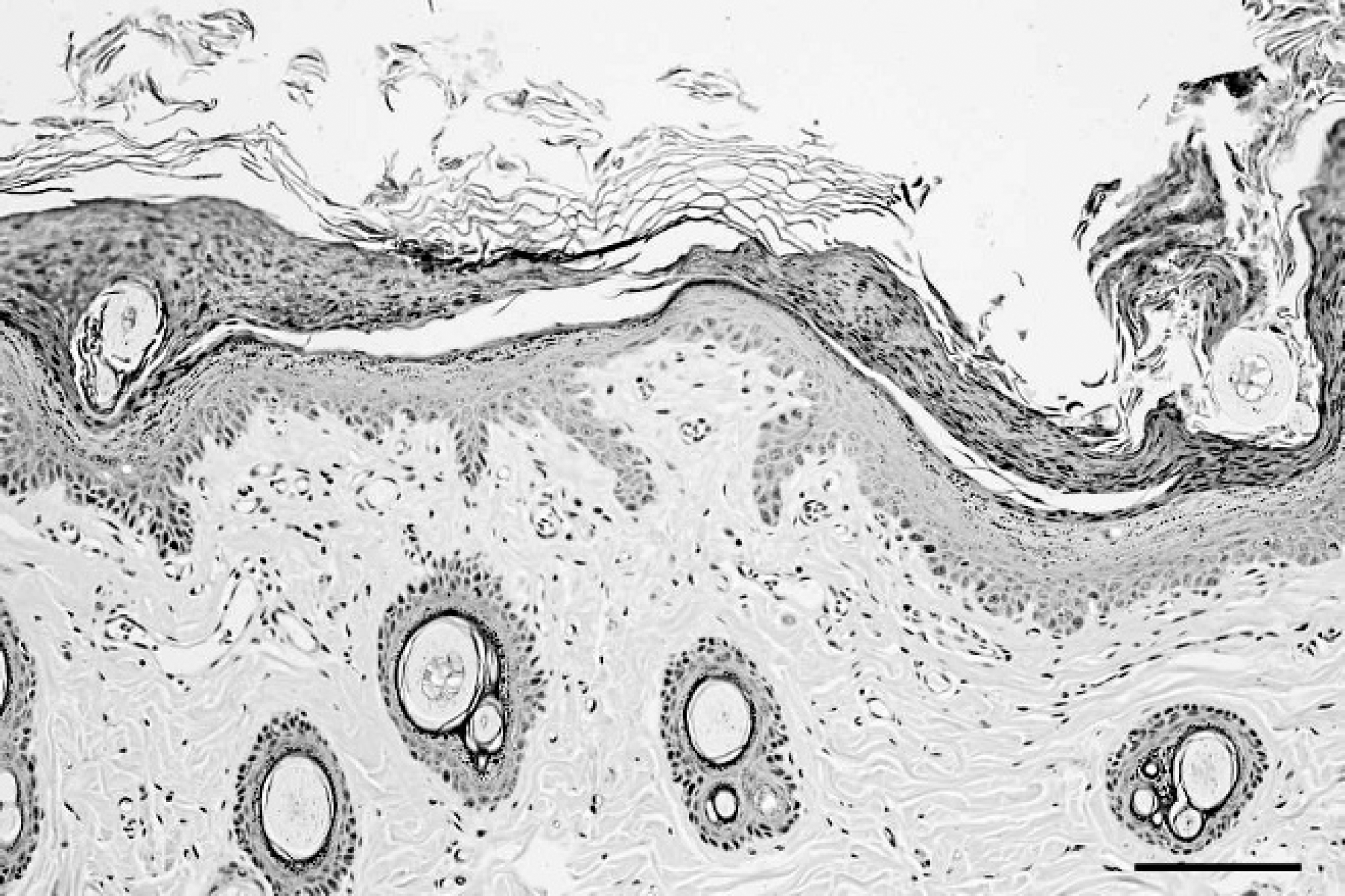
Five punch biopsies were performed on affected areas of skin from one of the male puppies. The fresh samples were immersion-fixed in 10% neutral buffered formalin and routinely processed. Histologically, the epidermis was hyperplastic and disorganized with marked parakeratotic to occasionally orthokeratotic hyperkeratosis (Fig. 2). Individually, keratinized cells were present at various levels of the epidermis, with occasional prominent pallor of keratinocytes within the stratum spinosum. The stratum granulosum was disorganized with variable size and shape of keratohyalin granules (Fig. 3). Marked surface parakeratotic to rarely orthokeratotic hyperkeratosis extended into follicular infundibula. Crusts contained scattered bacterial cocci and bipolar yeasts.
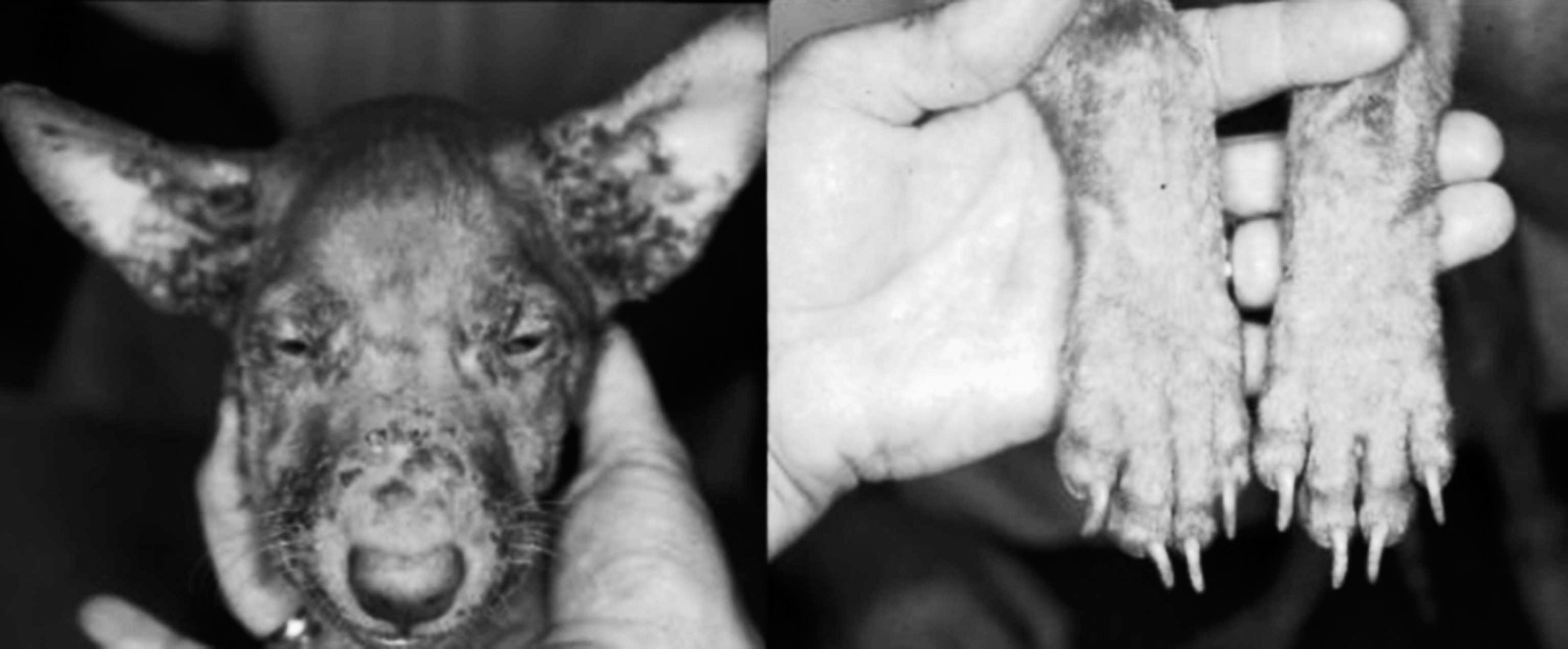
At initial presentation, severe crusting, exfoliative lesions involving face, pinnae, and extremities were observed.
Blood from the 2 affected male puppies was taken, and blood zinc concentrations were measured (<0.1–0.6 ppm, normal: 0.8–2.0 ppm). Skin lesions and constitutional signs partially improved following treatment with zinc gluconate (10 mg kg−1/day orally) and markedly improved after treatment with zinc sulfate (10 mg kg−1 intravenously). Blood zinc concentration normalized following initial intravenous administration then decreased to subnormal levels despite continued oral zinc supplementation.
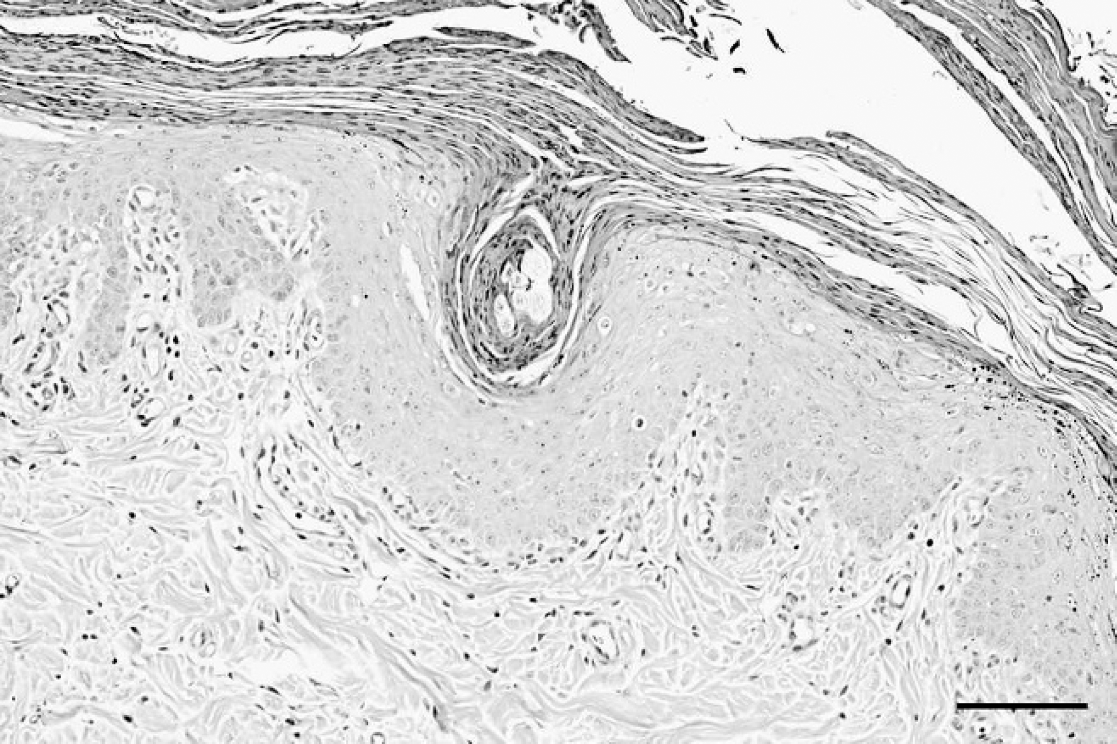
The epidermis appears hyperplastic and disorganized with marked parakeratotic hyperkeratosis. Mild perivascular to diffuse lymphocytic inflammation is present in the superficial dermis. Bar = 100 μm.
One of the male puppies developed an intestinal intussusception following 2 intravenous zinc sulfate treatments and was euthanized. The animal was submitted for complete necropsy examination. On gross examination, there was severe generalized crusting dermatitis that especially involved the head, pinna, and feet (Fig. 4). The animal was small for its age and thin. The thymus and lymph nodes were markedly smaller than expected for the age. There was moderate subaortic stenosis with post-stenotic and left ventricular dilation. The common bile duct was markedly dilated. Moderate internal hydrocephalus was present. Histologically, the epidermis and mucosal epithelium were markedly hyperplastic and moderately dysplastic with marked parakeratotic hyperkeratosis (Fig. 5). Thymus, lymph nodes, and bone marrow were markedly hypoplastic with a marked decrease in lymphocytes (Fig. 6).
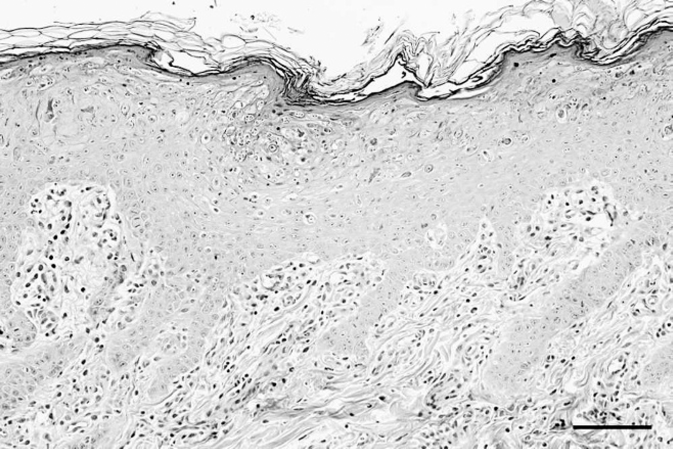
Individually keratinized cells are present at various levels of the epidermis with occasional prominent pallor of keratinocytes within the stratum spinosum. The stratum granulosum is disorganized with variable size and shape of keratohyalin granules. Mild perivascular to diffuse lymphocytic inflammation is present in the superficial dermis. Bar = 100 μm.
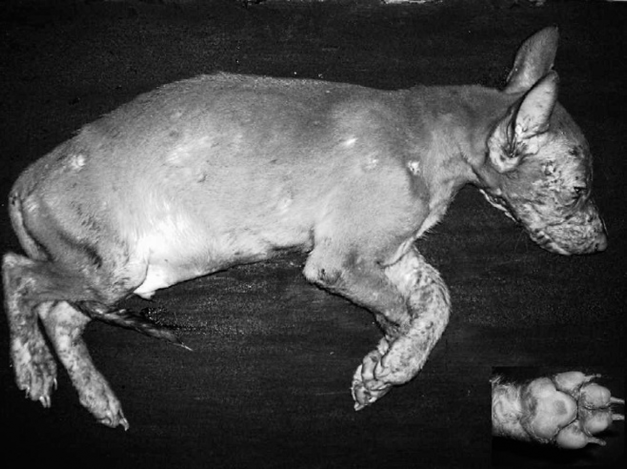
Postmortem presentation. Severe generalized crusting dermatitis is present with the most severe lesions located on face, pinna, distal limbs, and feet (inset).
The remaining 4 puppies in the litter were maintained in the same household. By 6 months of age, marked retardation of growth and severe enamel hypoplasia of erupting permanent dentition were observed in the affected animals compared with unaffected littermates (Fig. 7). At 7 months of age, the affected male was 11.4 kg and 50.8 cm at the withers; the unaffected male was 17.8 kg and 62.5 cm; the affected female was 10.4 kg and 50.8 cm; and the unaffected female was 15.5 kg and 53.4 cm.
Over the follow-up period, the dogs were administered intravenous zinc at regular intervals approximating 3-4 weeks dependent on developing skin lesions. Blood zinc concentrations remained subnormal or in low normal ranges despite therapy. Dermatologic lesions remained at an acceptable level throughout the follow-up period.
Postmortem skin sample. Although marked parakeratotic hyperkeratosis remains, there is a decrease in epidermal thickness and more orderly appearance of epidermis following intravenous zinc administration. Bar = 100 μm.
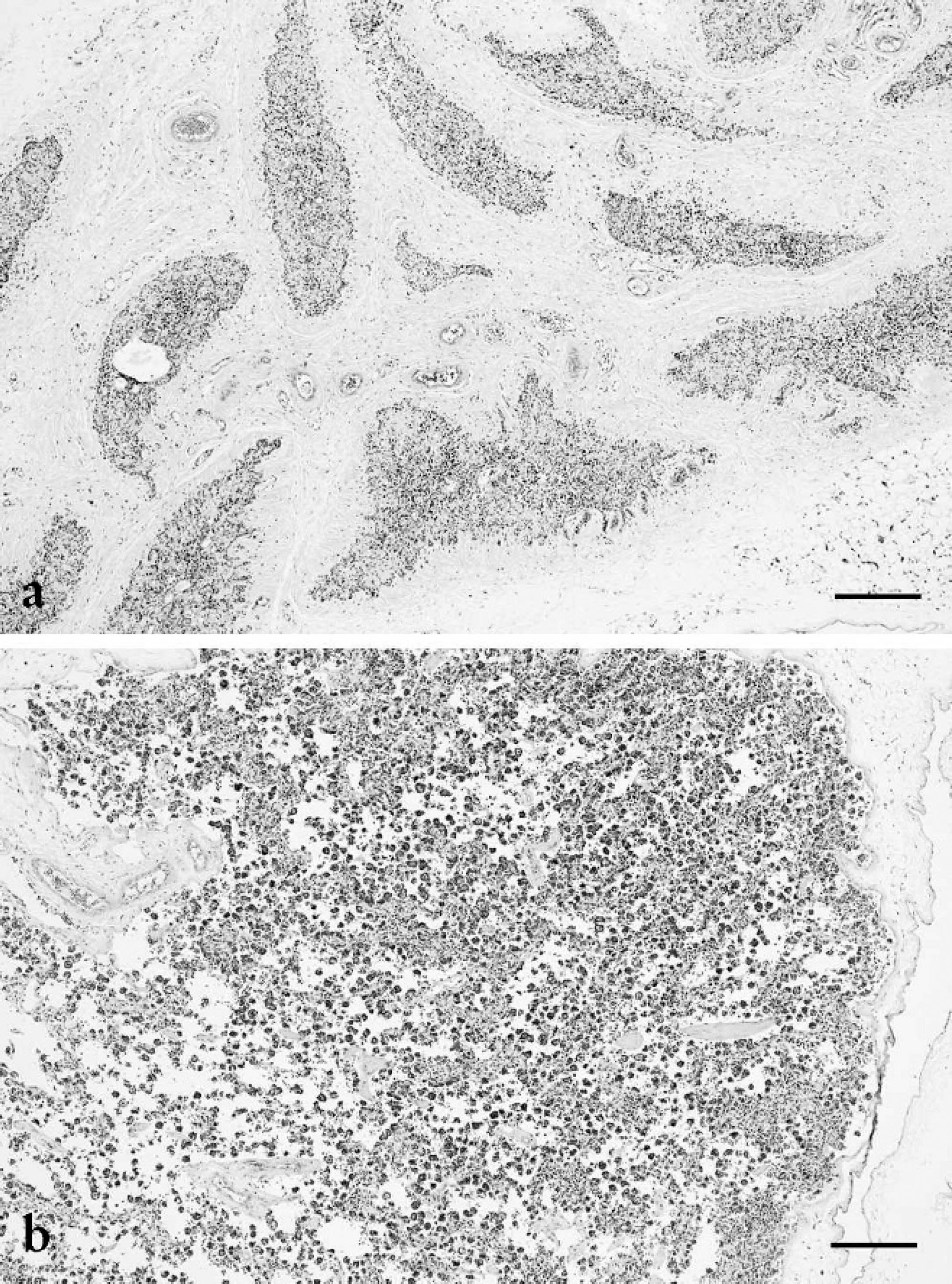
A marked paucity of lymphocytes is present in the (
Approximately 2 months later, while on zinc supplementation, the affected female littermate was presented with 1-week duration of subcutaneous to intradermal nodules in the perivulvar area, caudal inguinal region, and right flank. Routine biopsy was performed, and samples were routinely processed. Histologic findings consisted of deep, multifocal to confluent, suppurative dermatitis, folliculitis, and hidradenitis. Gram-positive cocci were observed in tissue Gram stains within deep suppurative foci, supportive of deep bacterial dermatitis. Histologic lesions associated with the zinc deficiency were much improved. At approximately 36 months of age, the remaining affected male developed renal failure and was euthanized. No postmortem examination was allowed, and the affected female dog was lost to follow-up.
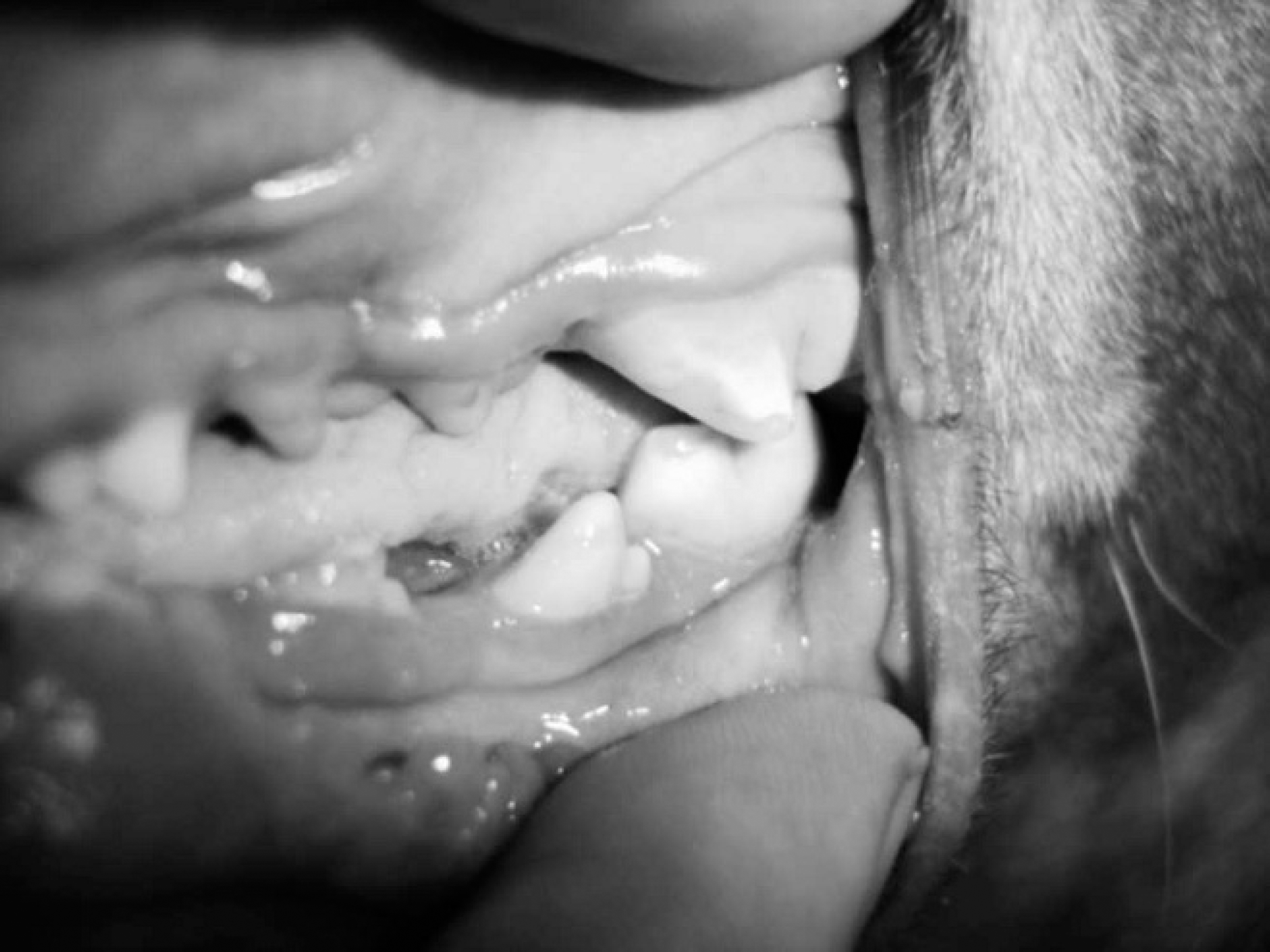
Severe enamel hypoplasia of permanent dentition was observed as affected animals matured.
Zinc-responsive dermatoses fall into the broad category of “nutritional” dermatopathies and are characterized by dietary deficiencies in zinc. Deficiencies in zinc can be the result of decreased concentrations in the diet or limitation of availability as a result of nutritional imbalance, or inherited abnormalities in absorption and/or metabolism, and have been well documented in the veterinary literature. 10 A third zinc-related syndrome, previously described only in Bull Terriers, is acrodermatitis. Acrodermatitis is a rare, inherited, autosomal recessive metabolic disease. This disease typically manifests as young as 6–8 weeks of age in both males and females. Affected Bull Terriers present with progressive crusting dermatitis, growth abnormalities, decreased immune function with chronic diarrhea, and bronchopneumonia 5,7,9 Survival to adulthood is rare.
The presentation, age of onset, profound serum zinc deficiency, gross lesions, and histologic findings in the Pharaoh Hounds presented in the current study are strikingly similar to acrodermatitis of Bull Terriers. In the Pharaoh Hound litter, both sexes were affected with clinical signs first noted at 3 weeks of age. The clinical response to intravenous zinc therapy in the Pharaoh Hounds is in contrast to previously described cases of lethal acrodermatitis in Bull Terriers. However, this response is similar to that reported in humans with acrodermatitis enteropathica. Acrodermatitis enteropathica has recently been shown experimentally to involve zinc transport mechanisms at the apical surface of enterocytes. 1
In conclusion, the present report describes a severe zinc-responsive dermatosis in Pharaoh Hounds that on initial presentation shared similarities with lethal acrodermatitis in Bull Terriers. However, the response of clinical signs to intravenous zinc therapy and long-term follow-up in this case provides evidence that the syndrome in Pharaoh Hounds may be more similar to acrodermatitis enteropathica in human beings.
Acknowledgements
The authors would like to sincerely thank Ms. Rebecca Stephens for allowing the examination and follow-up of this case. The authors are grateful for the support of the Oklahoma Animal Disease Diagnostic Laboratory.