
Abstract
Six cases of acquired duodenal diverticulitis (pseudodiverticula) were found in a flock of sheep over a short period of time. All the animals had duodenal lesions characterized by the presence of multiple saccular dilations filled with feed material. The mucosal surface was elevated by multiple small nodules that histologically corresponded to cystic dilatations of the duodenal glands, which likely caused the displacement, atrophy, and disappearance of the muscular layer, leading to pseudodiverticula. The gross appearance, microscopic findings, and epidemiological characteristics suggest that this is a different pathological process to that described for diverticula in animals to date.
Diverticula are epithelium-lined cavities of the intestine that are derived from the mucosal epithelium and extend through the muscularis mucosa, submucosa, and muscularis, and often reach the serosa, where they sometimes rupture, causing peritonitis. 2 Two types of intestinal diverticula have been described in the literature: congenital and acquired. Congenital diverticula, which resemble intestine in having all layers of the bowel in cross-section (mucosa, submucosa, and muscularis), are very infrequent (e.g., Meckel's diverticulum). Congenital diverticula are acquired and do not have muscular layer, or it is very attenuated. This last type of diverticula, characterized by the herniation of the mucosa and submucosa through the muscularis layer, is referred to as “pseudodiverticula.” 5
Diverticula have been well described in humans, where this condition is known as colonic diverticular disease and affects approximately 50% of individuals over 60 years of age in Western countries. 5 In humans, acquired diverticula are most frequently located in the left side of the colon, especially the sigmoid colon. 5 However, they can also occur in the esophagus, stomach, or duodenum. In animals, diverticulosis is a rare condition and has been reported only in horses, pigs, and sheep. 1 A cluster of 6 cases of acquired duodenal diverticula, or pseudodiverticula, in a flock of sheep is described in the current study.
The sheep belonged to a flock from Valencian Community (eastern Spain), which included 200 crossbred ewes that grazed extensively on natural pastures. The flock was not regularly vaccinated or dewormed, and the general hygienic conditions on the farm were poor (such as dirty floors and the presence of rats in the sheds where the animals were housed at night). All 6 animals were culled from the flock over a 3-month period because of their age (more than 6 years) and donated for educational purposes to the authors' university. The animals were euthanized by an intravenous injection of barbiturate, a and complete necropsies were performed immediately after death.
The carcasses were in poor nutritional condition, with inadequate fat reserves. Gross lesions were similar in all 6 animals and consisted of 0.1-8 cm in diameter, multiple, saccular dilations (diverticula), in the first 15 cm of the duodenum (Fig. 1). These diverticula were located in both the mesenteric and antimesenteric borders, and contained a moderate amount of foul-smelling feed material in their lumens. Interestingly, the mucosal surface between the diverticula was elevated by multiple, small, 0.2-0.5 cm in diameter nodules filled with mucus (Fig. 2). In addition, the animals had other gross lesions. Sheep no. 1 had a chronic, segmental thickening of the ileum and enlarged mesenteric lymph nodes with mesenteric lymphangitis and lymphangiectasia. The lungs of sheep nos. 2, 5, and 6 failed to collapse when the thorax was opened; were increased in size; were rubbery, heavy, and pale; and had costal imprints on the pleural surface. The tracheobronchial lymph nodes also were enlarged. Sheep nos. 3 and 5 had severe caseous lymphadenitis in the mediastinal, mesenteric, and periportal lymph nodes. Lastly, all 6 sheep had numerous parasitic lesions in the esophagus, lungs, liver, and abomasums, although these changes were more severe in sheep no. 4. Samples of gastrointestinal tract (abomasum, duodenum, and ileum), lungs, and lymph nodes were routinely processed and stained with hematoxylin and eosin, periodic acid-Schiff, Ziehl-Neelsen, and Masson trichrome stains for histological examination.
Microscopically, the duodenal lesions were characterized by the presence of a diffuse lymphoplasmacytic infiltrate in the lamina propria, hyperplasia of the goblet cells in the crypts of Lieberkühn, and moderate to severe dilatation of the duodenal glands (Fig. 3). This dilatation was associated with the displacement and atrophy of the muscular layer and the formation of diverticula with no muscular layer (pseudodiverticula; Fig. 4).
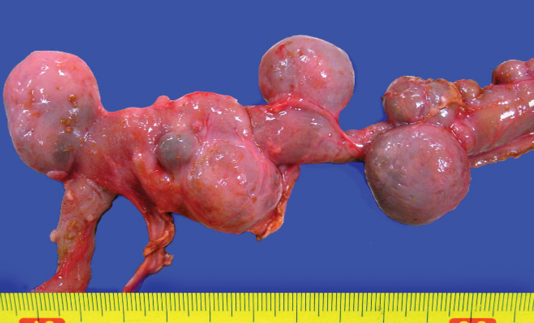
Multiple differently sized diverticula in the mesenteric and antimesenteric borders.
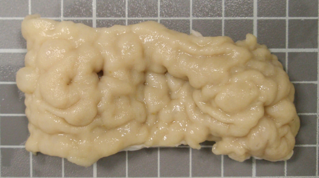
Mucosal surface elevated by the presence of multiple small nodules. Formalin-fixed tissue.
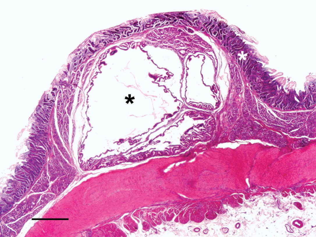
Photomicrograph of a nodule from Figure 2 where a severe dilatation of the duodenal glands (black asterisk) caused the displacement of the mucosa (white asterisk). Hematoxylin and eosin stain. Bar = 500 μm.
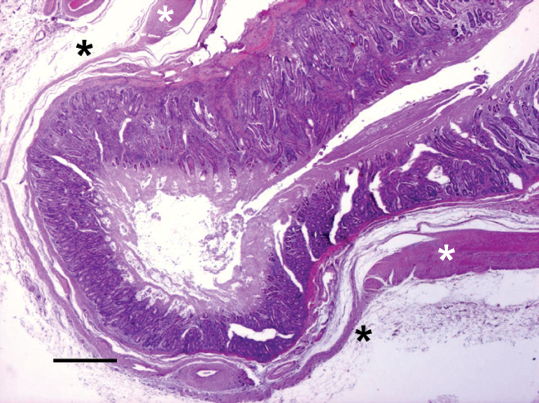
Photomicrograph of a small pseudodiverticulum with no muscular layer (white asterisk = muscular layer; black asterisk = no muscular layer, only serosa). Hematoxylin and eosin stain. Bar = 500 μm.
Aerobic and anaerobic bacterial cultures of samples of the diverticular content of 2 sheep yielded
Diverticula affect around 10% of people in Western countries, 7 and they have been documented in horses, pigs, and sheep. 4 To date, no epidemiological studies have been published on animal diverticulosis. The 6 cases reported herein occurred over a period of 3 months in the same flock of sheep. This would appear to be the first description of this lesion in a group of sheep from the same flock where a histopathological investigation of the intestinal lesions was performed.
The sheep in the current study had no clinical signs associated with diverticulosis. This also seems to be the case in humans, in which diverticula have remained clinically undetected in most cases. 7 Clinical signs are evident only when the diverticulum perforates and causes peritonitis. 1
In humans, diverticula occur most commonly in the colon 5 ; in horses and pigs, the most frequent location is the duodenum and the terminal ileum, respectively. 4 In sheep, diverticula have been described in both the duodenum and the colon. 1 Diverticula tend to follow the pathway of blood vessels and are mainly located close to the mesenteric attachment, probably starting at the point where mesenteric blood vessels penetrate the intestinal musculature. 1 In the cases presented in the current study, the diverticula were present in both the mesenteric and antimesenteric borders of the duodenum. The reason for this distribution is unknown but suggests that the development of diverticula in the sheep may not necessarily follow the vascular pathway.
Diverticula in horses and pigs have been generally characterized by hypertrophy 2 and hyperplasia 1 of the tunica muscularis. Muscular hyperplasia in the small intestine of horses and pigs may be a compensatory mechanism when imperfect development of the tunica muscularis occurs in young animals. Diverticula are probably the result of a hernia of the mucous membrane through larger than normal interruptions in the continuity of the hyperplastic muscular layers. 1 This hypothesis has also been proposed in sheep where gaps in the tunica muscularis could constitute a congenital defect. 3 However, no defects in the tunica muscularis were observed in any of the sheep in the present study. Other reports of duodenal diverticula in elk have indicated that there is a relationship between these lesions and biliary tract disease. 6 No biliary tract lesions were observed in the 6 sheep of the present study.
Because the cases described in the current study appear to differ epidemiologically (6 animals with pseudodiverticula from the same flock in a short lapse of time) and pathologically (pseudodiverticula were observed in mesenteric and antimesenteric borders, and evidence of gaps in the muscularis layer were not detected) from previously described diverticula in sheep, it is likely that their pathogenesis differs from that of previously reported cases. The findings suggest that the origin of the diverticula reported in the present study could be the dilatation of the duodenal glands leading to the displacement, atrophy, and, finally, the disappearance of the muscular layer, causing pseudodiverticula through an accumulation of feed material. The cause of this dilatation of the glands is unknown. The hypersecretion of mucus caused by the hyperplasia of the goblet cells in the crypts of Lieberkühn could contribute to the obstruction of the duodenal glands and to the resulting dilatation. It has been reported that human patients with gastric or duodenal ulcers have mild goblet cell hyperplasia in the duodenum. 8 However, only 1 sheep out of 6 in the current study had abomasal ulcers. The observed inflammation in the lamina propria could be related to the accumulation of feed material in the diverticula 7 and could contribute to early obstruction. In conclusion, the gross and microscopic findings and the epidemiological characteristics of this cluster of cases of pseudodiverticula suggest that this is a different pathological process from cases previously described.
Acknowledgements
This study was supported by Copernicus Program (CEU-UCH and Banco de Santander).
Footnotes
a.
Dolethal®, Vétoquinol SA, Lure, France.