
Abstract
An 18-year-old Arabian stallion was presented for recent onset of stranguria. Physical examination of the distal portion of the glans penis revealed multiple, smooth, glistening, grayish-pink, variably sized, exophytic, nodular masses circumferentially surrounding the external urethral orifice. Partial penile amputation was performed, and the entire specimen was submitted for histological evaluation. Microscopically, the masses consisted of abundant amounts of loosely arranged fibrovascular stroma with low numbers of spindloid to stellate fibrocytes. The overlying epithelium was mildly to moderately hyperplastic with short anastomosing rete ridges (pseudoepitheliomatous hyperplasia). The lesion was diagnosed as fibropapilloma because of features similar to bovine penile fibropapilloma including anatomical location, gross appearance, and histological characteristics. A sarcoid was considered but negated as the lesion lacked the classical streaming and interlacing spindle cell population, “picket-fence” appearance at the epithelial interface, and long, thin, dissecting rete ridges typical of most equine sarcoids. Polymerase chain reaction for the Bovine papillomavirus-1 and Bovine papillomavirus-2 E5 gene and for Equine herpesvirus 1, 3, and 4 was negative on formalin-fixed tissue specimens.
An 18-year-old Arabian stallion was presented to the Colorado State University Veterinary Teaching Hospital (Ft. Collins, CO) for recent onset of stranguria and numerous masses of unknown duration associated with the glans penis. The horse had been pastured in California for several years but was transported to Colorado just before examination. Physical examination of the distal portion of the glans penis revealed multiple, smooth, glistening, grayish-pink, 0.5–2.0-cm-diameter, exophytic, nodular masses circumferentially surrounding the external urethral orifice (Fig. 1). Partial penile amputation was performed, and the entire specimen was submitted for histological evaluation (Fig. 2a, 2b). Castration was also performed at this time. Microscopically, the penile masses consisted of an abundant amount of loosely arranged fibrovascular stroma with low to moderate numbers of spindloid to stellate fibrocytes admixed with low numbers of lymphocytes and fewer plasma cells (Fig. 3a, Fig. 3b). The overlying epithelium was mildly to moderately hyperplastic with short anastomosing rete ridges (pseudoepithelioma-tous hyperplasia) consisting of well-differentiated and well-organized epithelial cells that demonstrated orderly maturation and lacked any evidence of dysplasia (Fig. 3b, Fig. 3c). Mitotic figures were occasionally noted associated with the basal epithelial cell layer. No viral inclusions were identified.
The lesion was diagnosed as a fibropapilloma. A sarcoid was considered but negated as the lesion lacked the classical streaming and interlacing spindle cell population, “picket-fence” appearance at the epithelial interface, and long, thin, dissecting rete ridges typical of most equine sarcoids. Differential diagnoses for proliferative lesions of the equine penis include squamous cell carcinoma (SCC), squamous papilloma, habronemiasis, and chronic balanitis. 5,6 Squamous cell carcinoma, habronemiasis, and balanitis are often ulcerative, and all 4 conditions may additionally involve the prepuce (i.e., balanoposthitis). In the current case, the proliferative nodules were nonulcerative, were limited to the distal portion of the glans penis without preputial involvement, and microscopically were not consistent with any of the above-mentioned differentials. Additionally, no debris, dirt, calculi, or inspissated smegma was located within the fossa glandis upon examination of the amputated glans penis nor during clinical physical examination, thus suggesting chronic balanitis was less likely to have caused the proliferative, exophytic masses. Previous or chronic trauma with subsequent granulation tissue was also considered; however, no known trauma was reported. Additionally, although the horse presented as a stallion, he had not been bred for more than 2 years, and microscopically, the lesion lacked the features consistent with granulation tissue (proud flesh). 6
Location, gross appearance, and microscopic features resembled bovine penile fibropapilloma, which is caused by Bovine papillomavirus-1 (BPV-1). 5,7 Bovine papillomaviruses are divided into 6 subtypes (1–6) and 2 groups, A and B. Viruses in group A are known to transform fibroblasts and epithelial cells and include BPV-1 and −2. 1 Fibropapillomas are benign masses composed predominately of fibroblast proliferation with variable overlying epithelial hyperplasia and hyperkeratosis. In horses, sarcoid is typically the term used for fibropapilloma because of the sarcoma-like proliferation of dermal fibroblasts. 5 Similar to bovine penile fibropapillomas, it has been demonstrated that equine sarcoids are also papillomavirus-induced, more specifically caused by both BPV-1 and −2. 15 Because of the similarities between the lesion in the present case and the penile fibropapilloma of the bull, including anatomical location, gross appearance, and histological features, and the fact that this lesion did not resemble a sarcoma (i.e., was not sarcoid in nature), fibropapilloma was deemed the appropriate diagnosis.
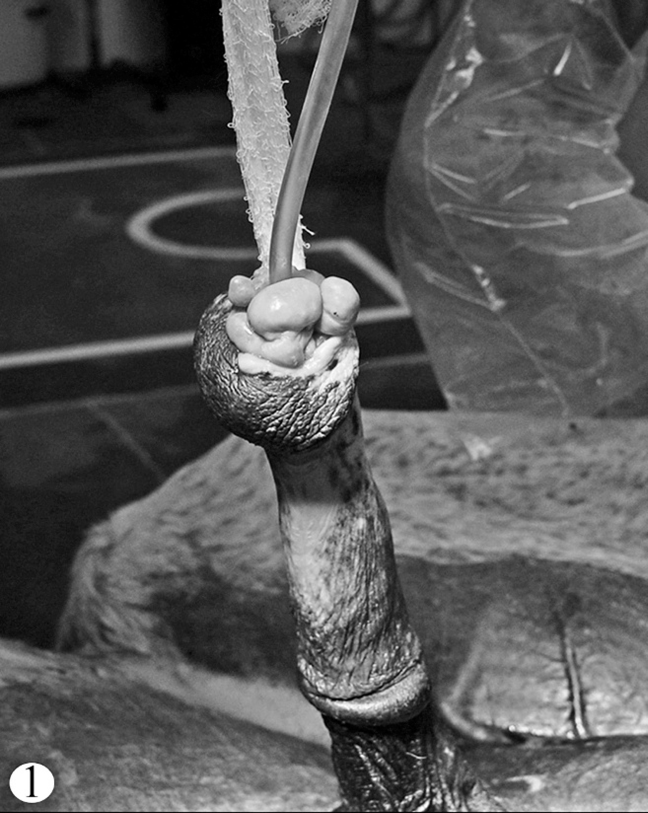
Digital image of the penis at the time of surgical preparation. The urethra is catheterized with plastic tubing, and the penis is tethered by white gauze. Note the numerous smooth, glistening, grayish-pink, variably sized, exophytic nodules circumferentially surrounding the external urethral orifice.
Sarcoids have been clinically categorized into several groups including verrucous, fibroblastic, mixed verrucous and fibroblastic, occult, nodular, and malevolent. 8,12 None of these subtypes is known to occur with any frequency on the penis. Histologically, several minor differences exist between groups but all groups share the dense fibroblast proliferation, a feature lacking in the penile lesion in the present case. Previous work has detected BPV DNA in sarcoids of zebras and DNA sequences similar to BPV in lesions analogous to equine sarcoids of domestic cats, a mountain lion, and a red-tailed deer. 11,14,16,21 Papillomavirus DNA has also been detected in mucocutaneous fibropapillomas in camelids. 17
In an attempt to identify a possible underlying etiology and further characterize the pathogenesis of the presented lesion, polymerase chain reaction (PCR) analysis to detect the highly conserved E5 gene of BPV-1 and −2 was performed on multiple sections of formalin-fixed, paraffin-embedded (FFPE) tissues from the penile masses as previously described. 11 Respective DNA amplification was not observed in any of the tested tissue. To more thoroughly investigate the potential involvement of other papillomaviruses, PCR for degenerate papillomavirus (degenerate PV-PCR) was additionally pursued on multiple FFPE sections as previously described. 11 Results, again, yielded no evidence of papillomaviral DNA. Because of the association of herpesvirus and fibropapillomas in sea turtles as well as herpesvirus and mucosal papillomas in psittacines, and as the cause of the lesion in the present case had yet to be determined, equine herpesvirus involvement was considered and investigated using PCR for Equine herpesvirus 1, 3, and 4 (EHV-1, −3, and −4) on formalin-fixed tissue. 4,9,19,20 Equine herpesvirus 1 and 4 are known to cause respiratory illness (rhinopneumonitis), occasional neurological disease, and abortion. 18 Equine herpesvirus 3 is the causative agent of equine coital exanthema, a venereal disease that can induce lesions in both mares and stallions. 6
Briefly, PCR testing for EHV-1, −3, and −4 was performed from deparaffinized, slide-mounted, FFPE tissues using 2 xylene washes followed by a series of 3 graded ethanol washes and 2 distilled water washes. The tissue was rehydrated in Tris-ethylenediamine tetra-acetic acid buffer before performing the DNA isolation technique. 2 DNA isolation from the rehydrated tissue was performed using the QIAamp DNA Mini Kit a with a prolonged incubation at 56°C and proteinase K digestion of 1-hr duration. 2,10 The PCR reactions were performed using High Fidelity PCR Master b on a Mastercycler EP thermal cycler c with primer sequences previously described. 3,22 Respective DNA amplification was not observed; however, formalin fixation of tissue may inhibit PCR.
Formalin fixation of tissue is known to cause cross-linking and degradation of nucleic acids through chemical modification. Isolation of DNA from FFPE tissues, however, can be achieved reliably by modification of the DNA isolation technique to include tissue digestion with proteinase K and prolonged incubation between 50°C and 60°C in a formalin-free buffer as was performed in the current case. 2,10,13 Additionally, amplification of DNA products less than 200 base pairs (bp) in length significantly increases the success of PCR reactions performed on DNA isolated from FFPE tissues. 10,13 The PCR assay for EHV-1 results in a 135-bp amplicon, thus reducing the potential for false-negative results on FFPE tissues. The amplicons in the EHV-3 and −4 assays are 520 and 326 bp, respectively, resulting in end products larger than typically recommended for PCR testing on FFPE tissues. In consideration of these factors, the authors recognize that PCR using fresh tissue is optimal (although no fresh tissue was available) and that PCR from FFPE tissues may result in false negatives.
In the horse, it is generally accepted that sarcoids have the potential to occur anywhere on the body; however, neither sarcoids nor fibropapillomas of the equine glans penis have been reported. Differential diagnoses including squamous papilloma, SCC, habronemiasis, balanitis, and trauma-induced granulation tissue were considered but ruled out because of the lack of appropriate microscopic features, respectively, and the absence of any history of trauma. Although this lesion was observed in a horse, a final diagnosis of fibropapilloma was made due to the anatomical location and marked similarities, both grossly and microscopically, with bovine penile fibropapilloma as compared with the typical microscopic features of equine sarcoids as previously discussed. Based on PCR results, the authors were unable to demonstrate the presence of papillomaviruses, especially BPV-1 and −2, or the herpesviruses EHV-1, −3, or −4. Thus, a definitive etiology remains undetermined. Further investigation may help to elucidate the pathogenesis of this unique lesion, which may involve a so far undetected equine or other papillomavirus or possibly an undetected herpesvirus. As of 11 months postoperatively, no masses have recurred, and the horse is reported to urinate without difficulty.

Approximately median sagittal section of the amputated formalin-fixed glans penis demonstrating the exophytic nodules surrounding the urethral orifice:

Photomicrograph of a single exophytic nodule on a narrow stalk;
Acknowledgements. The authors would like to thank Dr. Laurie Goodrich from the Colorado State University, College of Veterinary Medicine and Biomedical Sciences, Department of Clinical Sciences, for submitting this case and for providing the gross images. The authors are additionally grateful for the excellent technical assistance provided by Gabriele Czerwinski.
Footnotes
a.
Qiagen Inc., Valencia, CA.
b.
Roche Diagnostics Corp., Indianapolis, IN.
c.
Eppendorf AG, Hamburg, Germany.