
Abstract
An immunochromatographic strip for discriminating Foot-and-mouth disease virus (FMDV) infected from vaccinated pigs was developed based on synthetic peptide. Five peptides designed from the amino acid sequences of nonstructural proteins (NSP) of FMDV were synthesized, and pep5 located in NSP 3B reacted strongly with serum from FMDV-infected pigs but did not react with serum samples from healthy vaccinated pigs. An immunochromatographic strip was developed by using colloidal gold labeled with pep5 as the detector. Staphylococcal protein A and rabbit against peptide-conjugated ovalbumin antibody immunoglobulin G were blotted on the nitrocellulose membrane for the test and control lines. In comparison with 2 commercial NSP enzyme-linked immunosorbent assays, the peptide-based strip showed good specificity and sensitivity. The apparent agreements of this new assay with Ceditest® ELISA and UBI® ELISA were 98.59% and 96.63%, respectively. These results indicate that the strip can be adequately used to discriminate FMDV-infected animals from vaccinated animals.
Foot-and-mouth disease (FMD) is a highly contagious and important viral disease of cloven-hoofed animals, which causes high productivity losses. 1,6 As control measures of the disease, extensive culling has been performed in those countries that have been free of the disease, whereas vaccination and movement restriction has generally been adopted in regions of endemic disease. 7 In such endemic areas, it is important to identify animals, whether vaccinated or not, in which replication of Foot-and-mouth disease virus (FMDV; order Picornavirales, family Picornaviridae, genus Aphthovirus) has taken place to eliminate potentially infective animals. 2,3,11 Recently, a number of in-house and commercial tests to identify infection within vaccinated livestock (i.e., the presence of viral carrier animals) were developed and evaluated. 4,10,12 These methods are theoretically based on the assumption that semi-purified, inactivated FMDV vaccines mainly consist of capsid (structural) proteins, and so they are less likely to elicit production of antibodies against nonstructural proteins (NSP). An antibody response against NSPs is only related to in vivo virus activity. 3,8
However, the NSPs used in these test procedures were primarily produced in expression systems by using either bacteria or baculovirus, and nonspecific reactions can be caused by the presence of antibodies in test animals against these expression vector antigens. 14,15 More recently, synthetic peptides that contained B-cell epitopes of FMDV NSP have been used. It is reported that some peptides located in 2C, 2B, and 3B can be used to distinguish FMDV-infected from vaccinated animals, and enzyme-linked immunosorbent assay (ELISA) methods based on these peptides were successfully developed. 5,9,13 Although these assays provide accurate and sensitive results, they require specialized equipment and technical expertise. For this reason, the authors have developed a simple and rapid assay (immunochromatographic test strip) based on these peptides for the detection of anti-FMDV NSP antibodies and compared the new assay with the available diagnostic peptide ELISAs.
Five peptides based on the sequence of FMDV O/Tibet/CHA/99 (GenBank accession no. CAD62370) were designed according to previous reports 5,9,13 and synthesized (Table 1) by using an automated peptide synthesizer. a The purity of peptides analyzed by C18 high-performance liquid chromatography column was maintained at >90%. They were individually conjugated to ovalbumin (OVA). b The conjugates were made up of 1 mg/ml of distilled water and stored at −20°C. The screening of peptides was performed by using 1 vaccinated, 3 infected (antibody titers: ≥1:128 determined by liquid-phase blocking ELISA c ) and 1 negative serum sample by indirect ELISA. The result showed that all peptides reacted to varying degrees with 3 positive sera and did not react with either vaccinated or negative animal sera. In particular, the pep5 provided a higher optical density value than other four peptides under the same conditions (Fig. 1). Thus, the pep5 (3B2) was selected as a candidate antigen and used to develop the immunochromatographic test strip.
Synthetic peptides sequence of Foot-and-mouth disease virus serotype O.

Amino acid number on polyprotein.
The strip was prepared according to a previously described method. 16 Briefly, 150-μl peptide solution (1 mg/ml) was incubated with 10 ml of colloidal gold solution (pH 9.0) for 30 min at room temperature. After the addition of 1 ml of 10% bovine serum albumin b (BSA; immunoglobulin G [IgG] free) solution in 20 mmol/l sodium borate (pH 9.0), the mixture was incubated at room temperature for another 10 min, and the labeled antigen was washed by repeated centrifugation (14,000 x g) at 10°C for 30 min with 20 mmol/l sodium borate (pH 9.0) that contained 1% BSA and 0.1% sodium azide. The precipitate was resuspended with 1 ml of washing buffer to a final concentration of 0.15 mg/ml. Then, conjugate solution was prepared by dilution of the colloidal gold—labeled antigen with 20 mmol/l sodium borate buffer (pH 8.0) that contained 8.75% (w/v) sucrose, 8.75% (w/v) BSA, 0.6 mol/l NaCl, 10 mmol/l ethylenediamine tetra-acetic acid, and 0.1% (w/v) NaN3 to a final concentration of 0.1 mg/ml, and dispensed onto the fiber-glass d at the speed of 7.5 μl/cm (about 0.3 μg per pad) by using a dispensing platform e to produce the conjugated pad. The staphylococcal protein A (1.0 mg/ml) b and rabbit anti-OVA-peptide antibody IgG (1 mg/ml, method of preparation not shown) were dispensed e onto cellulose ester membrane d as the test and control lines, respectively. Then, the sample pad, conjugate pad, blotted membrane, and absorbent pad were assembled sequentially on the plastic backing support board with a 1–2-mm overlap. The master card was cut e to 4.080-mm wide strips. Each assembly was housed in a plastic case that then was sealed in a plastic bag in the presence of desiccant gel and stored at 4°C.
To use the immunochromatographic test strip, a serum sample diluted 1:20 with normal saline solution was applied to the sample pad. With a positive sample, the antibody binds to the antigen conjugate forming a gold-antigen-antibody complex, which binds to staphylococcal protein A and gives a red-colored band at the test-line region. The absence of this band suggests a negative result. To serve as a procedural control, a red band at the control-line region always appears regardless of the presence of anti-FMDV antibodies.
The specificity of the test strip was evaluated with standard negative serum samples from 50 naive pigs, 10 serum samples from pigs positive for a variety of pathogens (Porcine reproductive and respiratory syndrome virus, Classical swine fever virus, Bluetongue virus, Porcine circovirus-2), 20 FMDV-infected pig serum samples (which were screened by a commercial liquid-phase blocking ELISA c ), and sera from 168 pigs immunized with FMDV vaccine (identified positive for FMDV structural antibodies by FMDV viral protein 1 ELISA f ). Approximately 100 μl of each diluted sample was added to the sample chamber and left to stand for 5 min. A result was considered positive when red-purple bands appeared at both the test line and the control line. A result was considered negative when a red-purple band only appeared at the control line. The result showed that all of naive pig sera, immunized sera, and the serum samples positive for other non-FMDV pathogens were found negative for anti-FMDV NSP antibodies with the test strip. This indicated that the specificity of the strip was 100%. Nineteen of the 20 FMDV-infected serum samples were positive with the test strip; therefore, the sensitivity of this strip was 95% (19/20).
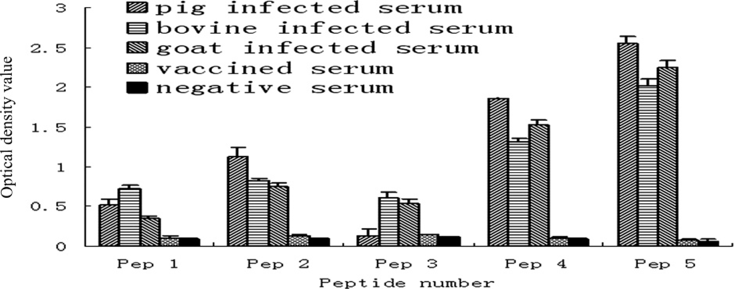
Identification of Foot-and-mouth disease virus (FMDV)-specific synthetic peptide. Pig, bovine, and goat positive sera taken from animals infected with FMDV serotype O. Vaccinated serum obtained from cattle vaccinated with FMDV serotype O inactivated vaccine. Negative serum: pig serum from a herd in Henan Province (China) with neither FMDV-infected nor FMDV-immunized pigs. The status of all the sera was confirmed by testing with a commercial liquid phase blocking enzyme-linked immunosorbent assay. All tests were performed in duplicate.

Then, the immunochromatographic test strip was used with 356 field pig sera collected from several FMDV-free herds. They were also tested by 2 commercially available NSP ELISAs (Ceditest® f and UBI® g ). The ELISAs were performed following the manufacturer's instructions. The immunochromatographic test strip and the 2 commercial ELISA kits provided similar results (Table 2). The agreement of the immunochromatographic test strip with the Ceditest ELISA and UBI ELISA for negative samples was 98.59% (351/356) and 96.63% (344/356), respectively. This indicated that the 3 test methods showed good correspondence.
In the prevention and control of FMDV, it is important to be able to determine whether animals have been infected with FMDV or vaccinated, and, most importantly, to be able to detect infected animals among those vaccinated. The ELISAs for detection of NSP antibodies have already proven to be very useful. They can be used to survey large numbers of sera to estimate the level of subclinical viral infection and to detect early incursion of FMDV into an animal population, regardless of serotype of the virus involved. 3,8,15 But ELISAs require specialized equipment and technical expertise, thus suspect samples must be transported to a laboratory for testing. The availability of a specific, simple, inexpensive, and disposable test that would be capable of providing results within minutes of taking a clinical sample and that could be used on suspect premises by a veterinarian would help to mitigate the spread of any infection. The immunochromatographic test strip described herein would meet this requirement.
It was known that, in contrast to the structural proteins, the amino acid sequences of FMDV NSPs are highly conserved, irrespective of different subtypes and serotypes. Because the new test strip based on the 3B peptide has been performed only with sera from animals infected with FMDV serotype O, some substitutions may affect the reactivity of peptides against sera from animals infected by other viral strains. Thus, further experiments are needed to investigate the relationship between the amino acid sequence substitution of synthetic peptide and its reactivity. Moreover, the identification of synthetic peptides and its diagnostic application has been done mainly with sera from pigs. Further experiments will help elucidate the exact reactivity of sera from other species with the 3B peptide-based immunochromatographic test strip.
Acknowledgements The study was supported by the Chinese National High Technology Research, Development Program (grant 2007AA100606), National Natural Science Foundation of China (30730068), and Major State Basic Research Development Program (2005CB523200). The authors thank Dr. Norman A. Gregson for editorial assistance. Suzhen Yang and Jifei Yang both contributed equally to this work.
Footnotes
a.
Protein Technologies Inc., Tucson, AZ.
b.
Sigma Chemical Co., St. Louis, MO.
c.
National FMDV Reference Laboratory, Lanzhou, China.
d.
HiFlow Plus Cellulose Ester Membrane, Millipore, Billerica, MA.
e.
XYZ-3000, Quanti 3000 Biojets, XYZ Biostrip Dispenser, CM-4000 Cutter; Bio-Dot, Irvine, CA.
f.
UBI® FMDV VP1 ELISA kit, UBI® NSP ELISA; United Biochemical Inc., Sanborn, NY.
g.
Cedi® FMDV-NS ELISA, Cedi-Diagnostics B.V., Lelystad, The Netherlands.