
Abstract
The antemortem biopsy records (5 cases) and necropsy records (34 cases) were reviewed from 39 kangaroo case submissions during a 14-year period to the Oklahoma State University and the Oklahoma Animal Disease Diagnostic Laboratory. The most common types of diseases in the kangaroos, based on necropsy examination, was disease of the alimentary tract (12 cases), pneumonia (6 cases), and disseminated toxoplasmosis (5 cases). The cause of disease was not determined in 6 case submissions. Based on histopathologic findings, clostridial enteritis/colitis was suspected in 2 kangaroos. Coccidian parasites were identified within histologic sections of the small intestine in 1 kangaroo. Pasteurella sp. (1 case) and Bacteroides sp. (1 case) were isolated from the lung in 2 cases of pneumonia. Most (77.3%) of disease in this study in the kangaroos with known ages occurred in animals older than 1 year. Two neoplasms were detected in the antemortem biopsy samples from 1 case.
Reports of natural disease in various species of kangaroos are limited in the veterinary literature, and retrospective studies regarding diseases of kangaroos are uncommon. 1,2,10,12 One retrospective study 1 reviewed hepatobiliary lesions in Australian macropods, and another retrospective study 12 was a survey of neoplastic disease in a zoologic collection of red kangaroos (Macropus rufus). Improving the health of kangaroos in captivity is the primary concern of veterinarians, owners, and caretakers of these animals.
The purpose of this report is to describe the causes of disease in captive kangaroos detected following necropsy examination and evaluation of antemortem biopsy samples examined by the Department of Pathobiology at the Oklahoma State University and the Oklahoma Animal Disease Diagnostic Laboratory (OADDL) during a 14-year period.
From January 1995 to July 2009, there were 39 case submissions from kangaroos for biopsy (5 cases) or necropsy (34 cases) examination. Clinicopathologic records were reviewed for all cases. For all cases, tissues were fixed in 10% neutral buffered formalin, embedded in paraffin, sectioned at 4–5 μm, and stained with hematoxylin and eosin. Gram, Grocott methenamine silver, and Ziehl–Neelsen acid-fast staining procedures were used in selected cases to detect bacteria, fungal organisms, and acid-fast bacteria in tissues, respectively. Bacterial cultures were performed on select cases at the discretion of the attending pathologist. Paraffin embedded sections from cases suspicious for protozoal disease were stained with anti–Toxoplasma gondii serum (caprine polyclonal antibodies, Veterinary Medical Research and Development, Pullman, WA) and anti–Neospora caninum serum (caprine polyclonal antibodies, Veterinary Medical Research and Development, Pullman, WA) by the avidin–biotin–peroxidase complex method.
Information regarding the age, species, and sex of individual animals was derived from the pathology records. The kangaroos examined ranged in age from 13 weeks to 12 years. The exact ages of 18 kangaroos were not determined. Nineteen females and 13 males were included in this study. Information regarding the sex of 7 kangaroos was not available. Submissions were from 24 red kangaroos (Macropus rufus) and 2 western grey kangaroos (Macropus fuliginosus); species was not identified in 13 animals. Thirty-two animals were from Oklahoma, 6 were from Arkansas, and a single animal was from Maryland.
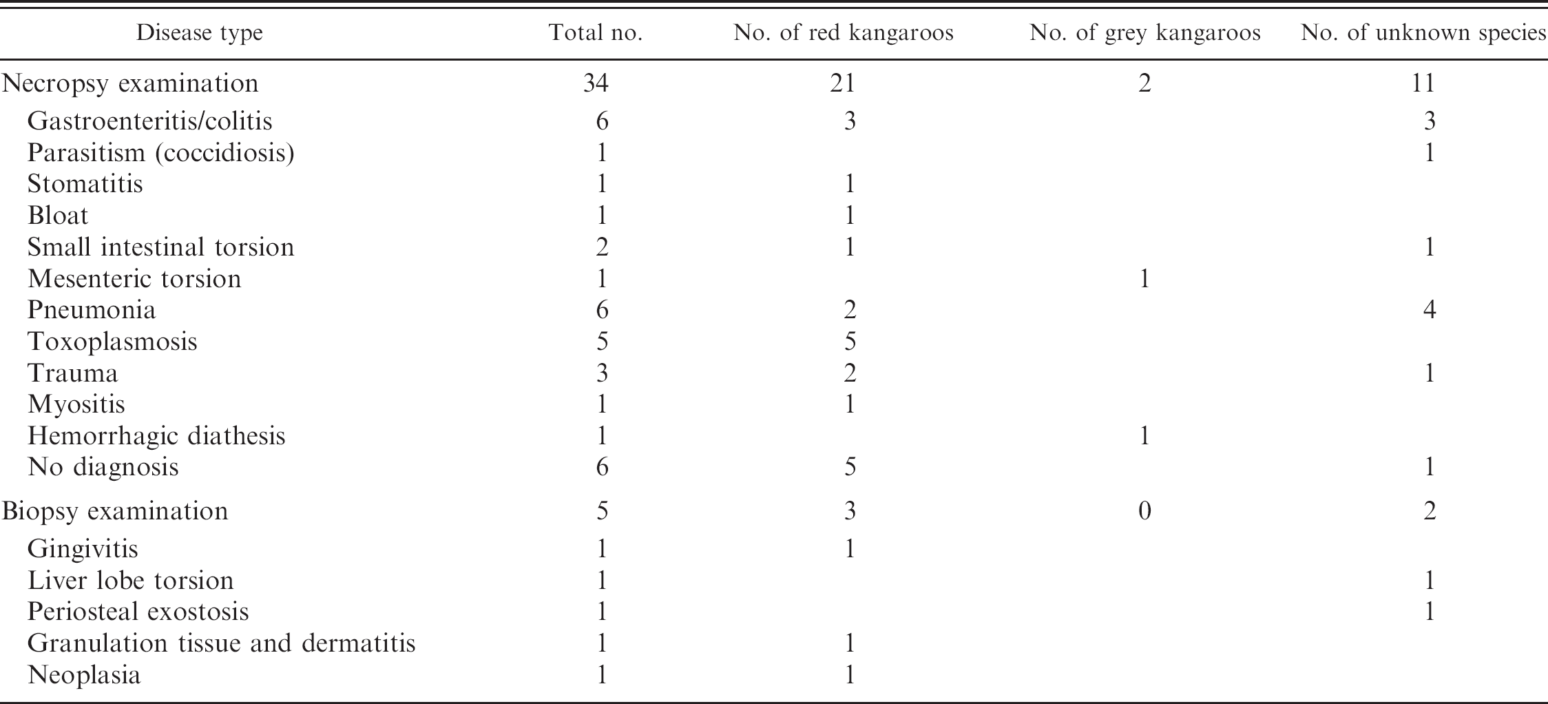
Various infectious and noninfectious disease processes were diagnosed in 28 kangaroos at necropsy (Table 1). A diagnosis was not obtained for 6 cases. Disease of the alimentary tract (infectious and noninfectious), pneumonia, and toxoplasmosis were most commonly diagnosed.
Disease of the alimentary tract was the leading cause of disease in kangaroos, being present in 12 kangaroos ranging in age from 8 months to 12 years. Necrotizing gastroenteritis or necrotizing colitis was diagnosed in 4 animals and 1 animal, respectively, with the kangaroos ranging in age from 8 months to 8 years. Using aerobic (2 of 5 cases), anaerobic (2 of 5 cases), salmonella (2 of 5 cases), and Campylobacter jejuni (1 of 5 cases) bacterial culture procedures, only Escherichia coli (2 of 5 cases) was recovered from intestinal tissues. No additional strain typing was performed, and the significance of the E. coli for these cases as a primary pathogen is not known. The microscopic lesions in 2 of these cases, necrotizing gastroenteritis (1 case) and necrotizing colitis (1 case) were consistent with clostridial disease similar to domestic ruminant species; however, neither bacterial cultures nor fluorescent antibody testing were performed for these cases. Parasitism was the cause of death in an 8-month-old kangaroo (species unknown). Microscopically, there was diffuse proliferation of sexual and asexual stages of coccidian parasites consistent with Eimeria sp. within the duodenum and jejunum. Neutrophilic and eosinophilic enteritis of unknown etiology was diagnosed in a female kangaroo (species unknown). Disease of the oral cavity was diagnosed in a euthanized, male red kangaroo (age unknown) at necropsy. Necrotizing stomatitis caused by Candida albicans was diagnosed in that animal. Oral candidiasis has been previously reported 7 in 4 hand-reared eastern grey kangaroos; antibiotic therapy and failure to thrive were suspected as predisposing factors to infection with C. albicans in that report. Noninfectious, diagnosed diseases of the alimentary tract included mesenteric torsion (1 case), small intestinal torsion with associated small intestinal infarction (2 cases), and gastric bloat (1 case). In a previous study on the digestive tract of the kangaroo, 10 the reported, noninfectious diseases included reactive gastritis from sand accumulation (1 case), trichophytobezoar (1 case), stomach rupture (3 cases), colon rupture (1 case), and jejunal incarceration (1 case).
. Summary of disease in 39 kangaroos based on biopsy and necropsy examination.
Pneumonia was the second leading cause of kangaroo disease and was present in 6 animals ranging in age from 7 months to 7 years. The age of 2 of these animals was not known. Fibrinosuppurative and necrotizing bronchopneu-monia (3 cases) was the most common form of pneumonia identified in this study and was diagnosed in both juvenile and adult animals. Aspiration pneumonia was diagnosed in 1 female red kangaroo. Pneumonic lungs from 3 cases were submitted for bacterial culture procedures, which included aerobic (3 of 3 cases) and anaerobic (1 of 3 cases) culture techniques. Aerobically, Pasteurella sp. (1 of 3 cases) and Bacteroides sp. (1 of 3 cases) were recovered. No significant aerobic bacterial growth was obtained from the lung of the third case. No bacterial growth was obtained from the anaerobic cultures. In 1 case, embolic pneumonia was diagnosed in a 6-year-old, male kangaroo (species unknown) and a large number of Gram-positive bacteria were identified; however, no fresh tissue was available for culture. Aspiration pneumonia (1 case) was diagnosed in a female red kangaroo (age unknown), and alveolitis (1 case) was diagnosed in a 7-month-old, female red kangaroo.
Toxoplasmosis was diagnosed in 5 red kangaroos that died without clinical signs from the same facility during a 5-month period. Infections occurred in both juvenile and adult animals. The youngest animal affected was 10 months old. Microscopically, there was a predominance of granu-lomatous to lymphoplasmacytic inflammation within visceral organs and lymphoplasmacytic inflammation within the brain. Toxoplasma gondii tachyzoites and bradyzoites were identified within the heart (4 of 5 cases), lung (2 of 5 cases), brain (1 of 5 cases), and liver (1 of 5 cases) of the affected animals. Positive immunoreactivity of the proto-zoal organisms to anti–T. gondii serum and negative immunoreactivity to anti–N. caninum serum was demonstrated in 2 kangaroos. Toxoplasmosis is a well-recognized disease in macropods and results in high morbidity and mortality. 2,4 Numerous reports of toxoplasmosis are in the literature, 1,2,4,8 with most outbreaks being associated to feral cat populations. A single report 4 suggests congenital infection occurred in a black-faced kangaroo (Macropus fuliginosus melanops).
Traumatic injuries in this study included cranial fractures (1 case), fracture of a cervical vertebra (1 case), and fracture of a lumbar vertebra (1 case). The causes of the fractures were not determined; however, they may have been due to predator–prey interaction because bobcats (Lynx rufus), coyotes (Canis latrans), and wild dogs are all present within the regions of Oklahoma where these animals were housed.
Myositis with associated hemorrhage of undetermined cause (1 case) was diagnosed in a female red kangaroo (age unknown), and no fresh tissue samples were submitted for ancillary testing, such as clostridial fluorescent antibody testing for this case. Hemorrhagic diathesis was diagnosed in an eastern grey kangaroo (age and sex unknown). Feed from this case was analyzed by mass spectroscopy for anticoagulant rodenticides; none was detected. A definitive cause of morbidity/mortality was not determined in 6 cases.
Antemortem biopsy samples were received for only 5 kangaroos, 3 red kangaroos, and 2 kangaroos of unknown species (Table 1). Histologic diagnoses included periosteal exostosis of undetermined cause (1 case), hepatocellular degeneration and microvascular thrombi because of torsion of a hepatic lobe (1 case), hyperplastic gingivitis (1 case), granulation tissue with a chronic dermatitis (1 case), and neoplasia (1 case). A 9-year-old, female red kangaroo was diagnosed with both a trichoepithelioma and a lipoma. In kangaroos, a trichoepithelioma has been reported 11 only once in an eastern grey kangaroo (Macropus giganteus).
Neoplasia in kangaroos is uncommonly reported. In a recent survey 12 of neoplasia in a captive group of red kangaroos, 6 of 28 necropsied animals (21%) were diagnosed with neoplasia. In the previous report, 12 it was postulated that because the facility housing the red kangaroos was constructed over an old landfill, the landfill may have played a role in the development of neoplasia through exposure to environmental carcinogens. Neoplasms in kangaroos are uncommonly reported and include mammary adenocarcinomas, 1,12 oral squamous cell carci-noma, 5,12 T-cell lymphoma, 12 pyloric submucosal lipoma, 12 chondrosarcoma, 6 trichoepithelioma, 11 seminoma, 13 basal cell epithelioma, 3 ovarian stromal tumor, 3 and hemangio-sarcoma. 1
According to the findings in the present study, based on necropsy and antemortem biopsy examination, disease of the alimentary tract represents most of the total disease diagnosed in this study (33.3%). In addition, 15.4% of animals had pneumonia and 12.8% of animals, all from the same facility, had disseminated toxoplasmosis. Although the ages of only 22 of 39 kangaroos (56%) were known, it is interesting to note that 17 cases (77.3%) of disease were in kangaroos with known ages older than 1 year. It is possible that young kangaroos may have a decreased incidence of disease compared with kangaroos older than 1 year because of their growth within the pouch of the dam. In one study, 9 the young kangaroos (joeys) left the pouch for the first time at mean ages of 283.86 ± 24.70 days (eastern grey kangaroo) and 298.40 ± 34.29 days (western grey kangaroo). Both species ultimately left the pouch at approximately 320 days old. 9 Unlike domestic animal species, the presence of joeys within the pouch of the dam may provide protection from temperature extremes and protect the joeys from feces-contaminated areas, thereby reducing their exposure to various pathogens, including bacteria, viruses, and parasites. In conclusion, the current study indicates that the most common causes of disease in kangaroos are disease of the alimentary tract, pneumonia, and disseminated toxoplasmosis.
Acknowledgements. The author would like to thank Mr. Curtis Andrew and the OADDL histotechnologists for their excellent technical assistance in preparation of the slides for this report and Mrs. Lori Bode for assistance with data retrieval.