
Abstract
The aim of the current study was to develop a nonradioactive in situ hybridization assay that can differentiate between genotypes 2a and 2b of Porcine circovirus-2 (PCV-2) in formalin-fixed, paraffin-embedded lymph node tissues from pigs with postweaning multisystemic wasting syndrome. Two different digoxigenin-labeled oligonucleotide probes were designed from the PCV-2 open reading frame 2 sequences. The PCV-2a–specific probe did not hybridize with formalin-fixed, paraffin-embedded lymph nodes from naturally PCV-2b–infected pigs and vice versa. Both PCV-2a–specific and PCV-2b–specific probes gave consistent negative signals in lymph nodes from naturally PCV-1–infected pigs. The in situ hybridization assay described in the present study represents a diagnostic tool that can differentiate between the 2 genotypes of PCV-2.
Porcine circovirus-1 and -2 (PCV-1 and PCV-2, respectively; family Circoviridae, genus Circovirus) 9 are the smallest, nonenveloped, single-stranded, circular DNA viruses. 8 Porcine circovirus-1 is not considered pathogenic, but PCV-2 has been implicated as the etiologic agent of Porcine circovirus–associated diseases, including postweaning multisystemic wasting syndrome (PMWS) and other clinical diseases. 3 Phylogenetic analysis has categorized PCV-2 into at least 2 major genotypes 5 : PCV-2a and PCV-2b. 10
The diagnosis of PMWS requires the presence of 3 criteria: 1) compatible clinical signs, 2) characteristic microscopic lesions, and 3) the presence of PCV-2 within the lesions (Sorden SD, Harms PA, Sirinarumitir T, et al.: 1998, Porcine circovirus and PRRS virus co-infection in pigs with chronic bronchointerstitial pneumonia and lymphoid depletion: an emerging syndrome in Midwest swine. In: Proceedings of the American Association of Veterinary Laboratory Diagnosticians, vol. 41, p. 75. Minneapolis, MN). In situ hybridization (ISH) and immunohistochemistry (IHC) have been the primary methods used for detecting PCV-2 in pathologic lesions. 2 However, there have been no reports of a monoclonal antibody that can specifically differentiate between the 2 genotypes of PCV-2 in formalin-fixed, paraffin-embedded tissues.
Although ISH and sequencing analysis of PCV-2 can differentiate between the 2 genotypes, there are advantages to using ISH over sequencing analysis to genotype virus associated with PMWS. Use of sequencing analysis of PCV-2 is largely restricted to diagnostic laboratories when only the formalin-fixed tissues are available. In addition, ISH provides cellular detail and histopathologic architecture so that PCV-2 genotype–infected cells and lesions may be diagnosed on the same section. Therefore, ISH should be considered a better technique than sequencing analysis of the PCV-2 genotype to diagnose PMWS because the characteristic microscopic lesions are important criteria for the diagnosis of PMWS. The aim of the current study was to develop a nonradioactive ISH assay that would allow differentiation between the 2 genotypes of PCV-2.
Formalin-fixed, paraffin-embedded lymph node tissues from pigs with PMWS were used in the present investigation. All 40 cases fall into the same category and were selected based on clinical signs, histopathologic lesions, detection of PCV-2 by IHC, and PCV-2 isolation. The main clinical signs of all 40 cases used in the present study were wasting or unthriftiness and pallor of the skin. Porcine circovirus-2a was isolated in 18 cases, and PCV-2b was isolated in 22 cases. The PCV-2 genotype was identified based on sequence analysis of the isolated PCV-2. Lymph node tissues from each of 5 pigs subclinically infected with PCV-2a or PCV-2b were also used. Negative tissue controls were from 1-day-old colostrum-deprived pigs not exposed to any types of virus, 7-day-old pigs experimentally infected with PCV-1, and pigs experimentally infected with Transmissible gastroenteritis virus (TGEV), Porcine epidemic diarrhea virus (PEDV), Porcine reproductive and respiratory syndrome virus (PRRSV), Classical swine fever virus (CSFV), Swine hepatitis E virus (SHEV), or Swine influenza virus (SIV).
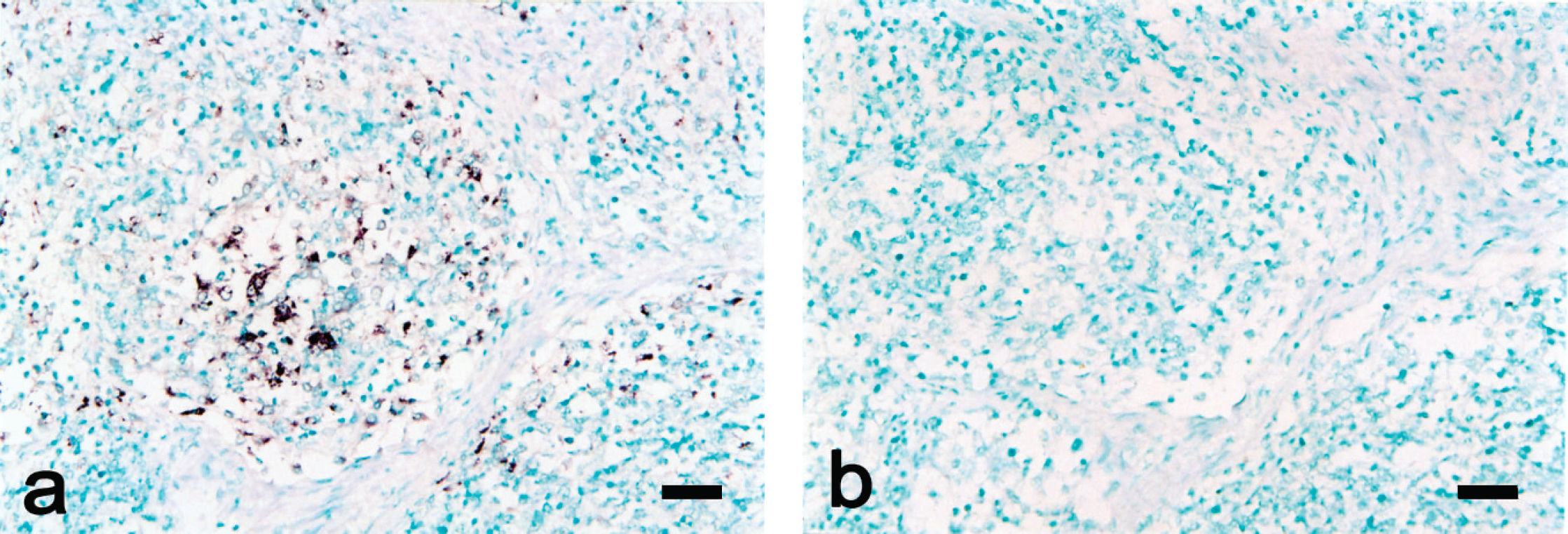
Serial sections of a lymph node from a pig naturally infected with Porcine circovirus-2a (PCV-2a).
PK-15 (porcine kidney epithelial cell line) cultures infected with PCV-1, PCV-2a, or PCV-2b were grown in 2-well chamber slides. After 5 days of incubation at 37°C, the medium was discarded, wells were rinsed with phosphate buffered saline (PBS; 0.1 M, pH 7.4), and the cells were fixed with PLP (4% paraformaldehyde, 100 mM
Two different digoxigenin (DIG)-conjugated oligonucle-otide probes were designed based on multiple alignments of open reading frame 2 sequences of PCV-2a and PCV-2b in GenBank (http://www.ncbi.nlm.nih.gov/Genbank/index.html) using MegAlign a and Oligo 4.0 b software programs. A 22–base pair (bp) probe was designed for detecting PCV-2a with the following sequence: 5'-GACCAACAAAATCTCTATACC-3' (nucleotide positions 1481–1460 on PCV-2a). A 20-bp probe was designed for detecting PCV-2b with the following sequence: 5'-GCTCAAACCCCCGCTCTGTG-3' (nucleotide positions 1481–1462 on PCV-2b). The oligonucleotide probes were labeled by random priming with DIG–2'-deoxyuridine 5'-triphosphate using a commercial kit. c Two sets of serial sections, 4 μm thick, were placed on positively charged slides d and stored at room temperature. One set of sections was processed for ISH of PCV-2a, and the other for ISH of PCV-2b. In situ hybridization was performed as previously described. 6
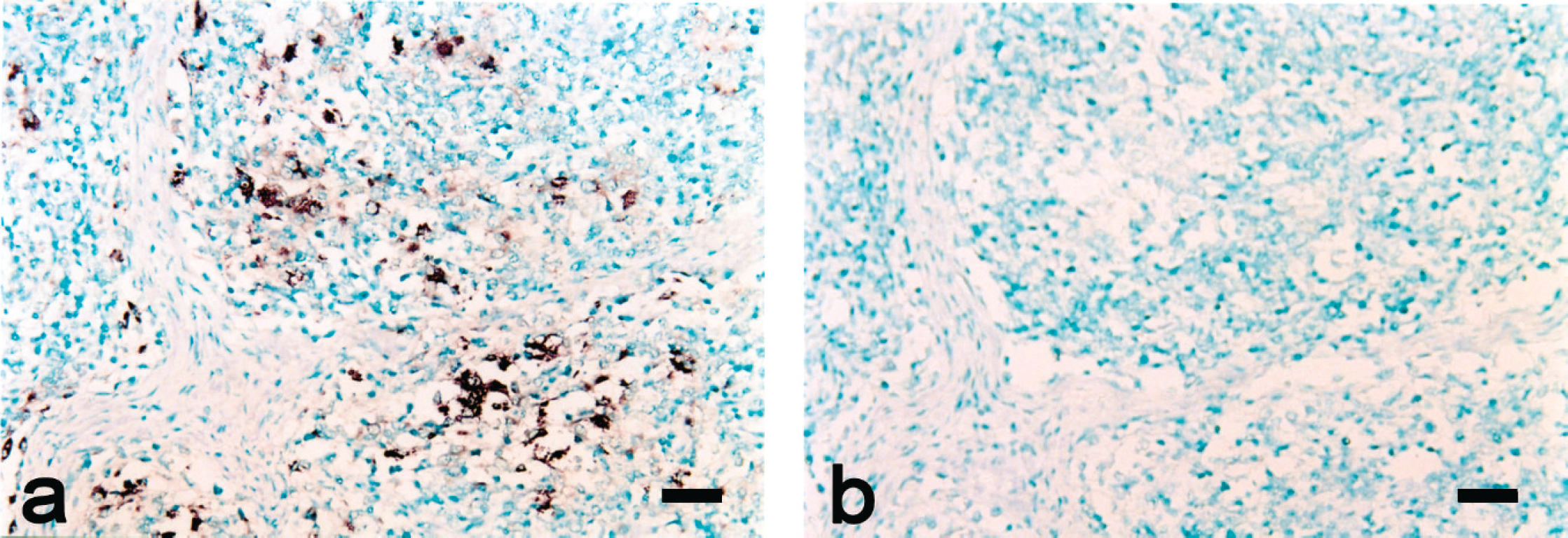
Serial sections of a lymph node from a pig naturally infected with Porcine circovirus-2b (PCV-2b).
The PK-15 cells were infected with PCV-1, PCV-2a, and PCV-2b and hybridized with the specific DIG-labeled PCV-2a and PCV-2b probes, respectively. DNA from both PCV-2a and PCV-2b was detected. The specificity of the probes was confirmed because 1) both probes showed no signal in mock-infected PK-15 cells and PRRSV-infected MARC-145 cells, 2) the PCV-2a probe did not hybridize with PCV-2b–infected PK-15 cells and vice versa, and 3) both PCV-2a and PCV-2b probes consistently showed no signal in the PCV-1–infected cell cultures.
The ISH assay was able to differentiate between the 2 genotypes of PCV-2. Porcine circovirus-2a DNA was detected in 18 PMWS cases (Fig. 1a). When lymph node tissues from these pigs were tested with the DIG-labeled PCV-2b probe, no hybridization signals were detected (Fig. 1b). Positive cells typically exhibited a dark-brown stain, primarily in the cytoplasm, but occasionally in the nucleus, with no background. In the inguinal lymph nodes, a strong hybridization signal for PCV-2a was detected in the cytoplasm of epithelioid macrophages and multinucleated giant cells. No hybridization signal was consistently seen in tissue sections pretreated with DNase A. Porcine circovirus-2a DNA was detected in 5 pigs subclinically infected with PCV-2a. Sections from the 2 negative control pigs showed no hybridization signals for PCV-2a.
Porcine circovirus-2b DNA was detected in 22 PMWS cases (Fig. 2a). When lymph node tissues from these pigs were tested with the DIG-labeled PCV-2a probe, no hybridization signal was detected (Fig. 2b). Cells that stained positive for PCV-2b included macrophages, epithe-lioid cells, and multinucleated giant cells. No hybridization signal was consistently seen in tissue sections pretreated with DNase A. Porcine circovirus-2b DNA was detected in 5 pigs subclinically infected with PCV-2b. Sections from the 2 negative control pigs showed no hybridization signals for PCV-2b. No hybridization signal was detected with the PCV-2a and PCV-2b probes on formalin-fixed, paraffin-embedded tissues from pigs experimentally infected with PCV-1, TGEV, PEDV, PRRSV, CSFV, SHEV, or SIV.
The results of the current study demonstrate the ability of the ISH assay described herein to detect and differentiate between PCV-2a and PCV-2b in formalin-fixed, paraffin-embedded tissues with nonradioactive DIG-labeled probes. Apart from the ISH assay developed in the present study, other studies have shown that polymerase chain reaction (PCR) methods can also differentiate between the 2 genotypes. 7 However, the diagnostic application of differential PCR is largely restricted because PCV-2 was also detected by PCR in lymph nodes from healthy pigs without clinical PMWS. 1 One of the important criteria for the diagnosis of clinical PMWS is to detect PCV-2 within the histopathologic lesions (Sorden SD, Harms PA, Sirinarumitir T, et al.: 1998, Porcine circovirus and PRRS virus co-infection in pigs with chronic bronchointerstitial pneumonia and lymphoid depletion: an emerging syndrome in midwestern swine).
A Canadian study suggested that microscopic lesions observed in PCV-2b–associated PMWS cases were more severe than those observed in PCV-2a–associated PMWS cases, 4 although no studies in the veterinary literature could be found to have investigated definitive differences in pathogenicity between PCV-2a and PCV-2b. Therefore, PCV-2 genotyping may be important in diagnosing PMWS cases. The ISH assay developed in the present study represents a diagnostic tool capable of differentiating between the 2 genotypes of PCV-2, and could be useful in surveillance programs for monitoring the 2 genotypes of PCV-2 in circulation or identifying those involved in PMWS outbreaks.
Acknowledgements. This research was supported by the Technology Development Program for Agriculture and Forestry, Ministry for Agriculture, Forestry, and Fisheries. The research was also supported by the Preparation Research Fund for WCU from Research Affairs of Seoul National University, the Brain Korea 21 Program for Veterinary Science, and a Korea Research Foundation Grant (KRF-2006-005-J02902) in the Republic of Korea.
Footnotes
a.
DNASTAR Inc., Madison, WI.
b.
Molecular Biology Insights Inc., Cascade, CO.
c.
Boehringer Mannheim Corp., Indianapolis, IN.
d.
Thermo Fisher Scientific Inc., Waltham, MA.