
Abstract
A 1.5-year-old, neutered, male ferret (Mustela putorius furo) was presented with sudden lethargy, anorexia, and diarrhea. Clinical and radiographic examinations revealed an intra-abdominal mass. An explorative laparotomy was performed. A neoplasm, located in the ileum wall, was submitted for histopathologic examination. The tumor consisted of weakly eosinophilic spindle cells arranged in a compact pattern with haphazardly interlacing bundles. Neoplastic cells labeled positively for KIT (cluster of differentiation 117, stem cell factor receptor) and vimentin. Based on histologic and immunohistologic results, this tumor was diagnosed as a gastrointestinal stromal tumor. Results suggest that this ferret tumor shares strong similarities with the canine and human counterparts.
Gastrointestinal stromal tumors (GISTs) are digestive mesenchymal neoplasms that are differentiated from neurogenous or myogenous neoplasms (schwannomas and leiomyomas or leiomyosarcomas, respectively) by the expression of KIT (cluster of differentiation 117, stem cell factor receptor). KIT is a protein kinase receptor encoded by the protooncogene Kit. Gastrointestinal stromal tumors are derived from interstitial cells of Cajal, also known as gastrointestinal pacemaker cells, or from their progenitor cells. 12
In veterinary medicine, spontaneous GISTs diagnosed by their immunopositivity for KIT have been described in dogs, 1,4,9,13 horses, 6,7 Spanish ibex, 15 a rat, 5 and nonhuman primates. 2,14 Due to the popularity of ferrets as pets, increased numbers of neoplasms in this species have been documented in the literature over the last few years. However, digestive neoplasms are uncommon. 8 The present report describes a mesenchymal digestive neoplasm in a ferret diagnosed as a GIST by KIT-positive immunohistochemistry (IHC).
A 1.5-year-old, neutered, male ferret (Mustela putorius furo) was presented to a local veterinarian with complaints of sudden lethargy, anorexia, and diarrhea. Clinical examination and radiographs revealed an intra-abdominal mass. In the course of an explorative laparotomy, a 3.5-cm diameter mass was found in the ileal wall. No similar nodular lesions were observed elsewhere in the peritoneal cavity.
The resected nodule was fixed in 10% neutral buffered formalin and submitted to IDEXX Laboratory Alfort (Alfortville, France) for histologic evaluation. The tissue samples were processed routinely, embedded in paraffin, sectioned at 4 μm, stained with hematoxylin eosin saffron, coverslipped, and examined microscopically. The immunophenotype of the neoplasm was investigated by IHC on additional sections using the streptavidin biotin peroxidase complex method. The following primary antibodies and dilutions were used: monoclonal anti-human vimentin, a 1:100; polyclonal anti-human KIT, a 1:50; monoclonal anti-human α-smooth muscle actin (α-SMA), a prediluted; polyclonal anti-human desmin, b prediluted; polyclonal anti-human S-100, b prediluted. Heat pretreatment for epitope retrieval by microwaving was performed in citrate buffer (pH 6) for vimentin and α-SMA detection and in an ethylenediamine tetra-acetic acid buffer (pH 9) for KIT detection. Normal smooth muscle within the vascular walls of the neoplasm served as a positive tissue control for vimentin, α-SMA, and desmin. For KIT immunodetection, normal interstitial cells of Cajal and mast cells present in the tissue section served as positive controls. For S-100 IHC, enteric neurons provided a positive control. Parallel tissue sections that lacked primary antibody application were used as negative controls.
Microscopically, the ileal mass was a nonencapsulated neoplasm located in the tunica muscularis. Multifocal infiltration of the serosa, inner muscularis mucosae, and mucosa (Fig. 1) by neoplastic cells was observed. In addition, the mucosa had focal ulceration. The neoplasm was highly cellular and composed of spindle cells arranged in a compact pattern or haphazardly distributed in interwoven bundles (herringbone pattern) and short fascicles (Fig. 2).
The collagenous stroma was minimal, and numerous congested blood vessels were present. Randomly distributed lymphatic vessels within the tumor had marked ectasia.
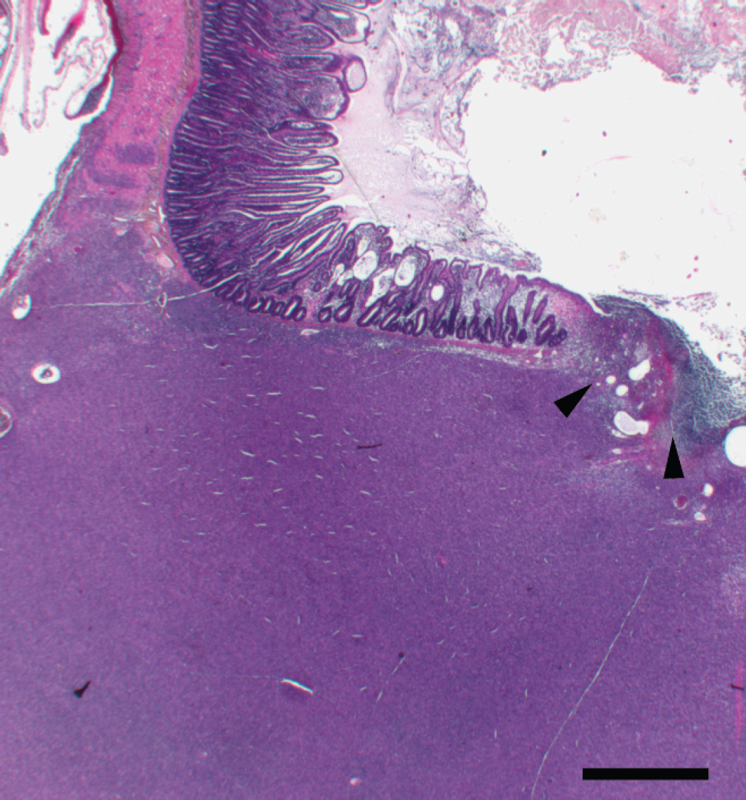
Ferret (Mustela putorius furo). Photomicrograph of an ileal neoplasm within the lamina muscularis with focal infiltration and ulceration of the tunica mucosa (arrowheads). Hematoxylin-eosin-saffron stain. Bar = 1,000 μm.
The neoplastic cells were monomorphic with indistinct cellular borders and a moderate amount of weakly eosinophilic to amphophilic fibrillar cytoplasm. Nuclei were round to oval and centrally located and had finely stippled chromatin with 1–3 small, eosinophilic nucleoli (Fig. 2). Although the mitotic rate was high, with 30 mitoses per ten 40 × fields of view, abnormal mitoses were not seen. Multifocal coagulative necrosis, neutrophilic and histiocytic infiltrates, hemorrhagic foci, and vascular thrombi were present within the neoplasm. Extensive myxoid degeneration was observed at the periphery of necrotic foci.
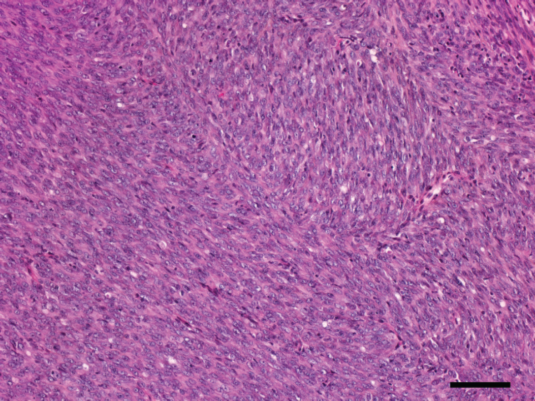
Ferret (Mustela putorius furo). Photomicrograph of interlacing fascicles of densely packed spindle cells. Hematoxylin-eosin-saffron stain. Bar = 100 μm.
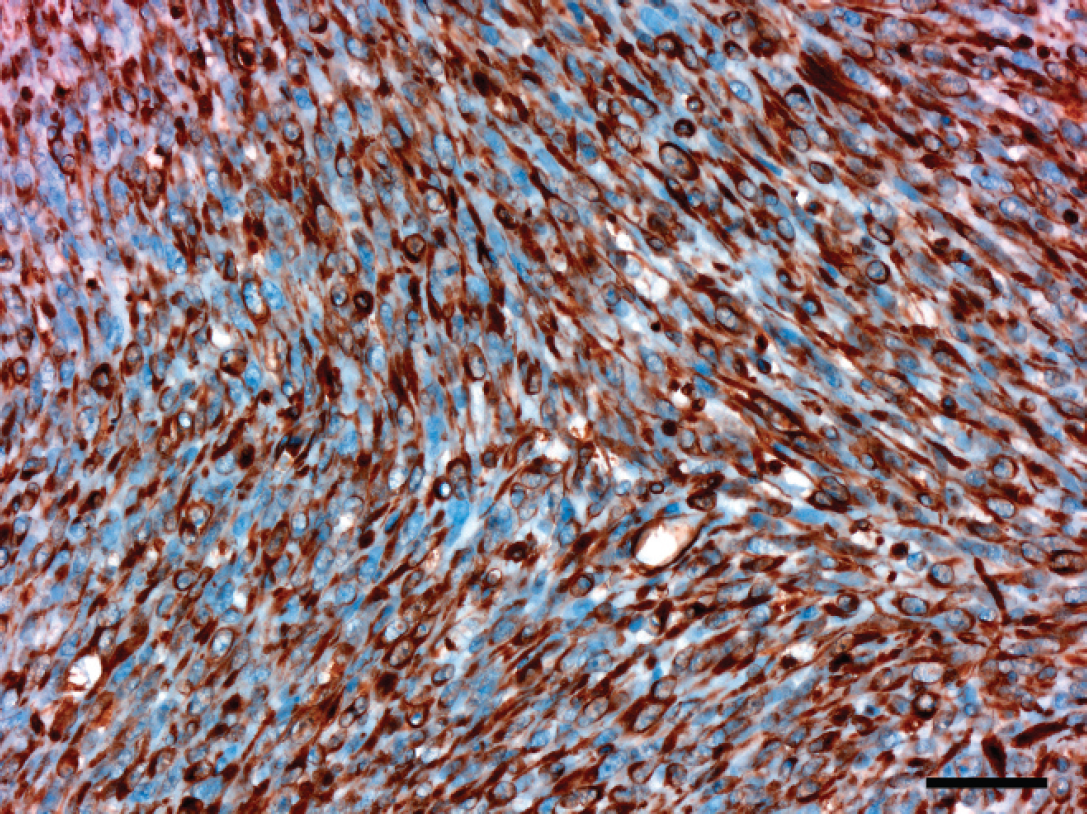
Ferret (Mustela putorius furo). Neoplastic cells stain positively for vimentin. Streptavidin-biotin peroxidase complex method; hematoxylin counterstain. Bar = 40 μm.
Histologic examination confirmed the presence of an intestinal mesenchymal neoplasm, but IHC analysis was necessary for a definitive diagnosis related to cellular differentiation. The differential diagnosis included GIST, leiomyosarcoma, schwannoma, liposarcoma, and undifferentiated sarcoma.
The neoplastic cells were diffusely and intensely positive for vimentin (Fig. 3). Labeling for KIT was extensive; more than 90% of the neoplastic cells had light brown cytoplasmic staining (Fig. 4). Neoplastic cells were negative for α-SMA, desmin, and S-100 (data not shown). Consequently, histopathologic and IHC characteristics of the neoplasm were consistent with a diagnosis of GIST.
Digestive mesenchymal neoplasms are rare in ferrets. An earlier review recorded only 16 neoplasms in the digestive tract (from the stomach to the anus), among 574 neoplasms in the ferret. 8 Only 2 of the neoplasms were of mesenchymal origin; 1 was a leiomyoma of the cecum and 1 a leiomyosarcoma of the rectum. 8 Recently, another intra-abdominal leiomyosarcoma was reported and probably was of intestinal origin. 11 However, KIT immunolabeling was not performed to differentiate GISTs from other mesenchymal neoplasms in these earlier reports. In human medicine, definitive diagnosis of GIST requires positive immunolabeling of tumor cells for KIT, as recommended by the National Institutes of Health (NIH) Consensus Conference. 3 Previously, publications indicate that IHC is the only histologic method to reliably distinguish KIT-positive GISTs from KIT-negative leiomyosarcomas in humans and dogs. 1,4,9 The GIST in the ferret of the current report was confirmed by positive immunoreactivity for KIT in the neoplastic cells compared with mast cells and interstitial cells of Cajal that served as internal positive controls. In human GISTs, the Kit gene is often mutated and Kit gain-of-function mutations are considered the major oncogenic event. 12 Point mutations in exon 11 (or more rarely in exons 9 or 13) lead to constitutive activation of the KIT receptor tyrosine kinase with subsequent signal transduction leading to cell proliferation. 10,12
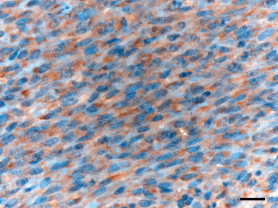
Ferret (Mustela putorius furo). Neoplastic cells have moderate, homogenous, cytoplasmic immunoreactivity for KIT. Streptavidin-biotin peroxidase complex method; hematoxylin counterstain. Bar = 20 μm.
The preferential site of GIST origin varies from species to species. Canine GISTs occur most commonly in the colon and small intestine, 4 whereas human GISTs arise mostly in the stomach. 10 Gastrointestinal stromal tumors are neoplasms composed of spindle to epithelioid cells that appear weakly eosinophilic to basophilic and have uncommon but prominent nuclear pleomorphism. In humans and dogs, the fusiform variant is more commonly observed. 4,9,10 Moreover, fusiform gastric GISTs in humans are subclassified as sclerosing, palisading vacuolated, hypercellular, and sarcomatous. 10 The latter subtype, which contains densely packed, uniform spindle cells, can have marked mitotic activity (>4 mitoses per ten 40 × fields of view) and diffuse nuclear atypia evidenced by nuclear enlargement and hyperchromasia without much pleomorphism. 10 The histomorphology of the neoplasm in the current case is consistent with that of an ileal GIST and resembles a human fusiform variant with a sarcomatous subtype.
Characterization of malignancy and prognosis of human GISTs is challenging, because even a tumor with a histologically benign appearance can metastasize. In 2001, the NIH Consensus Conference provided a simple scheme to assess the risk for aggressive behavior of GISTs based on mitotic rate, tumor size, and location. 3 In animal GISTs, a similar classification scheme has not been defined. In canine neoplasms, mitotic activity can reach 19 mitosis per 10 high-power field. 1,4 However, the neoplasm in the current report had a much higher mitotic index (30 mitosis per ten 40 × fields of view). The high mitotic rate, infiltrative nature, and extensive necrosis are suggestive of a neoplasm with an aggressive behavior. However, this animal remains free of local recurrence and metastatic disease 4 years after initial diagnosis.
Acknowledgements. The authors acknowledge Drs. Olivier Marie and Jean-Michel Trompe for the medical history and collection of clinical samples, Jean-Luc Servely for illustrations, and Dr. Marc Chodkiewicz for his critical review of the manuscript. The authors are indebted to Professor Jean-François Emile (Department of Pathology, Hôpital Ambroise Paré, Boulogne, France) for his advice regarding pathologic investigations, and Professor Jean-Jacques Fontaine in the Pathology unit and Dr. Laurent Tiret for supporting this work under a CentroNuclear Myopathy project. Anne Girard-Luc was supported by a grant from IDEXX Laboratory Alfort.
Footnotes
a.
Dako Denmark A/S, Glostrup, Denmark.
b.
Thermo Scientific, Runcorn, United Kingdom.