
Abstract
Serum thymidine kinase (TK) activity has recently been evaluated as a serum marker for human and canine hematopoietic neoplasms. The purpose of the current study was to establish the significance of serum TK activity in the diagnosis of bovine leukosis. The discrimination value for TK activity was set at 5.4 U/l based on the receiver operating characteristic curve. In the group of clinically healthy cows, only 2 out of 83 cows (2.4%) had serum TK activity above the discrimination value. In contrast, 19 out of 20 cows (95.0%) with bovine leukosis showed serum TK activity above the discrimination value, although only 7 of 79 (8.9%) cows diagnosed with diseases other than bovine leukosis showed elevated serum TK activity. Thymidine kinase activities of all Bovine leukemia virus-positive cows with or without lymphocytosis were below the discrimination value. Sensitivity and specificity of measuring serum TK activity as a diagnostic tool for bovine leukosis was 95.0% and 95.9%, respectively. Results indicate that serum TK activity may be a marker for bovine leukosis.
Bovine leukosis is one of the most common neoplastic diseases of cattle. This lymphoproliferative disease has been classified into 2 types based on clinicopathological, epizootiological, and pathological findings: enzootic bovine leukosis (EBL) associated with Bovine leukemia virus (BLV) infection and sporadic bovine leukosis (SBL), for which the cause or causes are unknown. 3,15 Cattle developing EBL or SBL often present with loss of condition, an abrupt drop in milk production, enlarged superficial lymph nodes, exophthalmos, and partial to complete anorexia, particularly with regard to grain or concentrates. 1 There is no cure for the disease, and death occurs 2–3 weeks following signs of clinical illness and development of neoplasms. 16
Swelling of superficial lymph nodes, lymphocytosis, and detection of neoplastic lymphocytes in peripheral blood are often found in typical cases of bovine leukosis and are sufficient to cause one to suspect this disease. 16 A definitive diagnosis is usually obtained by cytology of aspirates from primary neoplasms or neoplastic lymph nodes; however, a recent study 21 showed that the sensitivity and specificity of fine-needle aspiration of enlarged peripheral lymph nodes ranged from 38% to 67% and from 25% to 80%, respectively. Definitive diagnosis can be difficult in cattle without typical signs of bovine leukosis. 19 Although an antibody test for BLV is useful as an adjunct to the diagnosis of EBL, 22 the rate of development of neoplasms among BLV-infected cattle is low, ranging from 0.1% to 10%. 18 It has been reported 9 that total lactate dehydrogenase (LDH), LDH2, and LDH3 activities increase, while LDH1 activity decreases in cattle with EBL. However, this may not be specific to EBL, as LDH activity increases with other diseases as well. 11 Thus, a reliable diagnostic marker for bovine leukosis is required in veterinary practice.
Thymidine kinase (TK) converts thymidine to thymidine monophosphate. Especially in rapidly proliferating cells, it serves as part of a salvage pathway for DNA synthesis. Thymidine kinase is activated during the gap 1-synthesis phase of the cell cycle, and its activity in plasma correlates with neoplastic cell proliferation. 2 Increases in serum TK concentrations are seen in human patients with acute and chronic leukemia, chronic lymphocytic leukemia, Hodgkin's and non-Hodgkin's lymphoma, multiple myeloma, and myelodysplastic syndrome. 4,6,13,14 Estimation of serum TK activity is useful as a tumor marker for detecting, grading, and monitoring tumors in patients with lymphoma and leukemia. Several recent studies have shown that plasma TK activity is also helpful in the diagnosis and monitoring of canine lymphoma and leukemia. 20 The present study attempted to establish the significance of serum TK activity in the diagnosis of bovine leukosis.
A total of 242 sera were divided into 5 groups: group A included 20 cows diagnosed with bovine leukosis; group B included 79 cows with various other diseases; group C included 33 clinically healthy, BLV-positive cows without lymphocytosis; group D included 27 clinically healthy, BLV-positive cows with lymphocytosis; and group E included 83 clinically healthy, BLV-negative cows. Ninetynine cows from group A and B were definitively diagnosed at the Obihiro University of Agriculture and Veterinary Medicine (Japan) from May 2006 to April 2009. Diagnosis was made based on postmortem examination and histo-pathological findings. For group A, immunohistochemical staining using monoclonal antibodies against bovine differentiation antigen of lymphocytes (i.e., cluster of differentiation [CD]3 against T cells and B-lymphocyte antigen [BLA]36 against B cells) was performed for some cows with bovine leukosis (Table 1). Breeds in group B included 75 Holstein-Friesian (HF), 3 Japanese Black (JB), and 1 hybrid of Japanese Black × Holstein-Friesian (F1). Diseases (and breeds) in group B included 28 inflammatory diseases (27 HF, 1 F1), 14 congenital diseases (14 HF), 6 amyloidoses (6 HF), 5 orthopedic disorders (4 HF, 1 JB), 4 nonhematopoietic neoplastic diseases (rhabdomyosarcoma, hepatic carcinoma, lipoma, and brain tumors; 4 HF), 3 metabolic diseases (1 HF, 2 JB), 3 cardiac myopathies (3 HF), 2 kidney failures (2 HF), 2 hemopericardiums (2 HF), and 12 other diseases (12 HF). All HF cows from groups C, D, and E were raised by private farmers in the Tokachi area of Hokkaido, Japan, and were randomly selected regardless of age and reproduction status. Cows that belonged to groups C and D had antibodies against glycoprotein 51 (gp51) of BLV, while cows from group E did not. An agar gel immunodiffusion kit was used to determine whether cows had antibodies against gp51. a In order to examine the association between persistent lymphocytosis (PL) and bovine leukosis, clinically healthy BLV-infected cows were divided into 2 groups based on the European Economic Community (EEC) key; peripheral lymphocyte counts were normal in group C but were high in group D. For groups C and D, blood lymphocytes were calculated based on a blood-smear differential count, and absolute leukocyte counts were determined with an automated hematology analyzer. b The EEC key was applied to the hematological criteria for lymphocytosis. This key has been used as an indicator of animals that are in the preclinical stages of leukosis. 3 The 83 cows of group E were used as a control group.
Hematological findings, anti-Bovine leukemia virus (BLV) antibody tests, and immunohistochemical findings in cattle of group A.*
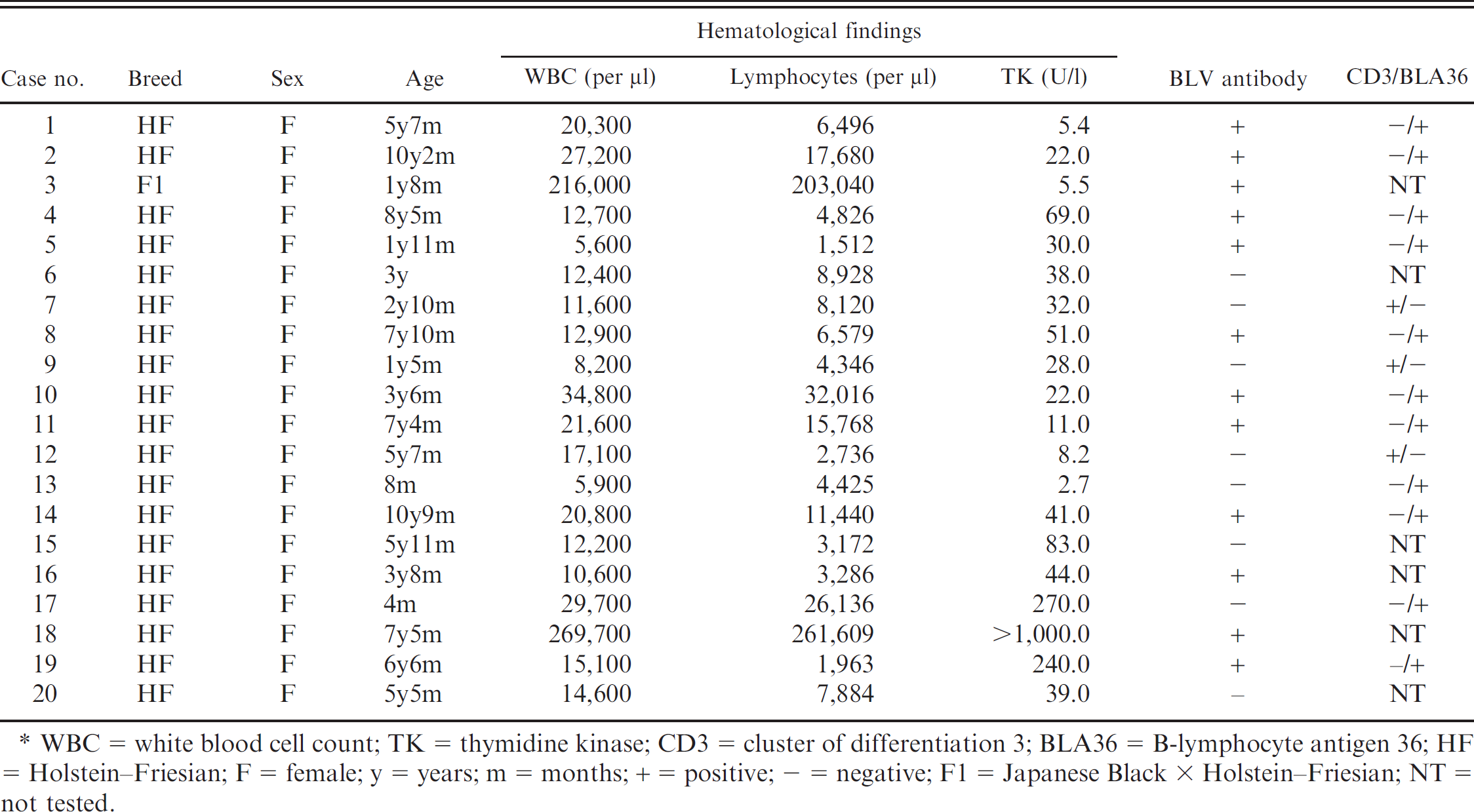
WBC = white blood cell count; TK = thymidine kinase; CD3 = cluster of differentiation 3; BLA36 = B-lymphocyte antigen 36; HF = Holstein-Friesian; F = female; y = years; m = months; + = positive; - = negative; F1 = Japanese Black × Holstein-Friesian; NT = not tested.
Serum samples were subjected to a TK activity assay, which was performed using a commercial radioenzyme TK assay kit with 125I-iododeoxyuridine as the tracer. c Thymidine kinase activity was expressed as units per liter (U/l). The reportable range of the assay was 1.0-1,000.0 U/l.
Prior to comparing serum TK activity among groups, an optimum discrimination value was established using the receiver operating characteristic (ROC) curve. Fisher's exact test was used to assess differences in TK activity between groups. Analyses were performed using the computer program R, d and P < 0.05 was considered significant. The discrimination value for TK activity was set at 5.4 U/l based on the ROC curve (area under the curve: 0.989). The sensitivity and specificity of measuring serum TK activity as a diagnostic tool for bovine leukosis were 95.0% and 95.9%, respectively. In the control group (group E), TK activity ranged from <1.0 U/l to 7.6 U/l. Fifty-five out of 83 cows (66.3%) in group E had TK activity measuring <1.0 U/l, and only 2 out of 83 cows (2.4%) had serum TK levels above the discrimination value.
Table 1 summarizes hematological findings, anti-BLV antibody tests, and immunohistochemical findings in cows from group A. Thymidine kinase activity in group A ranged from 2.7 U/l to >1,000.0 U/l. Median TK activity in group A was 32.0 U/l (97.5 percentile: 256.5 U/l); 1 cow from group A (no. 18) was excluded because TK activity exceeded the reportable range. Nineteen out of 20 cows (95.0%) with bovine leukosis had serum TK activities above the discrimination value. Elevated serum TK activity was observed independent of neoplastic cell immunophenotypes and presence of anti-BLV antibodies. Seven out of 20 cows (nos. 1, 5, 8–10, 12, and 13) with bovine leukosis did not manifest typical symptoms of bovine leukosis, including external lymphadenopathy and lymphocytosis with atypical lymphocytes in peripheral blood. Among these 7 cows, all except one (no. 13) presented with high TK activity (5.4–51.0 U/l).
Thymidine kinase activity in group B ranged from <1.0 U/l to 30.0 U/l, and median TK activity was 1.7 U/l (97.5 percentile: 9.91 U/l). Sixteen out of 79 cows (20.3%) in group B had TK activity of <1.0 U/l, and only 7 out of 79 cows (8.9%) had serum TK activity above the discrimination value (5.9–30.0 U/l). Diseases in these 7 cows included bone dislocation, postcaval thrombosis, nephritis, tetralogy of Fallot, internal hydrocephalus, suppurative pleuropneumonia, and myorrhexis. Serum TK activity was below the discrimination value (1.6–3.6 U/l) in 4 cows with non-hematopoietic neoplasms. In groups C and D, TK activity ranged from <1.0 U/l to 2.3 U/l and from <1.0 U/l to 2.2 U/l, respectively. Only 3 out of 33 cows in group C and 4 out of 27 cows in group D had TK activity >1.0 U/l. The proportion of cows with increased serum TK activity (>5.4 U/l) was significantly higher in group A (P < 0.01) and lower in groups C and E (C: P < 0.05; E: P < 0.01).
In the present study, 95.0% of cows with bovine leukosis had elevated serum TK activity (>5.0 U/l). In contrast, only 8.9% of cows diagnosed with various diseases other than bovine leukosis had serum TK activity above the discrimination value. These results indicate that serum TK activity might be a marker for bovine leukosis. Only 1 out of 20 cows with bovine leukosis (no. 13) showed serum TK activity below the discrimination value. This was an atypical case: the cow had a mass of neoplastic B cells that compressed the spinal cord and caused ataxia. The cause of decreased serum TK activity within the reference interval was unclear. Among the 7 cows with atypical bovine leukosis, 6 presented with increased TK activity, indicating that serum TK activity measurements should be useful in diagnosing atypical cases of bovine leukosis. In addition, since elevated serum TK activity occurred independently of neoplastic cell immunophenotypes and anti-BLV antibodies, serum TK activity may be a potential marker for both EBL and SBL.
Enzootic bovine leukosis is divided into 3 stages: serologically positive, but negative for lymphocytosis; serologically positive and positive for PL; and leukemia. Approximately 30% of infected cattle progress to PL, which is characterized by polyclonal expansion of B cells, while only a small percentage of BLV-infected cattle develop malignant lymphosarcoma. 18 Given the relationship between PL and bovine leukosis (see above), hematological keys such as Bendixen's key and the EEC key have been used as indicators of animals in the preclinical stages of leukosis. 3
Serum TK activities were measured in groups C and D, but none of the cows in these groups had activity levels above the discrimination value. It has previously been reported 17 that TK activity reflects tumor volume and cell proliferation, and several studies have shown that host immune responses are associated with disease progression of BLV infection. 10 In cows with lymphocytosis (group D), host immune responses might have suppressed cell proliferation to a certain degree and consequently repressed TK activity. Further studies that measure serum TK activity and lymphocyte counts serially during the course of EBL are needed in order to determine the stage at which serum TK activity begins to increase and in order to investigate the relationship between EBL stages and serum TK activities.
Serum TK activity was elevated in 7 out of 79 cows diagnosed with various diseases other than bovine leukosis. Serum TK activity is sometimes elevated in subjects with nonhematopoietic neoplasms (such as breast carcinoma, epithelial ovarian cancer, and lung cancer), 5,8,12 infections with some viruses (Measles virus, Rubella virus, Varicella virus, Human herpesvirus 1, and genus Cytomegalovirus), and in pregnant women. 7 In the current study, postmortem examination did not reveal neoplastic disease in the 7 cows with elevated TK activity, and the causes of increased activity are as yet unknown.
Because postmortem examinations were not performed in groups C, D, and E, a limitation of the present study is that the 143 clinically healthy cows may be affected by atypical leukosis, reproduction status, and/or viral infection. Further investigation is needed to elucidate the specificity of serum TK activity in cattle, and more cases, including those involving nonhematopoietic neoplasms, should be examined.
Acknowledgements. This study was supported in part by the Japan Livestock Technology Association. The authors thank Drs. Mistuo Ishii, Hidefumi Furuoka, and Takane Matsui for their technical assistance and valuable discussion.
Footnotes
a.
Kitasato Institute Research Center for Biologicals, Saitama, Japan.
b.
Nihon Kohden, Tokyo, Japan.
c.
Kishimoto Clinical Laboratory, Inc., Obihiro, Japan.
d.
R Foundation for Statistical Computing, Vienna, Austria.