
Abstract
Bovine herpesvirus 1 (BoHV-1) is an infectious agent of concern in the international export of bovine products; it is endemic in the United States, but it has been eradicated in many countries of the European Union (EU). For export of semen to the EU, accurate assessment of BoHV-1 status of the bull is required and is usually accomplished by measuring the level of antibody to the virus. The gold standard is virus neutralization (VN) using overnight incubation with the virus, a test approved by the World Organization for Animal Health (OIE). Enzyme-linked immunosorbent assay (ELISA) is also approved for international trade. The lone U.S. Department of Agriculture–approved commercial ELISA was compromised with specificity problems, which necessitated the development of a different ELISA. Of 4 monoclonal antibodies evaluated, 1 directed against glycoprotein C of BoHV-1 was found to be the most reliable. One hundred twenty-eight characterized positive samples and 334 negative serum samples were tested. The blocking ELISA showed 97.4% sensitivity and 99.4% specificity as compared with OIE VN. The Wisconsin Veterinary Diagnostic Laboratory ELISA fulfills the OIE requirement for a blocking or competitive ELISA to qualify animals for export to BoHV-1-free countries.
Bovine herpesvirus 1 (BoHV-1) is a double-stranded DNA virus of the genus Varicellovirus in the Alphaherpesvirinae subfamily of the Herpesviridae family. Bovine herpesvirus 1 is endemic in the United States and infects cattle primarily through aerosol inhalation (upper-respiratory infection) or genital contact. Clinical signs are often mild, and although BoHV-1 has not been shown to be a cause of high mortality, infection results in a latent state and lifelong infection. 6,12 Reactivation of latent BoHV-1 infections can occur due to corticosteroid treatment or stress resulting from transportation, overcrowding, and inclement weather. 6,12
Bovine herpesvirus 1 can be shed in semen and transmitted during natural and artificial insemination and has been shown to induce abortion as well as to decrease milk production. 6 Bovine herpesvirus 1 has been eradicated in Austria, Denmark, Finland, Norway, Sweden, and Switzerland; because of this, export of bull semen must be accompanied by a certificate that assures that the animal has tested negative for antibodies to BoHV-1. 6,16 Virus neutralization (VN) and enzyme-linked immunosorbent assay (ELISA) are the conventional methods of detecting BoHV-1 antibodies.
Several blocking ELISAs have been developed by European laboratories, with sensitivity and specificity comparable to that of the World Organization for Animal Health (OIE)–approved VN test. 9 For regulatory reasons, the monoclonal antibodies (mAbs) and their respective commercial ELISA kits are not available in the United States. A competitive ELISA was developed in the United States using the D9 mAb, 14 which recognizes glycoprotein B. In the original publications describing the D9 mAb, 3 other monoclonal antibodies were also described. 4,5 F2 and G2 are mAbs specific for glycoprotein C, and H2 is specific for glycoprotein B. Glycoprotein B is required for virus penetration, 8 whereas glycoprotein C is a nonessential protein that has been shown to play a role in virus replication and attachment. 1,15 Glycoprotein C mediates the virus's initial interaction with cell receptors, which is followed by subsequent reaction with glycoproteins B and D. 10 Sera positive for BoHV-1 antibodies react strongly to glycoprotein C constituents. 1 Furthermore, the D9, F2, G2, and H2 mAbs have been shown to react and neutralize BoHV-1 2–5 and will not cross-react with other bovine respiratory viruses. 5
Bovine blood samples submitted to the Wisconsin Veterinary Diagnostic Laboratory (WVDL; Madison, WI) from diagnostic and export cases were used in test development. Tubes were spun to remove serum from the clot. Each serum sample was divided into 2 aliquots: 1 for ELISA and another for VN, which was heat inactivated at 56°C for 30 min and then stored frozen at −20°C. The OIE-approved VN assay was performed with a 24-hr incubation of virus and sera as prescribed 16 using titered Colorado strain of BoHV-1. a The BoHV-1 concentration was adjusted to achieve 30–300 (TCID50) per well. 13
A sandwich ELISA was developed to assess available mAbs prior to development of the blocking ELISA. To produce antigen for both ELISAs, the Colorado strain of BoHV-1 a was propagated in Madin-Darby bovine kidney (MDBK) cells b at a multiplicity of infection of 0.1. After an adsorption period of 1 hr, the cells were incubated at 37°C in Minimal Essential Medium c containing 5% fetal bovine serum d (FBS). At first observation of cytopathic effect, the flasks were frozen at −70°C. After thawing, the cells were sonicated for 1 min, and the supernatant was clarified at 30 × g for 10 min. The virus supernatant was then filtered using a 0.45-μm syringe filter and layered on a 30% sucrose cushion. The virus was spun at 100,000 × g for 2 hr, and the virus pellet was suspended in 1 mmol Tris buffer c (pH 8.0) and frozen at −70°C. A mock infection was performed in an identical manner to produce cell lysates to determine the optical density (OD) baseline in uninfected cells.
Four commercially available mAbs were assessed by a sandwich ELISA to determine reactivity to BoHV-1. All 4 mAbs were isotype immunoglobulin G (IgG) 2b except for G2, which was an IgG1 isotype. e Unpurified mAb (1 mg/ml) was titrated in sodium bicarbonate/sodium carbonate buffer c (pH 9.6) using dilutions between 1:50 and 1:6,000. Monoclonal antibody dilutions were adsorbed to microtiter plates d and incubated overnight at 4°C, then blocked with 300 μl fish-derived, protein-blocking buffer f for 1 hr at 37°C. Plates were washed 2 times with phosphate buffered saline (PBS) g with 0.05% Tween 20. c Plates were incubated with alternating rows of BoHV-1 antigen and mockinfected antigen diluted to a concentration of 1 μg/well for 1 hr at 37°C. Washing was repeated as described above. Undiluted bovine test sera were added to the plates and incubated for 1 hr at 37°C. Plates were washed 4 times and incubated with a 1:1,000 dilution of rabbit anti-bovine IgG conjugated to horseradish peroxidase (HRP). h Hydrogen peroxide combined with 3,3′,5,5′-tetramethylbenzidine (TMB) h in a 1-component format was used as substrate and chromogen for development of the assay. The reaction was terminated with 1% HCl. h Optical density was measured at 450 nm.
The WVDL blocking ELISA was developed with the F2, G2, and H2 mAbs that were identified by the sandwich ELISA as suitable antibodies for the blocking ELISA. The mAbs were purified and conjugated to HRP using commercial laboratories. i j Each conjugated antibody was titrated, and the dilution, which yielded an OD of 0.6–1.0 at 450 nm, was selected. The Colorado strain BoHV-1 antigen a at a concentration of 1 μg/well was adsorbed to microtiter plates d and blocked with fish-derived, protein-blocking buffer f for 1 hr at 37°C. Plates were washed twice using a manual plate washer and PBS 0.05% Tween 20 as described above. Adsorbed and blocked plates were stored for a minimum of 24 hr to a maximum of 1 week at 4°C. Fifty microliters of undiluted bovine test sera was added in duplicate to the wells containing BoHV-1 antigen and mock-infected antigen and incubated for 30 min at 37°C. Plates were not washed prior to the addition of conjugated mAb i j (50 μl), which was diluted in fish-derived, protein-blocking buffer. f The HRP-conjugated mAb and test sera were incubated for 1 hr and 30 min at 37°C, after which the plates were washed 4 times with PBS Tween 20. A 1-component solution (100 μl) of substrate (H2O2) and chromogen (TMB) h was added, and the assay was developed at room temperature for 15 min. The reaction was terminated with 1% HCl, h and the OD was measured at 450 nm.
Results of the commercial indirect enzyme-linked immunosorbent assay (ELISA) compared with those of the World Organization for Animal Health (OIE) virus neutralization (VN) assay.*
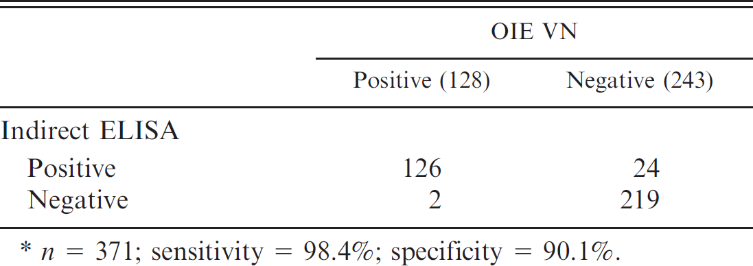
n = 371; sensitivity = 98.4% specificity = 90.1%
All OD readings of samples and controls were adjusted by subtracting the background OD level in the corresponding mock-infected well. The percentage inhibition (%I) was calculated using the following formula:

Samples were considered to be positive at 50–100% inhibition and negative below 50%.
The current study describes the development of a blocking ELISA to detect antibodies to BoHV-1. Blocking ELISAs are a prescribed test in the OIE manual to qualify animals and animal products for export; however, no U.S. Department of Agriculture (USDA)–approved blocking ELISAs are commercially available in the United States. Blocking assays developed in Europe approach 100% sensitivity and specificity but cannot be used in the United States without USDA approval. A commercial USDA-approved indirect ELISA was available until 2005, but the low specificity (Table 1) of the ELISA prompted the development of the WVDL blocking ELISA.
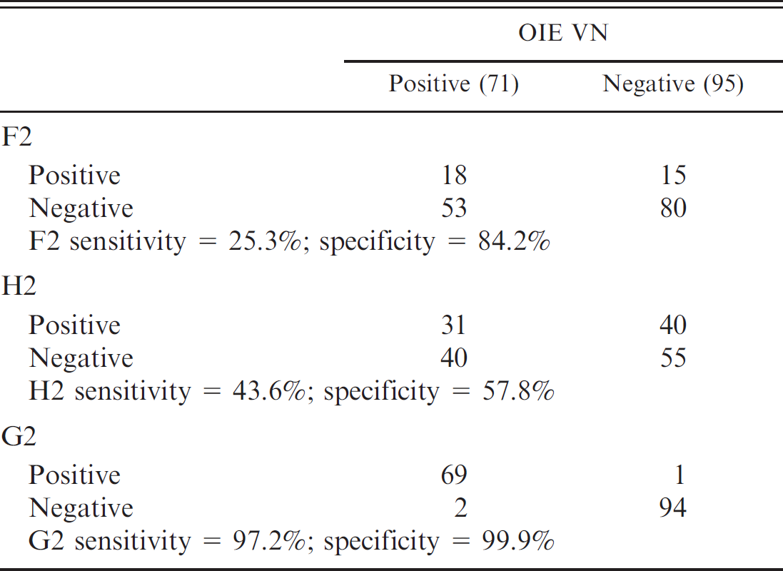
The sandwich ELISA was used as a screening tool to determine reactivity of each mAb with BoHV-1. Of the mAbs available for use in the United States, D9 and H2 are specific for gB. The D9 antibody was reported to be effective in a blocking ELISA format with 100% sensitivity and specificity on a limited number of samples. 7,8 Unfortunately, this hybridoma cell line no longer produced high levels of antibody and reacted too weakly in the sandwich ELISA to warrant further evaluation (data not shown). The weak signal could presumably be due to minimal amounts of D9 antibody relative to the other proteins in the ascites fluid. Monoclonal antibodies G2, F2, and H2 reacted with sufficient strength in the sandwich ELISA and were conjugated to evaluate in a blocking ELISA format with 71 well-characterized positive and 95 negative samples. Although the gB epitope has been shown to be the preferred target in European ELISAs, 7,9 the conjugated H2 directed to gB showed poor sensitivity and specificity when compared with OIE BoHV-1 VN (Table 2). The F2 and G2 mAbs recognize the gC protein of BoHV-1. The F2 mAb performed poorly in the blocking ELISA, whereas the G2 mAb gave better correlation with OIE BoHV-1 VN (Table 2) and thus was chosen for further evaluation with a larger set of samples.
Comparison of conjugated monoclonal antibodies F2, H2, and G2 to World Organization for Animal Health (OIE) virus neutralization (VN) results.
Results of the Wisconsin Veterinary Diagnostic Laboratory blocking enzyme-linked immunosorbent assay (ELISA) using G2 as the conjugated antibody compared with those of the World Organization for Animal Health (OIE) virus neutralization (VN) on a larger set of samples.*
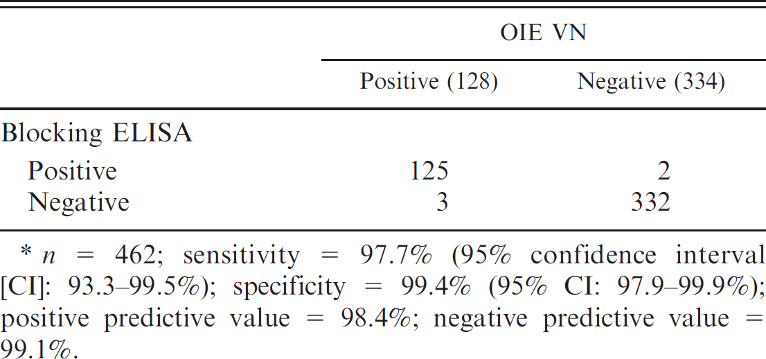
n = 462; sensitivity = 97.7% (95% confidence interval [CI]: 93.3–99.5%); specificity = 99.4% (95% CI: 97.9–99.9%); positive predictive value = 98.4% negative predictive value = 99.1%.
Bovine serum samples (462) were tested using the G2 blocking ELISA, with a cutoff of 0–49% inhibition for negative samples and 50–100% inhibition to denote positive samples. The results were compared with the OIE-approved VN assay as the gold standard. Of these, 128 were positive as determined by the OIE VN and further confirmed by another laboratory using a kinetic ELISA k ; 334 were negative by OIE VN. Sensitivity, specificity, and predictive values were calculated using previously published methods (Table 3). 11 The G2 blocking ELISA was unable to detect antibodies specific to BoHV-1 in 3 samples that were determined to be positive by OIE VN. The blocking ELISA detected BoHV-1 antibodies in 2 serum samples that were determined to be negative by OIE VN.
The discrepant samples (3 false-negative and 2 false-positive) were sera submitted by the genetics industry firms. Samples submitted from the bovine genetics industry fall into 2 categories. The first is prepurchases; these animals are tested at about 6–9 months of age prior to shipment to a bull stud premise. The other category is composed of animals admitted to a bull stud that are tested in semiannual surveys and before exportation of product. These bulls range in age from 1 year to 10 years. The false-negative samples were all in prepurchase status and gave percentage inhibition values from 0% to 37%. The corresponding OIE VN titers were 16 or lower, indicating that a low level of antibody was present and could reflect the presence of maternal antibody presumably too low to be detected by the ELISA. Of the 2 false-positive sera, 1 was from an older bull, which initially tested negative but then showed 75% inhibition for 2 successive tests and then reverted to negative status and remained so on all subsequent tests including the WVDL ELISA. This may reflect a nonspecific reaction due to possible environmental conditions such as change in diet. The second false-positive serum consistently tested positive in a borderline range. In serological assays, cross-reactions with other viruses can sometimes explain weak reactions; however, no cross-reactions were reported for the G2 mAb with other respiratory viruses or BoHV-2 or −4. 5 The G2 mAb is known to react with BoHV-5 according to vendor literature, e although this virus is not commonly found in the United States. Cross-reactions of the G2 mAb to other ungulate herpes viruses have not been tested.
The goal of the current study was the development of a blocking ELISA with a sensitivity and specificity that would be acceptable for the bovine genetics industry, with the restriction of using mAbs available in the United States. BoHV-1 can have a large economic impact on the livestock genetics industry, owing to the export restrictions of countries where the virus has been eradicated. Consequently, an assay to detect BoHV-1 that has high specificity and sensitivity such as the WVDL G2 ELISA is of utmost importance to the livestock genetics industry.
Footnotes
a.
National Veterinary Services Laboratories, Ames, IA.
b.
American Tissue Culture Collection, Manassas, VA.
c.
Minimal Essential Medium, Tris buffer 8.0 pH, sodium bicarbonate/sodium carbonate buffer pH 9.6, Tween 20; Sigma-Aldrich, St. Louis, MO.
d.
Fetal bovine serum, Immulon II microtiter plates; Fisher Scientific Co., Chicago, IL.
e.
H2 (glycoprotein B, IgG2b isotype), G2 (glycoprotein C, IgG1 isotype), F2 (glycoprotein C, IgG2b isotype), and D9 (glycoprotein B, IgG2b isotype); VMRD Inc., Pullman, WA.
f.
SeaBlock-fish derived protein blocking buffer, Pierce Biotechnology Inc., Rockford, IL.
g.
FTA Hemagglutinin PBS, BD, Chicago, IL.
h.
Rabbit anti-bovine IgG horseradish peroxidase antibody, TMB 1-component substrate and HCl stop solution; KPL Inc., Gaithersburg, MD.
i.
Conjugated G2, F2 monoclonal antibodies; American Qualex, San Clemente, CA.
j.
Conjugated H2, F2 monoclonal antibodies; Chromaprobe Inc., Maryland Heights, MO.
k.
Kinetic ELISA, Animal Health Diagnostic Center, Cornell University, Ithaca, NY.