
Abstract
A sarcomatoid carcinoma was diagnosed in the lung of a 10-year-old captive Egyptian fruit bat (Rousettus aegyptiacus). Both carcinomatous and sarcomatous cytologic phenotypes were identified histologically. Cells of both types stained positive for pancytokeratin and S-100. Stromal cells stained positively for muscle actin. No staining for vimentin was noted in either neoplastic or normal internal control tissues. To the authors' knowledge, this is the first report of a pulmonary sarcomatoid carcinoma in a bat, and only the third report of sarcomatoid carcinoma outside the human literature.
A 10-year-old, castrated, male Egyptian fruit bat (Rousettus aegyptiacus) was found to be weak and on the floor of its enclosure. The exhibit housed approximately 30 Egyptian fruit bats and 15 straw-colored fruit bats (Eidolon helvum). Clinical signs included hypothermia, severe dyspnea, and taut abdominal musculature. Diagnostic imaging revealed fluid in the abdomen, and 32 ml of serosanguineous fluid was removed via abdominocentesis; no subsequent analysis was done on this fluid. Supportive care was instituted, but the animal died several hours later.
At necropsy, the carcass weighed 154 g and was markedly dehydrated. Muscle mass and fat stores were reduced. The penis was extruded, swollen and bruised at its tip. A solid 7 mm × 3 mm cream-colored mass was adherent to the base of the heart and adjacent lung. A small amount of serosanguineous fluid was present in the abdominal cavity; there was no fluid in the pleural or pericardial spaces. All other tissues appeared normal. Differential diagnoses for the thoracic mass included a heart base tumor and pulmonary carcinoma. Tissue samples were fixed in 10% neutral buffered formalin, processed for histology by routine methods, and stained with hematoxylin and eosin.
Histologically, the mass was composed of neoplastic cells and was contained largely within the lung. There were two distinct regions with differing cytologic phenotypes. The first was dominated by large, round epithelial cells that tended to form packets (carcinomatous component; Fig. 1). Nuclei were round to ovoid with scant chromatin and variably 0–2 nucleoli. There was 4-fold anisokaryosis and occasional multinucleate cells were evident. Mitotic figures were rare. The cytoplasm was abundant with a nucleus:cytoplasm ratio varying from approximately 1:1 to 1:10.
The second region made up approximately 60% of the section examined and was comprised of spindle-shaped cells arranged in irregular swirls (sarcomatous component; Fig. 2). Nuclei of these cells were variably plump and oval to elongate, with finely stippled chromatin and occasionally a single nucleolus. Mitotic figures were rare. In a few intervening regions, cells displayed features of both phenotypes (transitional region; Fig. 3). There were several foci of necrosis within the mass. A large organizing thrombus occluded much of a large vein intimately associated with the tumor. Metastases were not observed.
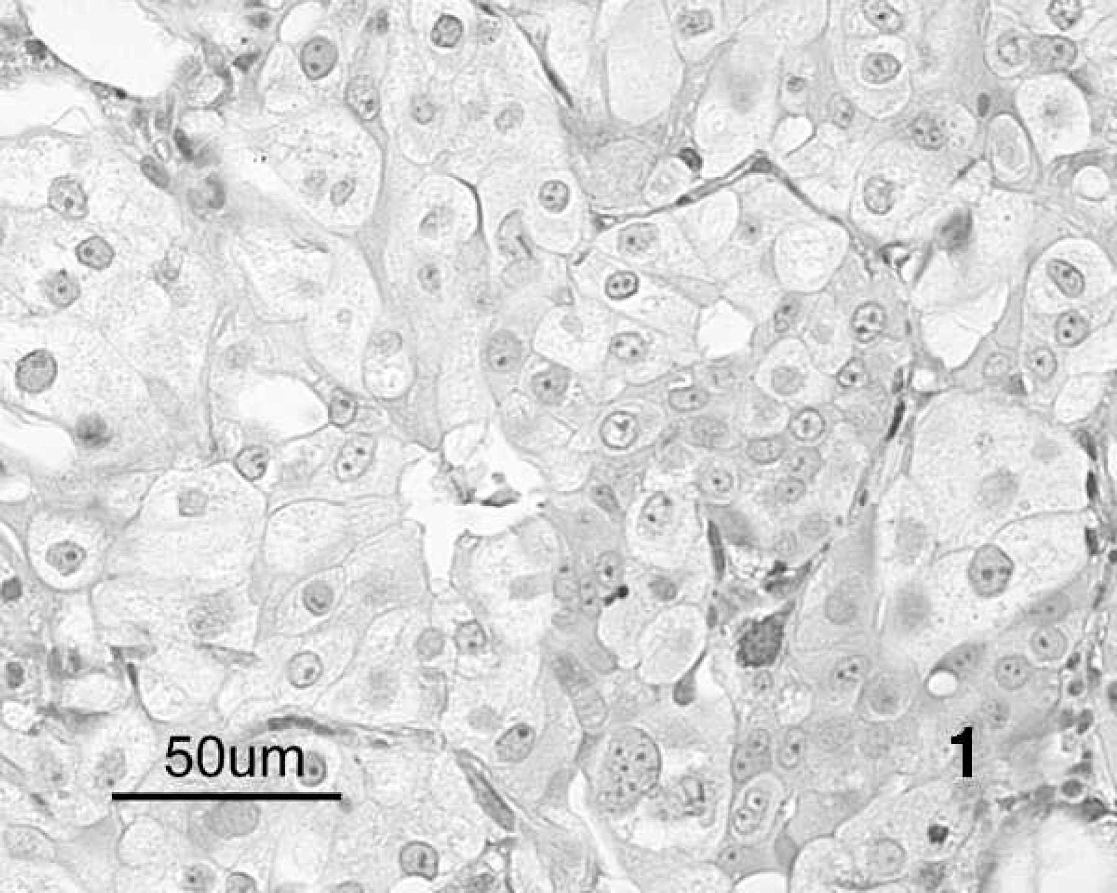
Irregular nests or packets of epithelioid cells with low nucleus:cytoplasm ratio in the epithelial region of a lung tumor from an Egyptian fruit bat. Hematoxylin and eosin. Bar = 50 μm.
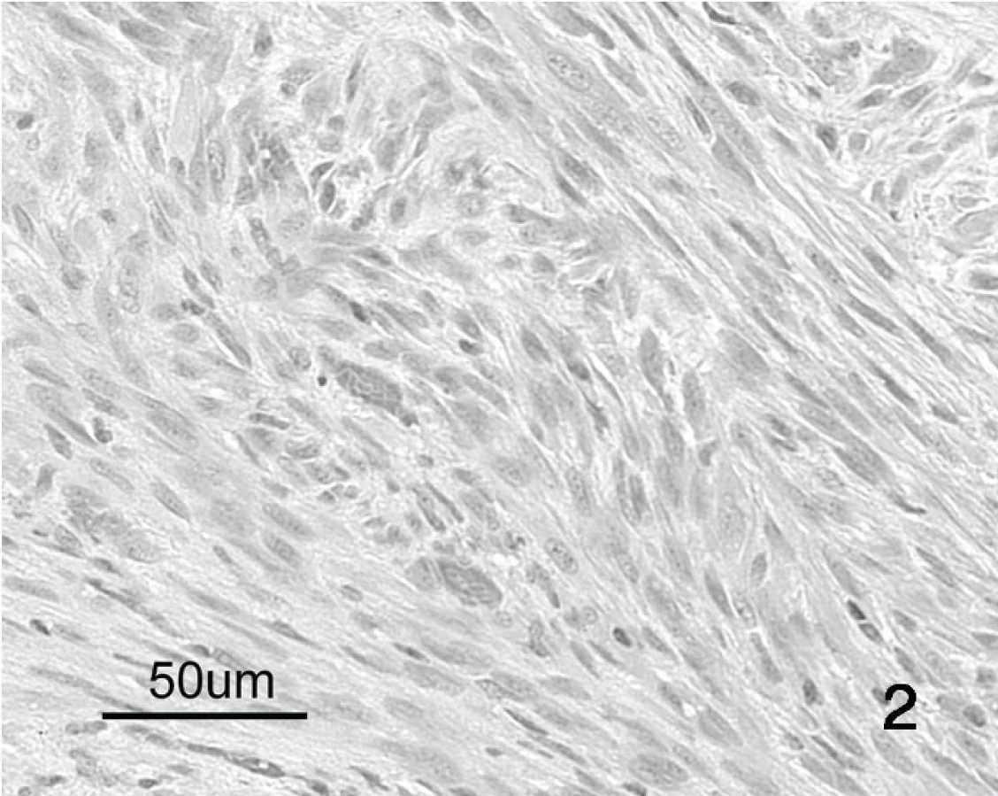
Irregular bands and swirling masses of spindle-shaped cells in the stromal component of a lung tumor from an Egyptian fruit bat. Hematoxylin and eosin. Bar = 50 μm.

Transitional region between the epithelial and stromal components of a pulmonary sarcomatoid carcinoma from an Egyptian fruit bat; masses of epithelial cells, several with areas of necrosis (right and mid-upper left portions of image), are intimately associated with a band of spindle cells (top center). Hematoxylin and eosin. Bar = 200 μm.

Strong pancytokeratin antigen–positive immunohistochemical staining of the cytoplasm of cells in the epithelial component of a pulmonary sarcomatoid carcinoma in an Egyptian fruit bat. Bar = 50 μm.

Weak pancytokeratin antigen–positive immunohistochemical staining of the cytoplasm of a few scattered cells in the stromal component of a pulmonary sarcomatoid carcinoma in an Egyptian fruit bat. Bar = 25 μm.

Strong S-100 antigen–positive immunohistochemical staining in the cytoplasm and nucleus of about 60% of the cells in the stromal component of a pulmonary sarcomatoid carcinoma in an Egyptian fruit bat. Bar = 50 μm.

Strong S-100 antigen–positive immunohistochemical staining in the cytoplasm and nucleus of the majority of nested epithelial cells in the epithelial component of a pulmonary sarcomatoid carcinoma in an Egyptian fruit bat. Bar = 50 μm.
Immunohistochemical staining of a pulmonary sarcomatoid carcinoma in an Egyptian fruit bat.*
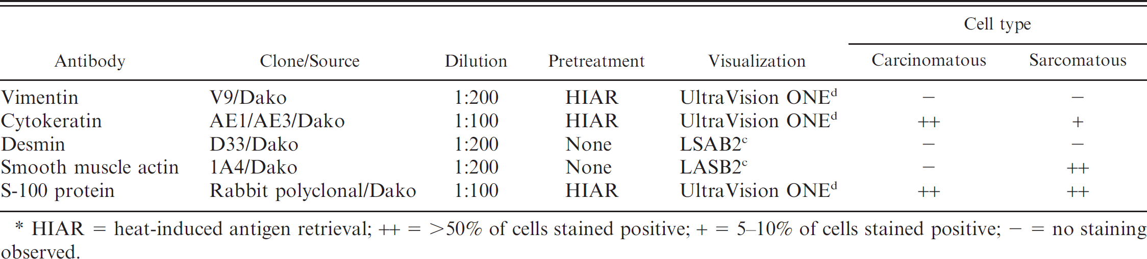
HIAR = heat-induced antigen retrieval; ++ = >50% of cells stained positive; + = 5–10% of cells stained positive; – = no staining observed.
Hemosiderin-laden macrophages were scattered in lung, liver, myocardium, and skeletal muscle. There was patchy atelectasis and mild pulmonary edema in the remaining lung. Congested pulmonary capillaries contained large numbers of neutrophils. Rare single cell necrosis was evident in the liver. Eosinophilic material (protein) was present in tubules of occasional nephrons, and there were rare mild lymphoplasmacytic interstitial infiltrates and mineralized tubules. Other findings included mild myocardial fibrosis, and atrophy of adipose tissue. Streptococcus sp. was cultured from the liver and lung.
A presumptive diagnosis of pulmonary sarcomatoid carcinoma was made. Ascites, pulmonary edema, and widespread hemosiderin pigment in macrophages suggested cardiovascular compromise, likely caused by pressure from the tumor on great vessels at the heart base, although chronic passive hepatic congestion was not noted. Terminal streptococcal septicemia, along with hypothermia and presumptive hypoglycemia, probably contributed to the death of this animal. Myocardial fibrosis was considered mild and subclinical.
Immunohistochemistry for vimentin, cytokeratin, smooth muscle actin, desmin, and S-100 protein was performed using an automated stainer a on 4-μm tissue sections mounted on charged slides. Table 1 outlines specific antibodies and their sources as well as pretreatment and visualization methods used for each antigen.
Anti-S-100 antiserum was raised in rabbits; the remaining antigens used were evaluated using mouse monoclonal antibodies. Following manual deparaffinization and rehydration, sections were treated with 3% hydrogen peroxide to quench endogenous peroxidase activity. Heat-induced antigen retrieval for vimentin, cytokeratin, and S-100 protein was accomplished using citrate buffer (pH 6) and a pressure cooker. b Sections were incubated with primary antibodies for 30 min, followed by a 30-min incubation with goat anti-mouse streptavidin-biotin (for smooth muscle actin and desmin) c or goat anti-mouse/rabbit polymer d visualization systems. NovaRed e was used as the chromogen.
Sections of normal canine tissues known to contain the antigen of interest were used as positive tissue (run) controls, and tissues on the test slides were used as species-specific internal controls. For negative reagent controls, duplicate sections of each control and test tissue were subjected to the same immunohistochemical procedure with substitution of antibody diluent alone for mouse monoclonal primary antibodies and substitution of non-immune rabbit serum at similar protein concentration for the rabbit polyclonal antisera.
Sarcomatoid carcinomas have been defined as exhibiting positive staining for keratin and vimentin, with inconsistent staining for other cytochemicals including S-100 protein, desmin, and smooth muscle actin. 6 Cells in carcinomatous and sarcomatous regions stained with variable intensity using antibodies to both pancytokeratin (staining the cytoplasm; Figs. 4, 5) and S-100 (staining the nucleus, cytoplasm, and intercellular material; Figs. 6, 7). Approximately 90% of cells in the stromal portion stained positively with antibody to muscle actin. Neoplastic cells did not stain with antibody to desmin. Vimentin was not expressed in either neoplastic or normal internal control tissues.
With morphologic and immunohistochemical staining characteristics of both epithelial and stromal malignancies, the neoplasm in the current study is best described as a sarcomatoid carcinoma. 6 Sarcomatoid carcinomas are generally considered to be epithelial neoplasms, which exhibit sarcomatous differentiation. 6 They have variably been described as carcinosarcoma, spindle cell carcinoma, blastoma, teratocarcinoma, pseudosarcoma, and squamous cell carcinoma with pseudosarcomatous stroma. 3,6 Pulmonary sarcomatoid carcinomas are primary neoplasms of the lung. The tumor is considered rare in humans, accounting for only 1 % of primary lung neoplasia in one review. 4 To the authors' knowledge, only 2 cases have been reported in animals, which described occurrences in a cat and dog, respectively. 2,5
There is significant homology of intermediate filament proteins, such as vimentin, across vertebrates, 1 yet the antivimentin antibody used in the present investigation apparently does not react with Egyptian fruit bat antigen. This underscores the difficulties of using diagnostic tests in species for which they are not validated. To the authors' knowledge, this is the first report of a pulmonary sarcomatoid carcinoma in a bat and only the third report of such a tumor in animals.
Acknowledgements. The authors thank Dr. Josepha Delay of the Animal Health Laboratory, University of Guelph, for assistance with immunohistochemical investigations.
Footnotes
a.
Dako Autostainer, Dako Canada Inc., Mississauga, Ontario, Canada.
b.
Decloaking Chamber, Biocare Medical, Concord, CA.
c.
LSAB®2, Dako Canada Inc., Mississauga, Ontario, Canada.
d.
UltraVision ONE, Lab Vision Corp., Fremont, CA.
e.
NovaRed™, Vector Laboratories Inc., Burlington, Ontario, Canada.