
Abstract
Urolithiasis has been reported in veterinary literature in some species of the Mustelidae family, including ferrets. In this species, the most common form of urolithiasis is that caused by struvite. The current study examined the case of mixed urolith in an 8-month-old female ferret (Mustela putorius furo) with antecedents of strangury and hematuria. A cystotomy was carried out to remove the urolith, which had a rough surface and a homogeneously porous interior and was formed by a mixture of struvite (60%) and calcium oxalate dehydrated (40%).
Urolithiasis has been observed in some species of the family Mustelidae. 7,11,14 Ferrets appear to be more sensitive to some of the predisposing factors that increase levels of magnesium and phosphates in urine (depending on the type of diet), thus developing an alkaline urinary pH (as in urinary tract infections). Thus, urolithiasis caused by struvite (phosphate, ammonia, and magnesium hexahydrate) is the most frequently reported in this species. 6–8,13 A case of cystine urolithiasis 3 and anecdotal references to calcium oxalate and calcium phosphate dihydrate uroliths (brushite) have also been reported. 8,13
An 8-month-old female ferret with a medical history of hematuria and stranguria lasting for 8 days was presented for examination. The animal's diet consisted of kitten food (with lower levels of protein, fat, and calcium than recommended for ferrets) for 6 months, and in the last 2 months, the diet was changed to a commercial ferret diet of unknown brand. During the physical examination, the ferret showed signs of abdominal pain, and a small hard and round structure was palpated in the caudal abdomen.
Analysis of a urine sample obtained from spontaneous micturition provided the following relevant information: specific gravity 1.032, pH 7, urinary protein 100 mg/dl (++), occult blood 80 RBC/μl (++), and sediment that contained 2–3 epithelial cells/per high-powered field (HPF), 5–6 struvite crystals/HPF, 5–6 erythrocytes/HPF, and a few bacteria. Unfortunately, it was not possible to perform a urine culture to confirm the suspicion of urinary tract infection in this voided specimen. Hematological and biochemical parameters were within reference intervals.
Radiographic examination clearly showed a radioopaque urolith in the urinary bladder with a diameter of 1 cm. A cystotomy was performed to remove the calculus. A dry food, formulated for the control of struvite, a was prescribed for 1 month, because the composition of the urolith was unknown but assumed to be a struvite calculus.
The urolith had a rough exterior surface and a porous interior material. Layers were not differentiated, and there was a tendency for the urolith to crumble (Fig. 1). Infrared spectroscopy b analysis was performed, and the spectrum clearly showed a complex in the region 3500–2000 cm-1, as well as the typical symmetrical and asymmetrical bands of struvite at 1434, 1010, and 570 cm-1 and the characteristic bands of calcium oxalate dihydrate at 1640, 1390, and 780 cm-1, as described in the literature 4,5 (Fig. 2). Composition of the urolith was 60% struvite and 40% calcium oxalate dihydrate.
As soon as the composition of the urolith was determined, the diet was changed to a commercial diet with adequate formulation for ferrets. Two years later, the patient has not shown signs of recurrent urolithiasis, and abnormalities have not been detected after urinalysis, with urinary pH remaining stable at around 5.
Urinary calculi are named according to the predominant mineral present in their composition, which is determined via quantitative analysis. In dogs and cats, uroliths that are without nidus or shells of different composition and that contain less than 70% of a particular mineral are classified as “mixed” uroliths. 9 Therefore, the urolith found in this ferret was classified as mixed.
Diet is an important factor involved in the creation of struvite uroliths in ferrets. As an obligated carnivorous species, urine pH should be acidic (∼6.0) 13 if the species is receiving a high-protein, meat-based diet. Struvite tends to precipitate in alkaline urine, whereas its solubility increases when urine pH decreases to 6.6. 10 Thus, struvite urolithiasis is rare in ferrets fed a high-quality cat food, which is based on animal protein. In contrast, struvite urolithiasis is a frequent condition in ferrets that are fed dog or cat food with a low content of protein. 2,7,8
Nevertheless, some ferrets fed on an adequate diet may also develop struvite uroliths. In these cases, the uroliths may be the result of kidney disease or bacterial infections caused by urease-producing bacteria, especially Staphylococcus, Pseudomonas, and Proteus spp. 8,13 The etiology of nonstruvite urolith formation in ferrets is uncertain but might involve metabolic changes or genetic variations. 8
The risk factors associated with calcium oxalate uroliths in humans include hypercalciuria, hyperoxaluria, hypomagnesaemia, metabolic acidosis, and a decrease in crystallization or aggregation inhibitors of calcium oxalate, such as citrate, in the urine. 1 Studies have not been carried out on these conditions in ferrets. However, studies made of captive normocalcemic otters (Aonyx cinerea) with calcium oxalate urolithiasis have shown that these animals can develop hypercalciuria through intestinal absorption, as urinary calcium excretion was increased up to 5 times during feeding periods compared with fasting periods. In the current study, although oxalate excretion was the same for both periods, the values were relatively high compared with those for dogs and humans without urolithiasis. 11
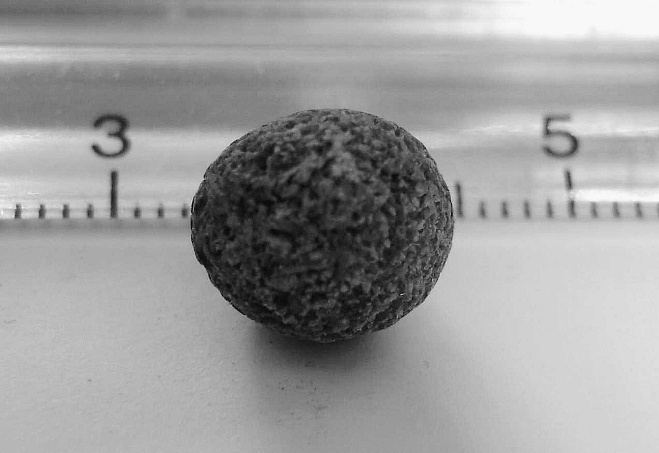
External aspect of the mixed urolith from the ferret showing its rough surface and porous structure.
Certain breeds of dogs form uroliths with different layers composed of both calcium oxalate and struvite. A struvite urolith usually forms as a result of urinary tract infection with urease-producing bacteria, whereas a calcium oxalate urolith forms due to such metabolic predispositions as hypercalciuria. 1
In the present case, the struvite component of the urolith could be related to a previous poor-quality diet and a possible dietary imbalance resulting from a change in the same, combined with a possible urinary tract infection. An explanation for the presence of calcium oxalate is more difficult. It is possible that hypercalciuria was involved, as in the case of the otters previously mentioned. As has been reported in humans with a normal excretion of citrate and a pH ≥ 6, the presence of hypercalciuria favors the formation of calcium oxalate dihydrate. 12 It was therefore hypothesized that this ferret was deficient in some inhibitors of crystal formation, aggregation, or both, which predisposed it to both urolith types. The lithogenic mechanisms involved in the formation and appearance of mixed uroliths in ferrets are not understood in detail, but the present case provides evidence for the existence of mixed uroliths in this species.
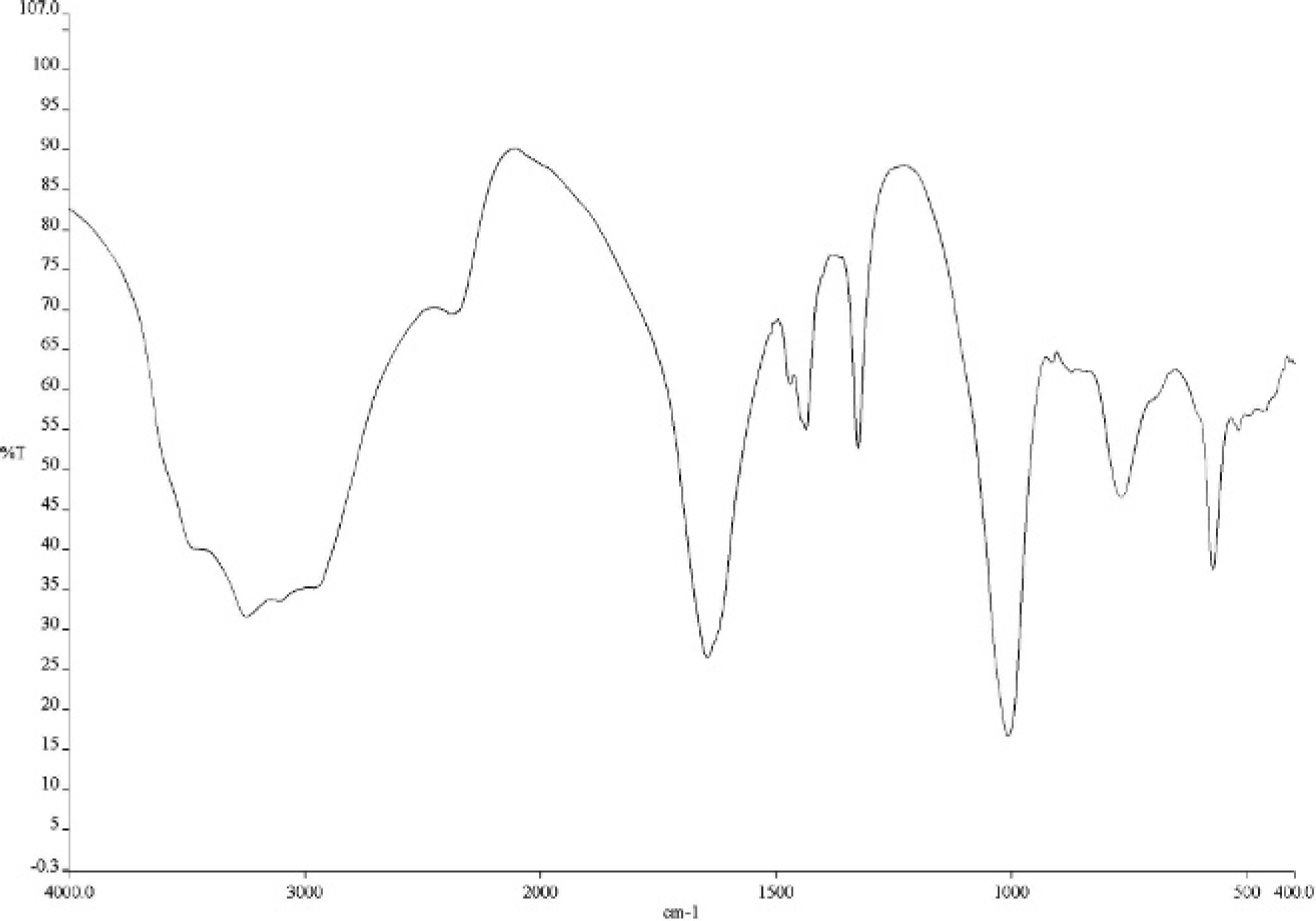
Infrared spectra of mineral composition of the mixed urolith from the ferret (see text for interpretation).
Footnotes
a.
Hill's Prescription Diet c/d Multicare Feline, Hill's Pet Nutrition, Inc., Topeka KS.
b.
FTIR 2000, Perkin Elmer Life and Analytical Sciences, Inc., Waltham, MA.