Abstract
The occurrence of neurological disease in cattle caused by Bovine herpesvirus in 11 farms from southern Brazil between 1987 and 2007 is described. Twenty-two animals were necropsied. Major clinical signs included excessive salivation, nasal and ocular discharge, circling, recumbency, depression, incoordination, grinding of teeth, and paddling movements. Necropsy findings in 10 of 22 cattle included hyperemia and softening of the rostral portions of the telencephalic cortex, with flattening of gyri, and malacia. Cattle in 10 cases did not show any gross lesions. Histological examination in most cases revealed nonsuppurative and necrotizing meningoencephalitis with acute neuronal necrosis, edema, eosinophilic intranuclear inclusion bodies in astrocytes and neurons, and infiltration of gitter cells. No histologic lesions could be detected in 4 cases. The initial diagnosis was based upon the clinical, epidemiological, and pathological findings. The diagnosis was confirmed by virus isolation in cell culture followed by virus identification by a glycoprotein C–based polymerase chain reaction. Seven isolates were identified as Bovine herpesvirus 5, and 4 were identified as Bovine herpesvirus 1.
Introduction
Bovine herpesvirus 1 (BoHV-1) and Bovine herpesvirus 5 (BoHV-5) are large, enveloped DNA viruses, belonging to the family Herpesviridae, subfamily Alphaherpesvirinae, and genus Varicellovirus. 18 Bovine herpesvirus 1 has a worldwide distribution and has been classically associated with respiratory disease (Infectious bovine rhinotracheitis virus), genital disorders (infectious pustular vulvovaginitis and infectious balanoposthitis), and abortion. 11 Bovine herpesvirus 5 infection has been associated with neurological disease and meningoencephalitis, conditions frequently reported in central–southern Brazil and Argentina. 3,7,14,15 Although BoHV-5 has been classically associated with neurological disease in calves, and less commonly in adult cattle submitted to stressing factors, 16 a few reports have described the isolation of BoHV-1 from the brain of cattle suffering from neurological disease or other conditions. 6,8,10,13,17 Both BoHV-1 and BoHV-5 are closely related genetically and antigenically, and the differentiation between them by routine diagnostic tests is difficult. 5 Cases of meningoencephalitis related to BoHV infection have often been attributed to BoHV-5 16 because this virus is far more commonly associated with neurological disease. Many of these reports, however, lack any virological characterization other than viral isolation and recognition of cytopathic effect. 3,7,15
The current study reports neurological disease in cattle caused by BoHV in 11 farms from southern Brazil. Cases occurred between 1987 and 2007. The diagnosis of BoHV was initially based on clinical signs, epidemiology, and characteristic encephalic lesions. Bovine herpesvirus infection was confirmed as the cause of neurological disease by viral isolation and by typing 11 isolates recovered from the brains of affected cattle using a glycoprotein C–based polymerase chain reaction (PCR). 19
Gross lesions in 10 of 22 affected animals that were necropsied were restricted to the brain and were characterized by hyperemia of the leptomeninges (cases 9 and 14) and hyperemia, softening, and hemorrhaging of the rostral portions of the telencephalon, flattening of gyri with segmental malacia characterized by brown–yellow discoloration of gray matter (Fig. 1), and occasional cerebellar herniation through foramen magnum (cases 1, 2, 10–13, 21, and 22). In 10 cases there where no gross lesions, and in 2 cases no information concerning necropsy findings was available.
Formalin-fixed, paraffin-embedded sections from brain and Gasserian ganglia (in 14 cases) were sectioned at 5 μm and stained with hematoxylin and eosin. Histologically, a nonsuppurative meningoencephalitis was observed in 18 of 22 cases (cases 1–16, 21, and 22). Lesions were characterized by acute neuronal necrosis (mainly in the telencephalic cortex and thalamus) and variable degrees of gliosis, edema, satellitosis, and neuronophagia. These lesions had variable distribution among the cases and among the various sections of brain regions in each case. Eosinophilic intranuclear inclusion bodies were observed in astrocytes and neurons in 6 cases (cases 1, 2, 6, 7, 11, and 21). In 10 of these 18 cases (cases 2, 6, 9–15, and 21), malacia was noted in the cerebral cortex and was characterized by necrosis of the neuroectodermal elements with maintenance of mesenchymal structures (vessels and microglia) and infiltration of gitter cells, sometimes with extensive hemorrhages. In 1 case (case 12), malacic lesions were observed also in basal ganglia and thalamus. In advanced cases, only vascular structures and a few gitter cells remained in the cortical area, leaving a cavity between white matter and leptomeninges (residual lesion). In 4 cases (cases 17–20) there were no histologic lesions. No lesions were found in Gasserian ganglia. Samples from the telencephalon from affected animals in each of the 11 farms (1 representative sample from each farm) were submitted for viral isolation. Virus isolates were typed using a previously described glycoprotein C–based PCR. 1,19 Of these, 7 isolates were identified as BoHV-5 and 4 isolates as BoHV-1 (Table 1). Even though additional animals in farms C, F, and H were not being tested by PCR, they were assumed to be infected by BoHV because they had shared identical epidemiological, clinical, and pathological findings.
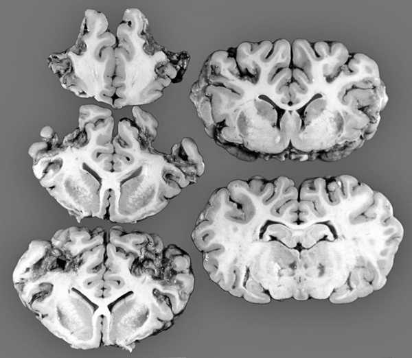
Cerebral coronal sections (case 12) showing segmental areas of cerebrocortical malacia, mainly in the rostral cortex. Basal nuclei and thalamic nuclei have softened gelatinous texture, indicating malacia.
Cases of meningoencephalitis due to BoHV have been reported more commonly in calves subjected to stressful factors, such as weaning, transport, crowding, introduction of cattle from other herds, grouping, and changes in feeding. 3,7,15 Similar stressing conditions were observed in 9 out of 11 farms in the current study Table 1. Bovine herpesvirus 1 and BoHV-5 are transmitted by direct and indirect contact and replicate in the oral, nasal, oropharyngeal, and ocular mucosa. 9 In 5 of 11 premises in the current study, cattle were kept in a feedlot situation, which provides close contact among susceptible cattle and favors the dissemination of virus within the herd. After replication in the site of entry, the virus invades nerve endings and is transported to sensory ganglia, where it replicates and establishes latency. Viral transport to the brain may result in massive virus replication and neurological disease or may result in subclinical infection. 2,12 This condition results in recovered and latently infected individuals, who may become a source of virus to other susceptible cattle in the case of virus reactivation. 9
Clinical signs were variable among the cases, but most affected cattle presented signs associated with viral replication in mucosae (nasal and ocular discharge) and with cerebrocortical lesions (paddling movements, depression, blindness, opisthotonus, and head pressing). 4 This presentation of clinical signs would be expected in cases of meningoencephalitis by BoHV, since affected cattle develop a more severe meningoencephalitis in the rostral portions of the cerebral cortex. 15,16 Excessive salivation, circling, incoordination, grinding teeth, muscle tremors, nystagmus, dysphagia, and protrusion of the tongue were observed less frequently and could be attributed to brainstem (and cranial nerves) or cerebellar lesions. 4
The characteristic distribution of the lesions in the brain of cattle suffering from meningoencephalitis due to BoHV is closely associated with the entry of virus into the central nervous system. 12 This pathway involves the axonal retrograde transport of the virus through the olfactory bulb after primary replication in nasal mucosa, invasion of cerebral cortex, replication, and production of neurological disease. 12,16
Meningoencephalitis by BoHV in cattle has some features that are similar to those of herpetic encephalitis in humans and other animal species. 20 Human herpesvirus 3 (commonly known as Varicella-zoster virus 1), herpes simplex viruses, and Equid herpesvirus 1 induce a vasculopathy and thrombosis that might lead to ischemic lesions and malacia. The pathogenetic mechanisms for the development of malacia in cases of meningoencephalitis by BoHV-1 and BoHV-5 have not been established. Despite viral infection in neurons and astrocytes, as indicated by the presence of inclusion bodies and absence of vascular lesions, malacia could be attributed to a secondary ischemic lesion caused by severe edema, which was seen in all cases in the current study. The absence of malacia in some cases may be attributed to differences in the neurovirulence of the virus or to individual susceptibility of cattle to infection, or both. 2 These differences could also explain the absence of histologic lesions in 4 calves, despite the development of characteristic clinical signs. Experimental infection in rabbits with highly pathogenic strains of BoHV-5 has consistently produced acute disease and clinical signs, many times without clear evidence of inflammatory or degenerative lesions in the brain (Flores, unpublished data).
Bovine herpesvirus 1 and BoHV-5 share similar structural, biologic, antigenic, and molecular characteristics, and differentiation by routine diagnostic assays is very difficult. 5 Therefore, cases of meningoencephalitis by BoHV are frequently reported to be caused by BoHV-5 based on the characteristics of virus growth and the cytopathic effect produced in cell cultures, as well as on the epidemiologic, clinical, and pathological findings. 3,7,16 In this sense, it is generally accepted that cases of meningoencephalitis by BoHV-1 are uncommon. 8,10,13,17 In 7 cases in the current study, PCR identified the virus as BoHV-5, and in 4 others the virus was identified as BoHV-1. These data show that cases of meningoencephalitis by BoHV-1 are not as uncommon as previously suspected, at least in southern Brazil. Similarly, analysis of 26 herpesvirus isolates from cattle with neurological disease from others regions of Brazil, Uruguay, and Argentina determined that 21 isolates were BoHV-5 and 5 were BoHV-1. 19 In summary, neurological disease in cattle caused by BoHV-1 and BoHV-5 infection share similar epidemiological, clinical, and pathological findings, characterized in most cases by nonsuppurative and necrotizing meningoencephalitis, mainly in the frontal telencephalic lobes. The differentiation of the causal agents can be difficult, but in the cases reported here, a glycoprotein C–based PCR provided good discrimination between BoHV-1 and BoHV-5.
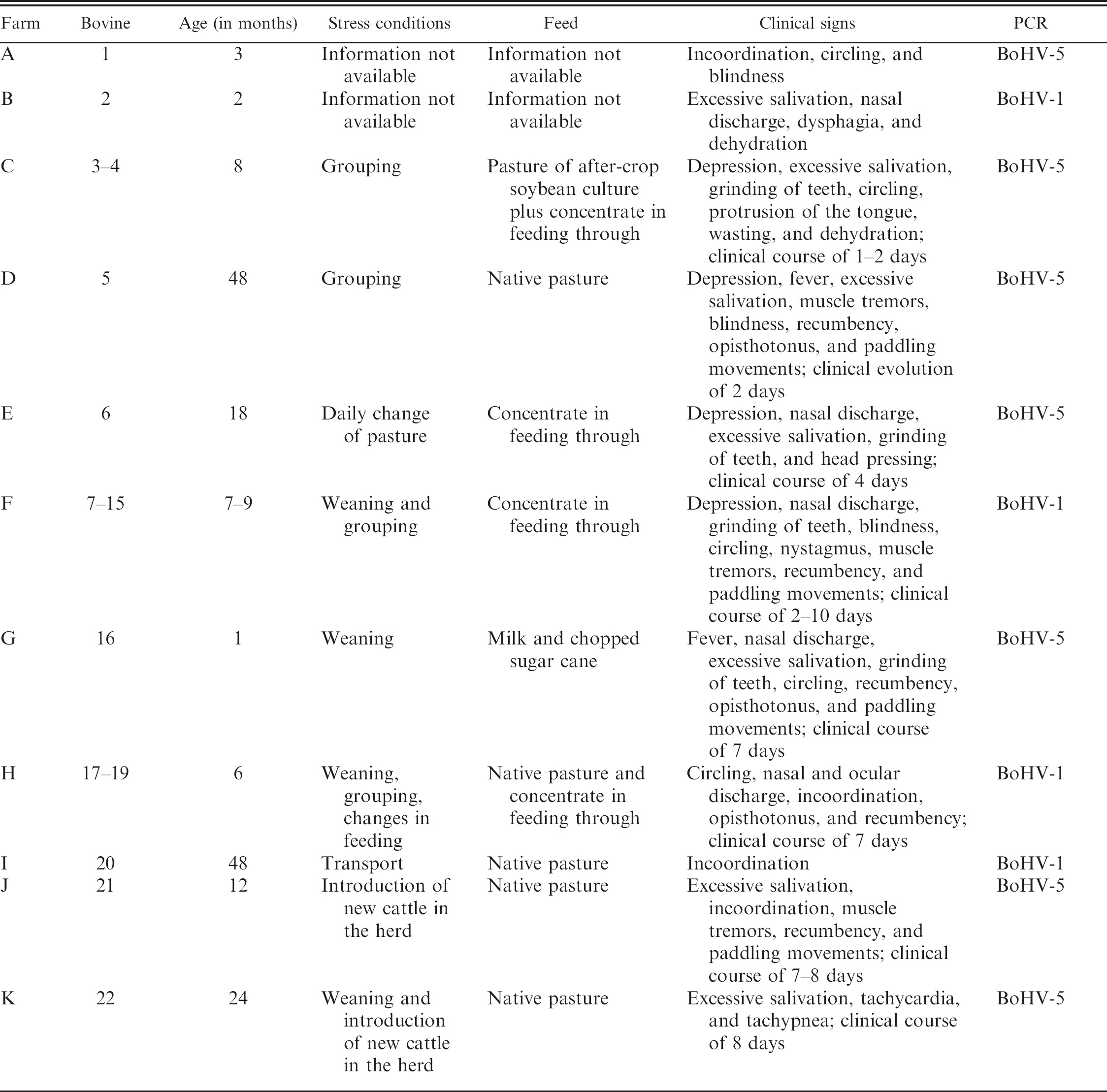
Epidemiological, clinical, and virological data of cattle affected by neurological disease caused by Bovine herpesvirus in 11 farms in southern Brazil.