
Abstract
In a review of 159 archived bovine autopsy cases submitted to the Georgia Veterinary Diagnostic Laboratory System from 2001 to 2017, we evaluated histologic lesions and other laboratory test results in cases diagnosed with central nervous system (CNS) disease to determine the most common disorders and the rate of definitive morphologic and etiologic diagnosis within the population. The most common breed was Aberdeen Angus or Angus (31%), and female animals comprised 60% of the overall submissions. Gross anatomic changes were described in 33% of cases that had histologic lesions. Suppurative meningoencephalitis was the most common diagnosis (28% of cases overall) and was frequently associated with bacterial sepsis. Lymphocytic meningoencephalitis without an identified etiology was diagnosed in 17% of cases. Polioencephalomalacia was the most commonly identified noninfectious diagnosis (17%). Hydrocephalus was the most common CNS comorbidity and potential congenital defect. Identification of specific agents was sporadic, and infectious disease testing was largely dependent on fluorescent antibody testing, almost all of which were negative. Our findings indicate the common differential diagnoses for cattle with neurologic signs in the southeastern United States, as well the need for a well-defined, methodical diagnostic approach, including autopsy, histology, and appropriate additional laboratory testing.
Neurologic disease is a common entity (~3–8% of cases) in beef and dairy cattle production medicine, and can be attributed to infectious, nutritional, metabolic, and genetic causes, among others.4,7,10,12,15 Affected cattle can develop a wide variety of clinical signs that can be nonspecific and subtle, making diagnosis difficult without a detailed autopsy and histologic examination, as well as microbiologic and toxicologic analysis. 17 Neurologic disorders can also be progressive, debilitating, and almost inevitably result in death or euthanasia. 4 Additionally, many neurologic diseases can be zoonotic, necessitating more thorough and rapid testing of potentially affected animals and assessment of human exposure, especially in rabies-suspect cases. 4 Although many neurologic diseases are well known to practitioners and producers, emerging agents, such as Schmallenberg virus and bovine astrovirus, have been identified in recent years and may pose a diagnostic challenge.8,11 Virus-induced neurologic disease is often characterized by lymphocytic or lymphoplasmacytic meningoencephalitis (LM; also referred to as nonsuppurative meningoencephalitis), but in many cases in which a cause cannot be established, LM cases remain idiopathic.14,16 Although studies investigating subsets of LM in cattle have identified specific infections associated with the inflammatory changes, the cause remains unknown in the majority of cases.14,16 Overall, accurate diagnosis of neurologic disease in cattle exemplifies the benefits of a systematic diagnostic process supported by valuable contributions from autopsy, histology, and other laboratory testing.
Herein we describe retrospectively cases of neurologic disease of cattle submitted for autopsy at the Athens Veterinary Diagnostic Laboratory (AVDL; Athens, GA) from 2001 to 2017 and at the Tifton Veterinary Diagnostic and Investigational Laboratory (TVDIL; Tifton, GA) from 2009 to 2017. Electronic web-based archives at both the AVDL and TVDIL were searched for bovine autopsy cases with a diagnosis of central neurologic disease using the following terms: abscess, amoeba, fungal encephalitis, Histophilus somni, hepatic encephalopathy, herpesvirus, hydrocephalus, Listeria, malignant catarrhal fever, meningoencephalitis, Mycobacterium, Neospora, polioencephalomalacia, pituitary abscess, porencephaly, rabies, salt poisoning, spinal, and Toxoplasma. Submission forms and autopsy reports from retrieved cases were reviewed, and cases were included in the study if a final gross and/or histologic morphologic diagnosis related to central nervous system (CNS) disease was achieved. All cases were reviewed by a pathologist (LL Clarke/DR Rissi at AVDL; IK Hawkins at TVDIL), and cases were excluded if no slides including CNS tissues were found to review. The breed, sex, age, type of submission (full or partial autopsy [only head submitted]), pathologic changes, and laboratory tests for each case were reviewed. The age was classified as <2 mo old (including fetuses and neonates), 2–12 mo old, and >12 mo old. Archived brain and/or spinal cord tissue sections were reviewed by all 3 authors to confirm the reported changes. Averages, percentages, and odds ratios were calculated using Excel (Microsoft, Redmond, WA).
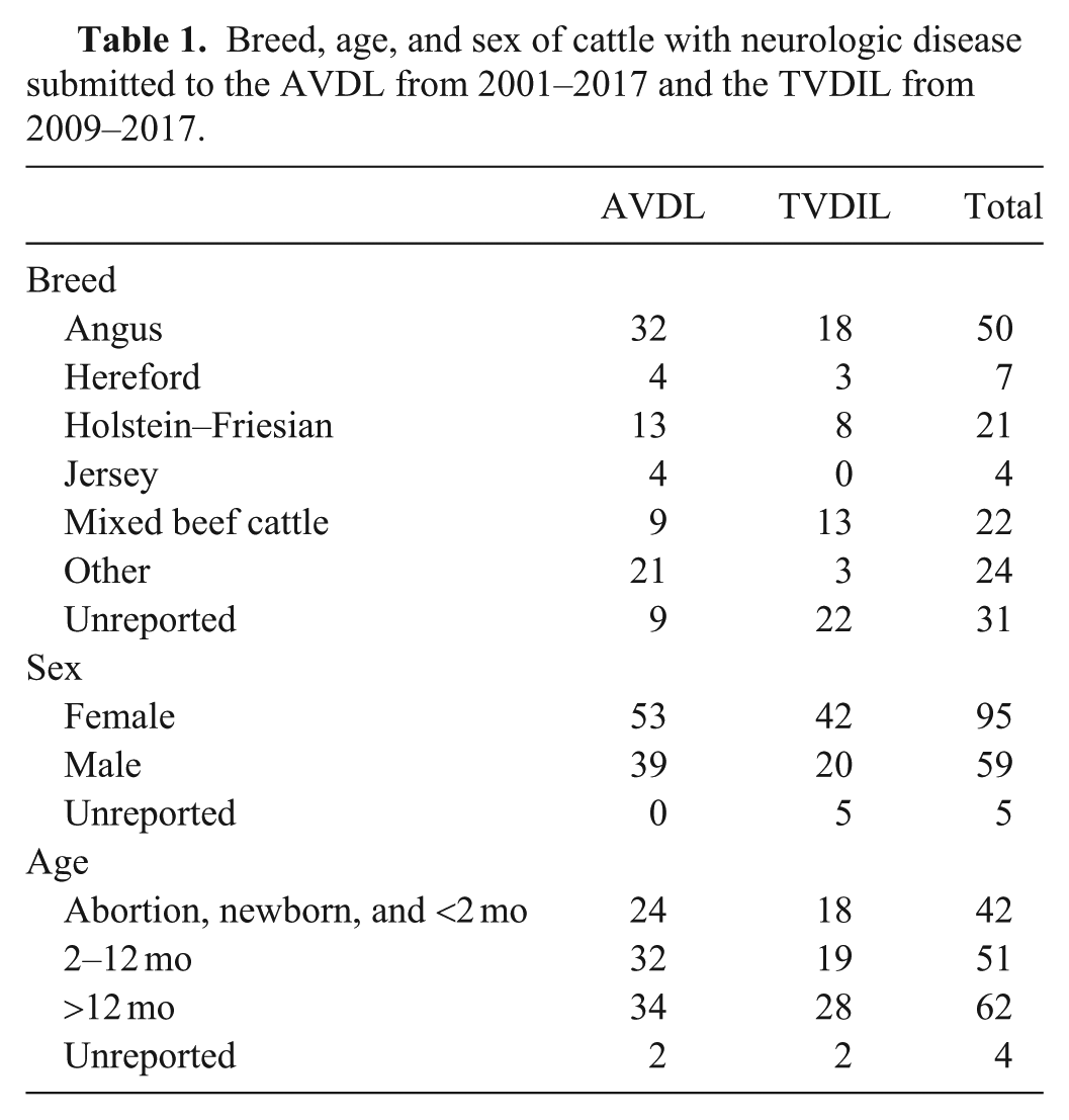
We identified 92 cases of neurologic disease in cattle from the AVDL (2001–2017) and 67 cases from the TVDIL (2009–2017) archives (Table 1). During the same time periods, 1,407 bovine autopsies were conducted at the AVDL and 1,657 bovine autopsies at the TVDIL, with neurologic diseases comprising 6% and 4% of submissions, respectively. Aberdeen Angus or Angus was the most common breed in both data sets and overall (31% of cases), followed by Holstein–Friesians (13%). This was similar to percentages of these breeds in overall bovine autopsy submissions (30% Aberdeen Angus and 19% Holstein–Friesian). Female cattle comprised 58% of AVDL cases and 63% of TVDIL cases (60% of overall cases). Cattle >12 mo old were the most frequently represented age group from the AVDL, the TVDIL, and overall (36–42%). Fourteen cases (9 from AVDL; 5 from TVDIL) were submitted as partial autopsies; the remainder were whole body autopsies. The spinal cord was examined in 14 cases (12 from AVDL; 2 from TVDIL).
Breed, age, and sex of cattle with neurologic disease submitted to the AVDL from 2001–2017 and the TVDIL from 2009–2017.
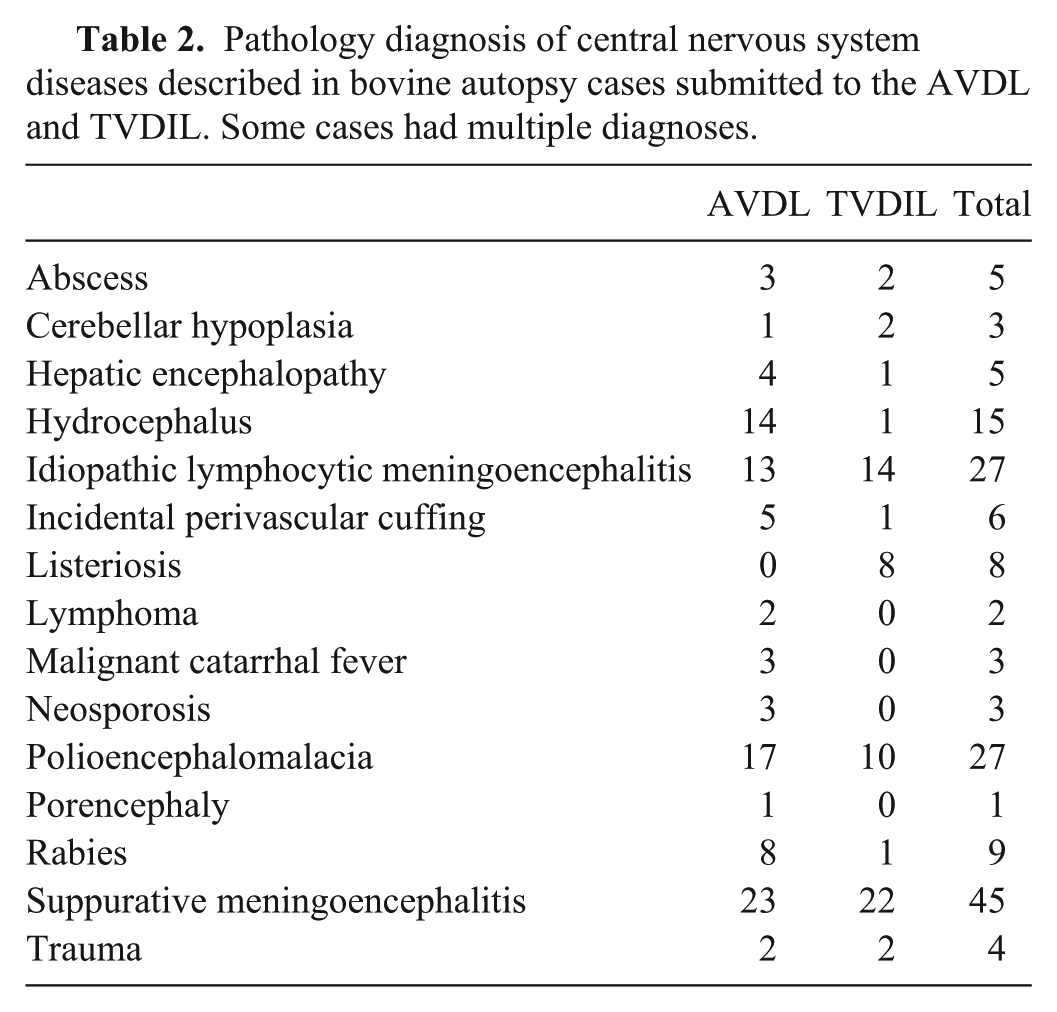
Some cases had multiple diagnoses (Table 2). The most frequent diagnosis in both datasets was suppurative meningoencephalitis (SME; Tables 2, 3). Gross anatomic changes in the brain were present in 56% of AVDL SME cases and in 36% of TVDIL SME cases, and included yellow cloudiness and thickening of the leptomeninges, leptomeningeal hemorrhages, dilation of the lateral ventricles (secondary hydrocephalus as a result of ependymal damage), and cerebellar herniation through the foramen magnum. Histologic changes consisted of perivascular infiltration of neutrophils, fibrin, and hemorrhage, and occasional bacterial organisms throughout the leptomeninges and neuroparenchyma. The main comorbidity associated with SME was septicemia, which was diagnosed histologically in other tissues (70% from AVDL and 53% from TVDIL of SME cases), and was observed primarily in cattle <2 mo old. Listeria monocytogenes infection was confirmed in 8 TVDIL cases via aerobic or Listeria-specific bacterial culture (66% of all TVDIL cases tested). The main histologic changes in listeriosis cases included microabscesses and lymphocytic perivascular infiltrates involving the neuroparenchyma, often with intralesional gram-positive bacterial rods. Lesions were most commonly observed in the brainstem and cerebellum. No cases of L. monocytogenes infection were identified at the AVDL, where a fluorescent antibody test (FAT) was the preferred confirmatory method (18% of all AVDL cases tested). Abscesses were identified in 3 AVDL cases and 2 TVDIL cases. Abscesses occurred in the pituitary gland (2 cases), cerebral leptomeninges, brainstem, and spinal cord, in which inflammation extended from the vertebral body (1 case each). Trueperella pyogenes was identified as the cause of 3 abscesses via bacterial culture; no bacteria were isolated from the remaining 2 abscesses. An additional subset of cases of meningoencephalitis that were not overtly suppurative or neutrophilic, but were histologically most consistent with a bacterial origin, were identified in both datasets (4 cases total), but no specific pathogens were identified in this subset by culture or FAT.
Pathology diagnosis of central nervous system diseases described in bovine autopsy cases submitted to the AVDL and TVDIL. Some cases had multiple diagnoses.
Suppurative meningoencephalitis in cattle submitted to the AVDL and TVDIL.
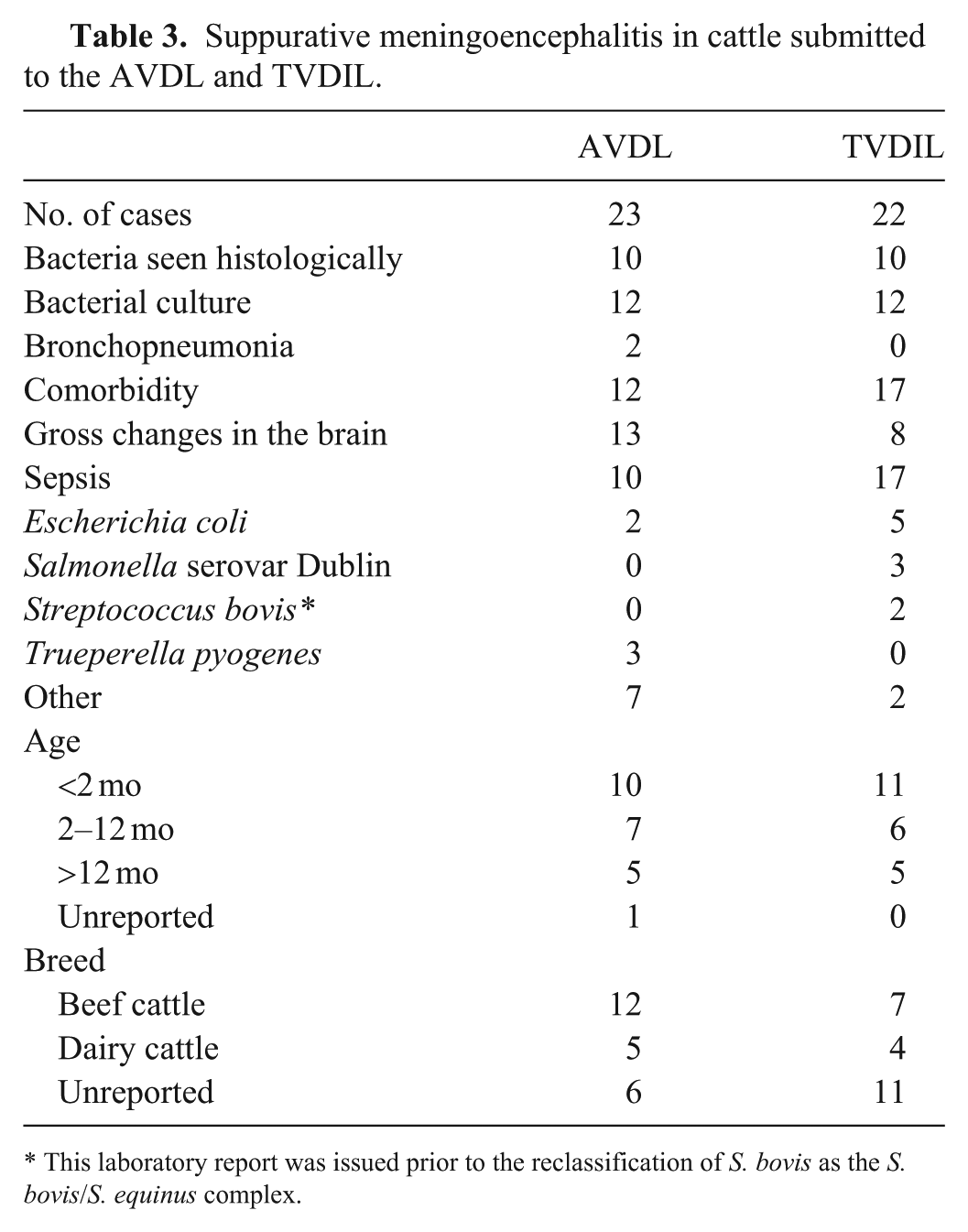
This laboratory report was issued prior to the reclassification of S. bovis as the S. bovis/S. equinus complex.
Twenty-seven cases from the AVDL (29%) and 15 from the TVDIL (22%) had histologic evidence of LM. Gross neuropathology changes were absent in all cases identified as LM histologically. The main established causes of LM were rabies, malignant catarrhal fever (MCF), and Neospora spp. infection. Rabies was diagnosed in 8 AVDL cases and 1 TVDIL case. Characteristic neuronal intracytoplasmic eosinophilic inclusions (Negri bodies) and lymphoplasmacytic meningoencephalitis were identified histologically in all cases diagnosed as rabies. Laboratory confirmation was achieved via FAT. Of all cases, 44 AVDL cases and 22 TVDIL cases were tested for rabies using FAT (48% of all AVDL cases tested; 33% of all TVDIL cases tested). MCF and Neospora spp. infection (3 cases each) were only identified in the AVDL dataset. MCF was characterized by LM and vasculitis. All 3 cases had characteristic lesions in other organs. MCF was confirmed by PCR specific for ovine herpesvirus 2 in 2 cases and by FAT in 1 case. Of all cases, 4 were tested for MCF using FAT, 2 using PCR, and 1 using virus isolation (3.6% of all AVDL cases tested). Neosporosis was characterized by LM with neutrophilic aggregates and neuronal necrosis associated with protozoal merozoites identified on immunohistochemistry (IHC), but these were less readily identified in hematoxylin and eosin–stained sections. Neospora spp. PCR was used to confirm histologic findings in one case and was positive. Myocarditis was described in all 3 cases. Of all of the cases, 5 cases from the AVDL (5.4%) and 4 cases from the TVDIL were tested using Neospora FAT (6.0%), none of which were positive. One case was positive on Neospora IHC but negative on FAT.
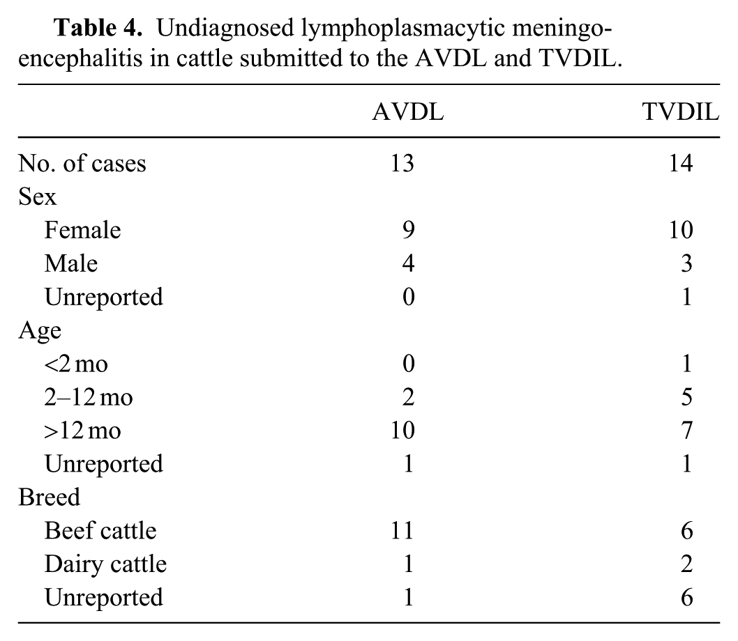
LM was identified via histology in 13 AVDL cases and 14 TVDIL cases without an identified etiologic agent (Tables 2, 4). Common histologic changes in these cases included infiltration of perivascular spaces in the leptomeninges and parenchyma with variable numbers of lymphocytes, macrophages, and plasma cells, with astrocytosis and astrogliosis, microglial nodules, neuronal necrosis, and neuronophagia. Gray and white matter were affected, but lesions were more evident in the white matter. No predilection for a specific neuroanatomic location was found; however, available tissue sections of brain were inconsistent among cases. In the AVDL cases, lesions were described in the cerebellum (5 cases), cerebrum (5), telencephalon (including the thalamus and hippocampus) (4), midbrain (2), and brainstem (3). In TVDIL cases, lesions were described in the cerebellum (5), cerebrum (5), telencephalon (5), and brainstem (5). Multiple unsuccessful laboratory assays were attempted in addition to histology to identify an infectious origin for the LM cases, varying by pathologist discretion and availability. Such tests included, in various combinations, FAT for rabies (18 cases), bovine herpesvirus 1 (9), bovine viral diarrhea virus (BVDV; 8), Listeria spp. (4), Toxoplasma spp. (4), Neospora spp. (2), MCF (2), bovine coronavirus (4), and bovine rotavirus (1); IHC for rabies (2), encephalomyocarditis virus (1), Listeria spp. (1), and Toxoplasma gondii (1); PCR for eastern and western equine encephalitis virus (2) and West Nile virus (1); aerobic culture (7) or Listeria spp. culture (5); and virus isolation (9). In 9 cases, LM was attributed in the report by the original pathologist to systemic changes secondary to non-neurotropic lesions, including clostridial myonecrosis, bacterial sinusitis, and bacterial endometritis. In addition to these pathology cases, there were 5 AVDL cases and 1 TVDIL case that were originally diagnosed as LM, but on histologic review, there was only minimal lymphocytic perivascular cuffing around a few vessels (Table 2). This change has previously been described as a histologic finding in nonclinical cattle and was considered incidental in these cases. 6
Undiagnosed lymphoplasmacytic meningoencephalitis in cattle submitted to the AVDL and TVDIL.
Degenerative or congenital conditions were also identified. Primary hydrocephalus was diagnosed in 9 AVDL cases, 7 of which had concurrent non-CNS congenital defects, including microphthalmia, thyroid hypoplasia, pulmonary artery stenosis, thoracic spinal scoliosis, unilateral cryptorchidism, patent foramen ovale, bilateral hydronephrosis and hydroureter, and partial palatoschisis. Two cases of hydrocephalus had other primary CNS lesions, one with concurrent porencephaly, and one with concurrent cerebellar hypoplasia. Histologic changes in primary hydrocephalus included thinning of the adjacent parenchyma and attenuation of ependymal cells, with occasional associated gliosis, neuronal necrosis, and infiltration with foamy macrophages. FAT on brain tissue for BVDV was negative in 7 primary hydrocephalus cases; one case was diagnosed as BVDV infection via a positive skin IHC, but no abnormalities other than hydrocephalus were noted. Other cases of hydrocephalus were considered secondary to SME (3 cases), cerebral abscess, LM, and lymphoma (1 case each), all with histologically evident damage to the ependyma. Three cases of cerebellar hypoplasia were identified (1 from AVDL, 2 from TVDIL), all late-term abortions. Histologic lesions included reduced Purkinje cells and a thinned molecular layer. All 3 cases were tested for BVDV by FAT (one additionally tested by BVDV ELISA), and all were negative.
Noninfectious disease was diagnosed in 38 cases. Polioencephalomalacia (PEM) was diagnosed in 17 AVDL cases and 10 TVDIL cases. Histologic lesions in all cases were similar, including multifocal-to-laminar cerebrocortical neuronal necrosis with edema and gliosis. Lead toxicity was confirmed by liver and kidney analysis in 1 of 6 tested AVDL PEM cases; sulfur toxicosis was confirmed by ruminal content analysis in 3 of 10 TVDIL PEM cases. The underlying etiology of the remaining 23 cases is unknown. Hepatic encephalopathy was diagnosed in 4 AVDL cases and 1 TVDIL case, characterized histologically mainly by cortical Alzheimer type II astrocyte formation and spongiosis of white matter tracts. These were attributed to severe hepatic fibrosis with destruction of the limiting plate; the cause was unknown in all cases.
Cerebral lymphoma was diagnosed in 2 AVDL cases. Histologically, large neoplastic lymphocytes expanded the leptomeninges diffusely, as well as surrounding parenchymal vessels, and extended onto the epidural surface of the dura mater in the spinal cord. In one case, B-cell lymphoma was confirmed via CD79a IHC; the second was not further characterized. Neither animal had evidence of lymphoma in any other organs.
Two cases from the AVDL and 2 cases from the TVDIL were diagnosed with trauma. Grossly evident subdural hemorrhage or yellow-brown discoloration of the meninges suggestive of xanthochromia was described in 3 cases. Histologic lesions included shrunken, necrotic neurons, multifocal areas of meningeal and parenchymal hemorrhage, and axonal spheroids in the cervical spinal cord. Extra-cranial lesions were not described in any case.
The neurologic diseases in our study are similar to the disorders reported in similar retrospective studies in the Americas and Europe. In Mexico, rabies, botulism, thrombotic meningoencephalitis caused by H. somni, and PEM were the most commonly identified neurologic diseases of cattle. 12 In a Brazilian study, the main bovine neurologic diseases were rabies, hepatic encephalopathy, bovine herpesviral meningoencephalitis, cerebral babesiosis, Solanum fastigiatum poisoning, MCF, and PEM. 13 An Italian study reported PEM as the most common cause of seizures in cows, 5 whereas a Scottish study reported listeriosis, vertebral osteomyelitis, and encephalitis of unknown cause as the main neurologic diseases. 7 A retrospective study using cerebrospinal fluid to investigate bovine neurologic diseases 15 identified listeriosis and neonatal bacterial meningoencephalitis, followed by PEM and hepatic or uremic encephalopathy, as the most frequent diagnoses. A British study concentrating on the surveillance of bovine spongiform encephalopathy identified cases of listeriosis, LM of unknown cause, thromboembolic or granulomatous encephalitis, neuroectodermal tumors, and PEM as the most common disorders. 9
Our study is likely more comprehensive than these previous investigations, given the larger sample size, greater retrospective time frame, and the variety of cattle operations that utilize the AVDL and the TVDIL. Overall, no particular breed, sex, or age demographic was statistically more likely to develop a specific neurologic disease when compared to selected neurologic cases overall. Similar to other studies,12,16 gross anatomic changes were reported in the minority of cases (42% of AVDL cases and 21% of TVDIL cases) and were often nonspecific when reported. Only 33% of all cases had gross anatomic changes reported that suggested a specific diagnosis, most frequently in cases of SME or hydrocephalus. A definitive etiology (bacterial, viral, or nutritional) was diagnosed in only 28 AVDL cases and 25 TVDIL cases, or 33% overall. We believe that this low diagnostic rate is the result, in large part, of the underutilization of laboratory testing or use of less sensitive testing methods.
Bacterial SME was the most frequent histologic diagnosis, and most affected animals had lesions outside the CNS, including sepsis and bronchopneumonia. The source of infection was not identified in all septic cases, but the majority were young calves with multiple organ involvement. Identification of common concurrent viral infections was not seen with these cases, indicating these were either primary bacterial infections or that laboratory methods failed to identify underlying viral infections. A diagnosis of listeriosis was more commonly achieved at the TVDIL than the AVDL, indicating possible geographic or environmental differences, regional differences in the management style of cattle operations, or problems related to sample submission and/or laboratory methods. Pituitary and meningeal abscesses were most commonly associated with T. pyogenes, which is consistent with previous reports. 4
Identified causes of LM in our case series were rabies, MCF, and Neospora spp. infection. The underlying cause of LM in a larger subset of our cases was undetermined. Although similar cases have been sporadically attributed to bovine astrovirus infection, 8 our findings are consistent with what has been reported in most other laboratories, in which LM remains an idiopathic disorder after tissues are tested unsuccessfully for multiple pathogens.14,16 Further characterization and diagnostic efforts for these cases are a valid priority, given that many of the potential etiologic agents, such as Schmallenberg, are, as of mid-2019, non-endemic to the United States and others, such as rabies, are zoonotic. New laboratory methods such as deep genomic sequencing, as well as more complete and systematic testing with existing assays, may be of use to identify potential causative agents in these cases. Idiopathic LM has been reported in other animal species and poses a diagnostic challenge for practitioners and neurologists alike. 2
In SME, LM, and congenital cases, FATs used to evaluate for potential viral causes were largely negative, and there was an overall lack of viral diagnoses. Higher rates of positivity were achieved when IHC, culture, and/or PCR was used in combination with the gross autopsy and histology. FATs for rabies in cattle have been shown to be less sensitive than IHC, attributing the discrepancy to sample integrity, individual reactions to disease, incubation period, and other influencing factors. 3 Likewise, the TVDIL had a much higher rate of diagnosing listeriosis, relying primarily on culture, than the AVDL, which primarily tested using FAT. In the case of congenital defects, although infection with BVDV is the most likely cause for these lesions in the United States, 1 this virus was infrequently identified in the congenital cases examined herein, which were almost all tested using FAT. This observation may help to guide testing protocols, given that several foreign animal diseases, including Schmallenberg, Aino, Akabane, and some serotypes of bluetongue, may look similar.
Several differences were found between our data and previous reports. We found fewer cases of CNS lymphoma or other types of neoplasia than would be expected from previous studies,9,15 and we identified no cases of distal lumbar spinal lymphoma. Additionally, we found no cases of fungal or amoebic meningoencephalitis, mycobacteriosis, toxoplasmosis, herpesviral encephalitis, or thromboembolic meningoencephalitis caused by H. somni. None of the SME or bacterial meningoencephalitis cases were associated with otitis media or interna, which was expected in a percentage of cases. We would speculate that, in many of these diseases, diagnosis can be achieved without a CNS examination, and because this examination is cost-prohibitive for producers and time-intensive for pathologists, it is frequently foregone in cases of systemic disease in which the etiology can be identified in other, more accessible, tissues. In other instances, such as H. somni, organisms can be difficult to isolate in culture or recognize on histology.
Potential biases in our study stem from case selection, given that we only included cases in which intracranial and spinal nervous tissue was included in the autopsy, thereby excluding cases in which a CNS examination was not specifically requested or not indicated by clinical signs. Our sampling was also biased by excluding cases in which clinical neurologic disease may have been reported, but no gross or histologic lesions were identified, as can be the case with numerous toxic or nutritional etiologies. Other limitations include the lack of uniform testing among cases, including the variation of histologic sections available for each case, the lack of fresh tissue given the retrospective nature of the study, and a lack of reasoning in reports behind the use of some of the laboratory tests and positive controls.
We confirmed that autopsy, histology, and other laboratory testing are important contributing components of a neurologic disease investigation, given that grossly evident lesions of disease are subjectively inconsistent and nonspecific. Zoonotic agents (rabies, Salmonella, Listeria) were identified in several cases that may require public health follow-up to protect exposed personnel. Bacterial agents were the most commonly identified cause of neurologic disease in cattle and one of the most readily diagnosed. Viral agents were less commonly identified, a result that may be due in part to heavy reliance on FATs, which are known to be less sensitive than other methods (culture, IHC, and PCR). Lastly, a significant proportion of cases with similar histologic changes were gathered under the descriptor LM, for which a definitive diagnosis has remained elusive. Renewed evaluation of existing laboratory methods and development of new laboratory tests may help to identify etiologies in these cases.
Footnotes
Declaration of conflicting interests
The authors declared no potential conflicts of interest with respect to the research, authorship, and/or publication of this article.
Funding
The authors received no financial support for the research, authorship, and/or publication of this article.