
Abstract
Piscirickettsia salmonis is the etiologic agent of piscirickettsiosis, an economically significant disease offish. Isolation of P. salmonis by culturing on fish cell lines has been the standard technique since the initial isolation of the organism. The ability to grow P. salmonis on artificial media would relieve facilities of the cost of maintaining cell lines, permit isolation at fish culture sites with fewer contamination problems, and allow easier transport of isolates to diagnostic facilities for confirmation assays. This report describes the successful culture of P. salmonis on enriched blood agar.
Keywords
Piscirickettsia salmonis causes a systemic infection (piscirickettsiosis) in salmonids 5,7 and seabass. 4 Epizootic mortality caused by piscirickettsiosis has ranged from less than 1% to greater than 90%. 13 Cell culture has been the gold standard for the isolation of P. salmonis since its characterization as the etiologic agent of piscirickettsiosis in the early 1990s. 11 The isolation of P. salmonis from fish tissues or water samples is challenging, because the bacterium is sensitive to the low levels of antibiotics routinely used in cell culture. With no antibiotics in the culture media, contamination is a major problem when inoculating with tissues collected from morbid fish. In addition, many of the fish culture facilities are in locations that do not have the capability of maintaining cell cultures. For this reason, preliminary diagnosis of piscirickettsiosis is normally made by examining Gram-stained, Giemsa-stained, or methylene blue-stained kidney imprints or smears. 6,11 Confirmation of piscirickettsiosis is by serologic methods (e.g., fluorescent antibody test 11 and enzyme-linked immunosorbent assays 1,9 ) or by molecular assay (e.g., polymerase chain reaction [PCR] 12 ). The ability to grow P. salmonis on artificial media would relieve facilities of the cost of maintaining cell lines, permit isolation at fish culture sites with fewer contamination problems, and allow easier transport of isolates to diagnostic facilities for confirmation assays.
In addition to the problems associated with diagnostics, research into virulence factors and the antigenic makeup of P. salmonis has been hampered by the need to purify the bacteria away from the cell culture host material. 2,3,8,10,15,16 The growth of P. salmonis on artificial media will eliminate the need for these protocols in many cases and will vastly simplify preparation of the bacteria.
Three isolates of P. salmonis were used to test the culture media: the original isolate from Chile, LF-89 7 (provided by J. L. Fryer, Oregon State University, Corvallis, OR). originally cultured from Coho salmon (Oncorhynchus kisutch); an isolate from Canada, ATL-4–91, isolated from Atlantic salmon (Salmo salar; provided by G. Traxler. Pacific Biological Station, Nanaimo, BC); and an isolate from Norway, NOR-92, also isolated from Atlantic salmon (provided by H. P. Melby, National Veterinary Institute. Oslo, Norway). Freshly thawed material from frozen Chinook salmon embryo (CHSE-214) cell culture super-natants, infected with P. salmonis, was streaked onto 5% sheep blood a agar plates that had the addition of 3% fetal bovine serum b (FBS), 0.1% cysteine, c and 1% glucose, d designated BFCG. At the same time, CHSE-214 cell cultures were inoculated with the frozen P. salmonis supernatants and incubated at 16°C.
The BFCG cultures were incubated at 16°C; after 6 days, small (0.1 mm) circular, entire, white, convex colonies were visible. A single colony was picked and restreaked for isolation on a BFCG plate and was incubated as before. BFCG plates without the added FBS, designated BCG, or without the cysteine, designated BFG, were inoculated and incubated as before. Colonies identical to those previously described were observed after 6 days on the BFCG and the BCG (Fig. 1) cultures, but no growth was observed after 3 weeks on the BFG cultures. This indicates that the addition of cysteine, but not FBS, is necessary for growth of P. salmonis. This is similar to the media used to isolate and culture a new Francisella sp. described from cod. 14 Eight days after inoculation, cytopathic effect (CPE) typical of P. salmonis was observed in the CHSE-214 cell cultures inoculated with the freshly thawed P. salmonis infected supernatant.
To confirm that P. salmonis was the organism growing, after 6 days of incubation, a single colony was picked from a BCG plate and suspended in 50 μl of sterile distilled water. In addition, 1 ml of the CHSE-214 P. salmonis-infected culture supernatant was removed 10 days after inoculation. The cell culture supernatant was centrifuged to pellet the bacteria and cell debris. The pellet was then resuspended in 100 μl of sterile distilled water. Five microliters of each suspension were used in a 50-μl P. salmonis-specific PCR. 12 The P. salmonis-specific PCR produced an amplicon of 467 base pairs (bp) with both the cell culture supernatant and the plate isolate suspension, confirming the cultured organism's identity as P. salmonis.
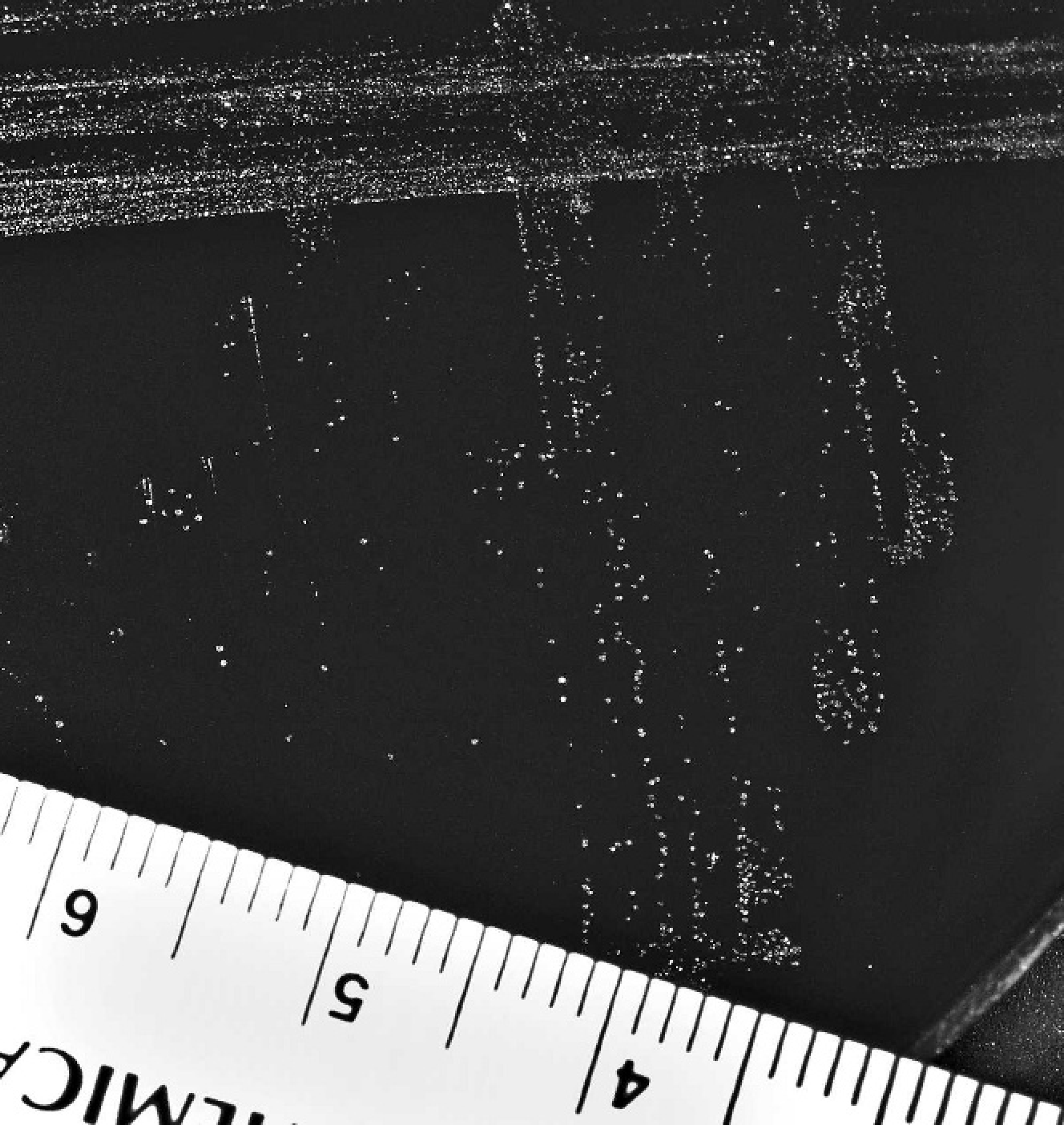
Colonies of Piscirickettsia salmonis, LF-89, after 6 days of incubation at 16°C cultured on 5% sheep blood agar with 0.1% cysteine and 1% glucose. Ruler denotes centimeters.
Single colonies of P. salmonis from a BCG plate were used to inoculate CHSE-214 cell cultures. After 8 days (16°C), typical P. salmonis CPEs were observed in the inoculated cultures. Supernatant was collected from the inoculated cell cultures on day 10 and a P. salmonis-specific PCR performed as described earlier. The PCR produced the 467-bp product as expected. Supernatant from the cell cultures showing CPE was streaked onto the BCG media and incubated as described earlier. After 6 days, small (0.1 mm) circular, entire, white, convex colonies identical to the colonies observed previously were visible.
The efficacy of the enriched blood agar media for primary isolation from fish tissues compared with cell culture isolations needs to be compared before this media can become a standard technique. However, the ability to isolate P. salmonis on artificial media will greatly simplify the identification of piscirickettsiosis in remote fish-culturing facilities. Correct and early identification of the agent will enhance treatment and prevention strategies. Using the ability to grow P. salmonis on artificial media as a standard protocol to determine the antibiotic sensitivity of the P. salmonis isolates can also be developed. The ability to grow P. salmonis in the absence of host materials may enhance the identification of virulence factors and vaccine candidate proteins. In conclusion, the ability to culture P. salmonis on artificial media should greatly improve diagnostics and research on this disease agent.
Footnotes
a.
Fisher Scientific, Sparks, MD.
b.
Invitrogen, Carlsbad, CA.
c.
Sigma-Aldrich, St. Louis, MO.
d.
Fisher Scientific, Fairlane, NJ.