
Abstract
Anatoxin-a, a toxin produced by several genera of blue-green algae, is considered a potent neurotoxin. Ingestion of water contaminated with the toxin results in acute neurological signs and often death. This report describes fatal cases of anatoxin-a ingestion in 6 dogs, with confirmation of anatoxin-a exposure by liquid chromatography/tandem mass spectrometry (LC-MS/MS/MS). In 1 outbreak, 3 dogs developed seizures and died within an hour after swimming in a river in California, while the other outbreak involved 3 dogs that died within 1 hour after swimming in a pond in Ontario. Anatoxin-a poisoning is rarely reported in dogs as a cause of acute neurological signs and death. However, increased occurrences of blue-green algae blooms in North America make this neurotoxin an important consideration in the diagnosis of sudden death associated with environmental water exposure. This brief communication reports on the isolation and detection of anatoxin-a from environmental water sources and the stomach contents of North American dogs dying of acute neurotoxicosis. This demonstrates the first documented cases of anatoxin-a poisoning in dogs in North America and the importance of LC-MS/MS/MS in identifying neurotoxins responsible for sudden death in cases of suspected blue-green algae toxicosis; especially those cases showing no gross or histological lesions.
Keywords
Reports of anatoxin-a toxicosis are less frequent than poisoning caused by the hepatotoxic blue-green algal toxins; however, poisoning has occurred worldwide. 1,5,6,9,10 Anatoxins are mainly produced by cyanobacteria in the Anabaena genus, 1 but also by other genera, such as Oscillatoria, Planktothrix, Microcystis, Aphanizomenon. Woronichinia, Cylindrospermum, and Phormidium. In dogs, anatoxin-a poisonings have been reported in Europe, 5,9,10,12 but to the authors' knowledge, this is the first documented report from North America. Anatoxin-a is also considered a contributing factor in the deaths of calves in the United States, 4 and Lesser Flamingos in Kenya. 13 Anatoxin-a poisoning may result in high mortality with clinical signs of muscle fasciculations, seizures, collapse, cyanosis, and death. 5,10 Although only a limited number of cases have been reported, it appears that treatment of anatoxin-a poisoning is of little or no benefit, and the outcome is usually lethal. Rapid death associated with anatoxin-a intoxication restricts pathological assessment as lesions are nonspecific or absent. Therefore, diagnostic approaches to confirm anatoxin-a poisoning need to be based on the identification of anatoxin-a in biological specimens. This report describes the diagnostic investigation of 6 fatal anatoxin-a poisonings in dogs, with confirmation of exposure through detection of anatoxin-a in stomach contents and water samples by liquid chromatography/tandem mass spectrometry (LC-MS/MS/MS).
In August 2002, 3 dogs died after swimming in the south fork of the Eel river in Humboldt County, California. The first 2 dogs developed seizures within 5 to 10 minutes of exposure to the water and died within 1 hour. An attempt at cardiopulmonary resuscitation (CPR) at the local veterinary hospital was unsuccessful. Stomach contents regurgitated during resuscitation attempts were collected. Although full necropsies were not performed, livers of both dogs were collected for further testing. The third dog began convulsing approximately 30 to 40 minutes after swimming in the same river at a slightly different location 12 days after the other 2 dogs had died. The dog was taken to the local veterinarian but was dead on arrival. A necropsy performed by the local veterinarian revealed a hemorrhagic liver as the only unusual finding. Samples were collected for toxicological evaluation. In May 2006, 3 dogs (2 Labrador retrievers and a Weimaraner), in a group of 11 dogs, died unexpectedly within 1 hour after swimming in a local pond at a dog sitter's farm in rural Ontario. The dogs had access to the pond, and several of the dogs had been observed swimming and eating the vegetation. The dogs spent approximately 5 minutes at the pond during a supervised walk around the farm. When the dogs arrived back at the house, 2 of the dogs became weak, collapsed with shallow breathing, and began twitching. Within minutes a third dog developed similar signs. A fourth dog, a Labrador retriever mix, subsequently developed similar signs, but survived. Hyperglycemia and acidosis were documented from 2 of the dogs from which antemortem blood was collected. The only dog submitted for postmortem examination displayed no gross or microscopic lesions in tissues.
Based on the rapid onset of severe neurological signs and suspected exposure to a neurotoxicant, in-depth toxicological analyses were performed. All 3 stomach contents from the California dogs were analyzed for the pesticide zinc phosphide by gas chromatography coupled with mass spectrometry (GC/MS). 14 Both livers of the California dogs that died within an hour of accessing the river were analyzed for strychnine and organophosphorus and carbamate insecticides by GC/MS. The stomach contents of the dog that died approximately 40 minutes after exposure to the river water were analyzed for strychnine and metaldehyde using GC/MS. Stomach contents from the Ontario dog that was necropsied were analyzed for organophosphorus and carbamate insecticides, strychnine, and the mycotoxins penitrem A and roquefortine. 15 None of the toxicants were detected in any of the specimens.
Water samples were collected and examined microscopically from the Eel river in California and the pond in Ontario. The California water samples were examined by a number of experts (Institute of Marine Sciences. University of North Carolina at Chapel Hill, Morehead City, NC; Section of Microbiology, College of Biological Sciences, University of California, Davis, CA; Mendocino County Environmental Health Division, Ukiah, CA) with various results, which illustrates the challenge of accurate algae identification in suspect cases. However, rapid and accurate work-up of this case was very important because water from the Eel river is used, after various preparation steps, as a drinking water source for state park employees. Initially, the samples were thought to contain Lyngbya spp., a freshwater alga that has become an increasing problem in recent years, forming dense mats in reservoirs and lakes mainly in the southeastern United States. In addition, the species Lyngbya wollei is known to produce paralytic shellfish toxins (PSTs). Paralytic shellfish toxins act by blocking neuromuscular transmission by binding to the voltage-gated sodium channels, leading to muscle weakness, incoordination, paralysis, respiratory distress, and death due to respiratory paralysis. 3
The initial identification of Lyngbya spp. in the Eel river water led to the analysis for PSTs by enzyme-linked immunosorbent assay (ELISA). a The water sample contained 0.01 μg/L PSTs and the stomach contents contained 1.6, 1.6, and 2.4 ng/g of PSTs, respectively, on a fresh weight basis (Dr. Wayne Carmichael, personal communication). These PST concentrations were considered incidental findings and unrelated to the acute deaths in the 3 dogs. Another sample collected from the Eel river a few days after the first 2 deaths was examined locally and suspected to contain Anabaena spp. and Lyngbya spp. However, the water samples received in the toxicology laboratory were found to contain filamentous, nonheterocyst-forming cyanobacteria mixed with a high number of diatoms. The filamentous algae were identified as Planktothrix spp. in the family Oscillatoreaceae, a benthic cyanobacteria shown to produce anatoxins and microcystins. 16 It becomes evident that in cases of suspected algal toxin poisoning, consultation with an experienced taxonomist is required for successful identification. The water sample from the pond in Ontario also contained Planktothrix when examined microscopically. It is important to note that the production of toxins by cyanobacteria is strain specific, and morphological observations alone cannot predict the hazard level. Thus, detection of toxins in both gastric contents and water is needed to confirm a suspect intoxication.
Based on the results of algae identification in water samples and the acute neurological signs observed in all dogs, the water samples and stomach contents were analyzed for anatoxin-a using a newly developed method. While there are various methodologies for the analysis of water samples, methods to detect anatoxin-a in biological specimens are limited. Liquid chromatography/mass spectrometry (LC-MS/MS) methods have been used for the determination of anatoxin-a in water and forensic samples. 2,7–9,11,17 LC-MS/MS techniques do not easily distinguish between anatoxin-a and phenylalanine, an essential amino acid present in biological material, as they have similar retention times and the same nominal mass. Multiple stage, tandem MS using an ion trap mass spectrometer (MS/MS/MS) technique is needed to prevent the misidentification of anatoxin-a due to interferences from phenylalanine, 7 which was applied in the presented cases. Briefly, samples (50 g) were sonicated in a glass 150-ml volume beaker for 5 minutes using an ultrasonic sonifier b set to 50% power. This was to facilitate cell wall breakage and the release of the toxins into the water. An aliquot was then filtered through a 0.45–um filter into an autosampler vial and injected into a linear ion trap mass spectrometer c coupled with an HPLC system. d The analytical column was a 150 mm × 4.6 mm × 4 μm Synergi Polar-RP e with a Polar-RP guard column cartridge. The mobile phase consisted of 0.1% formic acid in water and 0.1% formic acid in methanol containing 0.01 M ammonium acetate at a flow rate of 500 μl/min under a linear gradient of 30% to 80% formic acid in methanol (containing 0.01 M ammonium acetate) over 11 minutes. Mass spectral data (MS/MS/MS) was acquired in the positive ion electrospray ionization (ESI) mode with the following scan event: m/z 166 → m/z 149 → m/z 100–160: MS/MS conditions were collision energy (CE) 38.0% and isolation width (IsoW) 1.5; MS/MS/MS conditions were CE 46.0%, and IsoW 1.5. ESI parameters were as follows: capillary temperature 295°C, capillary voltage 32 V, spray voltage 4.0 kV, tube lens voltage 80 V. Ten microliters of standards in matching matrix or sample extracts were injected into the system above. Each set of samples contained a reagent blank, control, and fortified samples. Quantification was by comparison with a 5-point calibration curve using external standards and nonweighted linear regression.
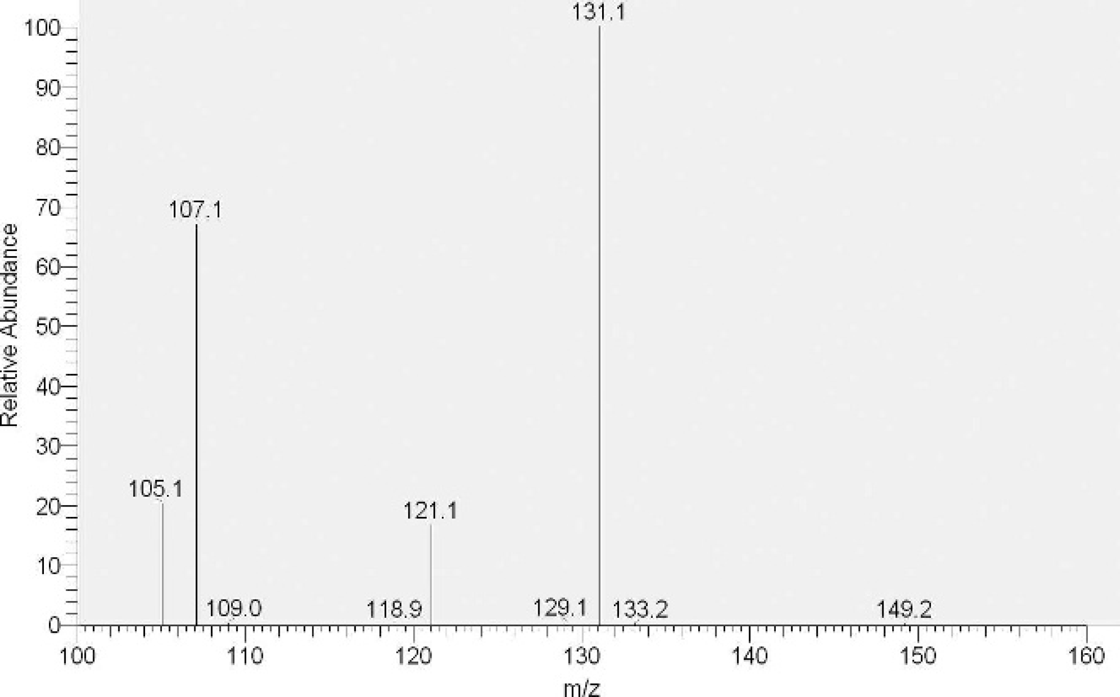
Positive ion ESI LC-MS/MS/MS product ion spectrum of anatoxin-a in pure analytical standard prepared in water. Anatoxin-a eluted from the HPLC column at 5.07 minutes.
Analysis of negative control water and stomach content samples for anatoxin-a by the LC-MS/MS/MS technique described above showed clean chromatograms with no background contribution from phenylalanine. Figure 1 shows the full-scan, positive ESI, MS/MS/MS mass spectrum of anatoxin-a in an analytical standard prepared in water. The spectrum is consistent with the ESI spectrum previously reported. 7,8 Figure 2 (top) shows a chromatogram of stomach content extract from an affected dog (Ontario) that was positive for anatoxin-a at 0.01 μg/g. Anatoxin-a was clearly identified in the sample by comparing the retention times and the MS/MS/MS product ion spectra (Fig. 2, bottom) to that of an analytical standard. The water sample from the Ontario case was positive for anatoxin-a at 1 μg/g. All water and stomach contents from the Californian dogs contained anatoxin-a: however, quantitation of the toxin was not performed at the time. The linear ion trap system provided consistent MS/MS/MS ion ratios, allowing for a high level of confidence in anatoxin-a identification. Peak area ratios for the product ions at m/z 107 and m/z 121 were each calculated against the peak area of the base peak at m/z 131. Ion ratios for anatoxin-a in diagnostic samples were within 20% of the ion ratios of the analytical standard. Anatoxin-a was stable during the sonication from 0 to 20 minutes under the sonication conditions described above. A sonication time of 5 minutes was chosen to minimize the sample preparation time. Seven replicate fortifications of water samples at 0.01 μg/g anatoxin-a gave an average recovery of 114% with 12% CV (relative standard deviation).
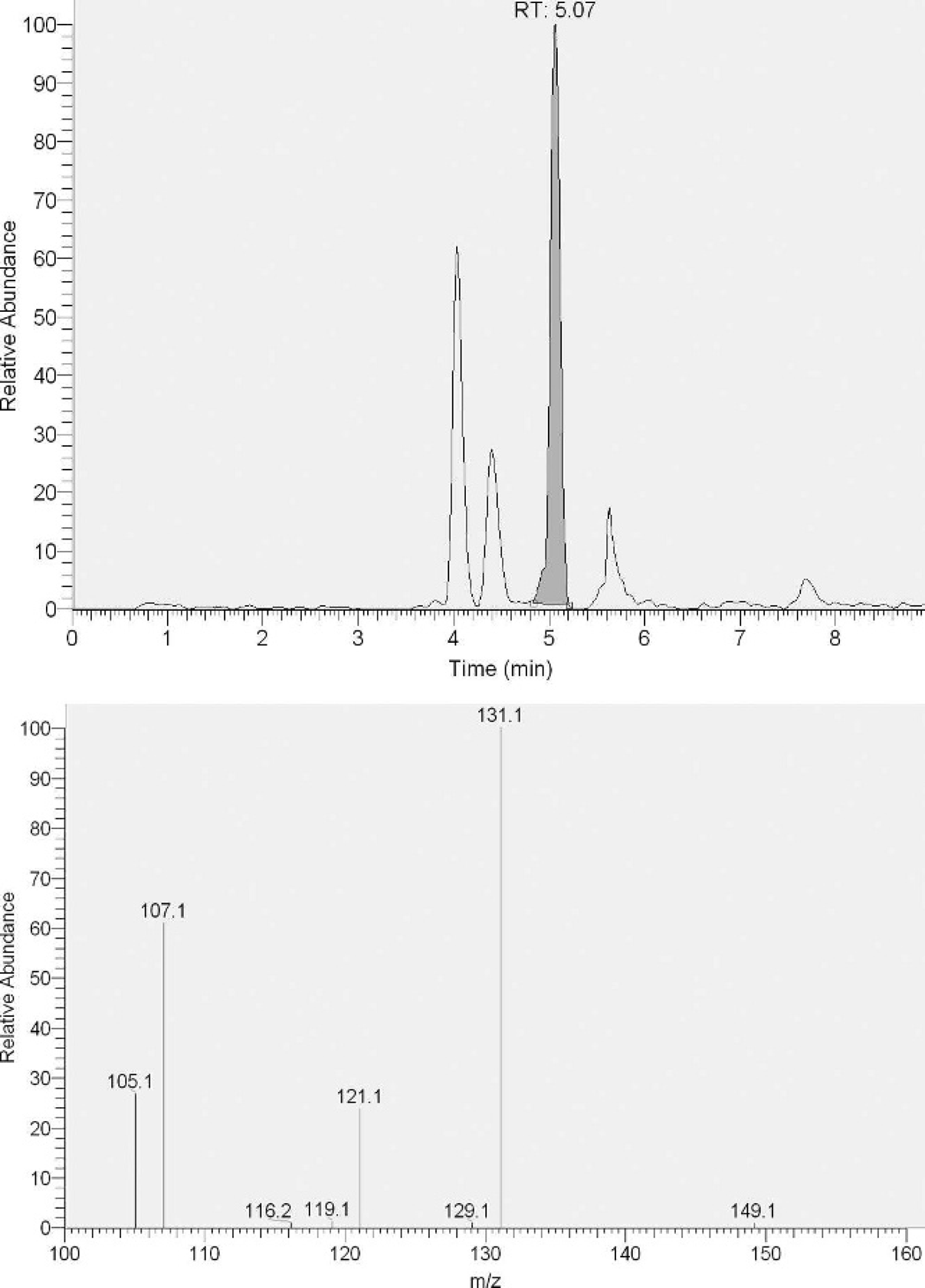
LC-MS/MS/MS product ion chromatogram
The diagnostic approach described in the present study provided crucial information in the diagnosis of anatoxin-a poisoning in dogs. It is important to consider anatoxin-a toxicosis when evaluating a dog that presents with seizures, especially if there was recent access to river or pond water. In suspected anatoxin-a poisoning cases, stomach contents are of the greatest diagnostic value and should be collected at presentation or during postmortem examination. Any concentration of anatoxin-a detected in a suspect case is of toxicological significance. The LC-MS/MS/MS procedure described here is especially suited to veterinary diagnostic laboratory situations for which rapid diagnosis of exposure to anatoxin-a is necessary. The method is a significant improvement over the existing LC-MS/MS methodology in that it provides a rapid and unequivocal determination of anatoxin-a in the presence of phenylalanine that is common in biological specimens and may otherwise give false-positive results. In addition, this method can be rapidly applied to evaluate the toxicity potential of suspect cyanobacteria proliferations in freshwater ecosystems that may threaten the health of animals and humans living in or using these systems for drinking water and/or recreational purposes.
Acknowledgements. The authors are grateful to Gene Whitehead for his valuable contribution to the analytical portion of this manuscript, Dr. Jack Meeks, Dr. Pia Moisander, and Jennifer Jendro for their assistance with the identification of the California algal bloom. The authors also acknowledge Drs. Joseph Humble and Judy Horvath for their help and timely assistance in providing the diagnostic specimens, and Dr. Wayne Carmichael in providing ELISA analyses.
Footnotes
a.
Ridascreen Saxitoxin Kit, R-BIOPHARM AG, Darmstadt, Germany.
b.
Sonifier, Model 450 with 1/2 inch biohorn, Branson Ultrasonics Corp., Danbury, CT.
c.
Linear ion trap mass spectrometer, Model LTQ, Thermo Finnigan Corp., San Jose, CA.
d.
Agilent, Model 1100 HPLC system, Agilent, Palo Alto, CA.
e.
Phenomenex, Inc., Torrance, CA.