
Abstract
During a period of 1.5 months, a newly established pig herd experienced a high number of mummifications and stillbirths, a high neonatal mortality rate, and many piglets with congenital tremors or hind leg ataxia. After clinical and histological investigations, the submitted animals were divided into 4 groups: mummified or stillborn (N = 6), live born with myocarditis (N = 5) (average age 22.8 days), live born without myocarditis (N = 14) (average age 20.0 days), and control animals from a different herd (N = 5) (newborn). Statistically significant differences were observed in the mean porcine circovirus 2 (PCV2) load among the 4 groups in the liver (P < 0.0001). The presence of PCV2 antigen within the myocardial lesions was confirmed by immunohistochemistry. A high load of PCV2 DNA was observed in myocardium, liver, and spleen from mummified or stillborn piglets (>1 × 10⁷ copies per 500 ng DNA), lower in piglets with myocarditis (>1 × 10⁵ copies per 500 ng DNA), and even further lower in pigs without myocarditis (<1 × 10⁵ copies per 500 ng DNA), whereas no PCV2 DNA was detected in the control animals. Myocardium, liver, and spleen were well suited for routine testing of fetuses and young piglets by quantitative real-time polymerase chain reaction. Neither porcine parvovirus nor encepaholomyocarditis virus was detected. These results indicate that the PCV2 infection might have been of etiological importance for the fetal deaths and piglet mortality observed in this herd.
Introduction
Porcine circovirus type 2 (PCV2) is widespread in the commercial swine population and has been associated with several disease complexes. 1,9,32,33 Naturally occurring reproductive diseases related to PCV2 infection have been reported earlier, 2,3,5,10,17,20,24,26,36 and experimental studies have shown an association between PCV2 and reproductive failure 12,22,28–31,37 that is consistent with the reported proposal of transplacental infection by PCV2. The results of a study of the distribution of PCV2 DNA in the reproductive tract, oocytes, and embryos of PCV2 antibody-positive pigs indicate that PCV2 may be associated with different tissues of the reproductive tract, but is unlikely to be associated with uterine-stage embryos. 4 Evidence of shedding of PCV2 in boar semen has been reported after both natural and experimental infection with PCV2. 16,18
Congenital tremor (CT) is characterized by tremor of the head and limbs of newborn piglets. CT type AII has traditionally been associated with an unidentified virus, and PCV2 has been proposed as this virus. 8 Whereas some authors have isolated PCV2 from piglets with CT, 7,34 other studies have been unable to verify those findings. 11,14
Although the presence of PCV2 has been demonstrated in cases of both congenital tremor and stillbirths, little is still known about the amount of viral DNA present in the affected animals. Therefore, the objective of this study was to quantify PCV2 DNA in different tissues from mummified and stillborn or weak born piglets as well as piglets suffering from congenital tremors and ataxia in a commercial herd.
Materials and methods
Animals. The materials for laboratory examination were submitted from a newly established integrated herd consisting of 36 first parity gilts originating from 4 different multiplier herds, 15 from one herd and 6–8 from the other 3 herds. All gilts were pregnant upon arrival and farrowed about 1 month later during a period of 14 days. Of the 15 gilts originating from a single multiplier herd, 12 gave rise to litters with mummified fetuses and/or stillborn piglets or piglets with CT or ataxia. In total, 20.8% (40/192) were mummified/stillborn, and 7.9% (12/152) of the live born had CT or ataxia. Twenty-one of the 152 live born piglets (13.8%) from these 15 gilts died or were euthanized before weaning. The gilts themselves showed no clinical signs of disease. The 21 other gilts in the farm had only 0.1 % (2/252) mummified or stillborn and a preweaning mortality of 0.3% (7/252). During a period of 1.5 months, 6 fetuses and 1 weak born piglet from 2 litters and 18 piglets (11-42 days of age) from 4 other litters were submitted for autopsy. The piglets were either euthanized because of severe signs of tremor or ataxia (6/18) or died of various causes (including respiratory problems and congenital deformities). Placental samples were available from 2 gilts.
Myocardium and liver tissue from 5 newborn piglets without any macroscopical and histological findings in the heart and liver were used as negative controls for the immunohistochemical examination. DNA isolated from myocardium and liver tissue samples from the same piglets was also tested by quantitative polymerase chain reaction (PCR). The piglets included as negative controls originated from a farm experiencing reproductive problems most likely caused by mycotoxins in the straw. After clinical and histological investigations, the submitted animals were divided into 4 groups; mummified or stillborn (N = 6), live born with myocarditis (N = 5) (average age 22.8 days), live born without myocarditis (N = 14) (average age 20.0 days), and control animals from a different herd (N = 5) (newborn).
Postmortem examination and immunohistochemical staining. At necropsy, tissue samples were collected from various organs, including brain and spinal cord, lung, myocardium, liver, spleen, kidney and mesenteric lymph node. The tissue samples were fixed in 10% neutral buffered formalin, processed routinely, sectioned at 5 μm and stained with hematoxylin and eosin (HE).
After clearing with xylene sections were rehydrated and stained with a polyclonal rabbit anti-PCV2 antiserum a at dilution 1:2,000 for immunohistochemistry. Immunohistochemical staining was performed on myocardium sections from 10 animals: 3 fetuses, 5 piglets with myocarditis and/or myocardial fibrosis, and 2 piglets without any histological lesions in the myocardium. Furthermore, immunohistochemical examination was performed on sections from the liver and mesenteric lymph node from 6 of these animals (Table 1).
Virological and bacteriological examinations. Sampling for virological investigations was performed from 25 animals from the case farm as follows. Myocardium (3/6), spleen (1/6), brain (3/6), liver (6/6), lung (5/6), kidney (1/6), and placenta (fetal part) (3/6) were sampled from the 6 fetuses, and the 19 live born piglets were sampled for myocardium (15/19), spleen (18/19), brain (17/19), mesenteric lymph node (18/19), tonsil (9/19), liver (19/19), lung (19/19), and kidney (18/19). DNA was isolated from 5–30-mg tissue using a commercial kit according to manufacturer's instructions b ; tissue weights and DNA concentrations were recorded. Quantitative real-time PCR was performed on the DNA samples as described previously 6 using primers and a probe designed to perfectly match the target sequence (PCV-C-1256U21 5'-ATA GCG GGA GTG GTA AGA GAA-3', PCV-F-1319L21 5'-GCA ACA GCC CTA ACC TAT GAC-3', TaqMan3-PCV2 5'-6-Fam-ATG TAA ACT ACT CCT CCC GCC ATA CCA TT-Tamra-3'). Results were recorded as the number of PCV2 copies per 500-ng DNA. When the DNA concentration in the eluate was less than 200 ng/μl, 2.5 μl of eluate was used as template in the quantitative PCR, and the resulting PCV2 estimate was adjusted to report the number of copies per 500-ng DNA. Nucleotide sequence analysis was performed on PCR-enhanced Cap gene (ORF2) using primers PCV-sense-Cap 5'-ATG ACG TAT CCA AGG AGG CG-3' and PCV-antisense-Cap 5'-TTA GGG TTT AAG TGG GGG GT-3' for amplification and additional internal primers published previously (5'-GAT TGT ATG GCG GGA GGA GT-3' and 5'-ATT GAC GAC TTT GTT CCC CC-3') 15 for sequencing.
Livers from 10 animals, all 6 fetuses and 4 piglets with myocarditis, were tested for porcine parvovirus (PPV) by hemagglutination (HA) test performed on red blood cells from hens. Briefly, the liver samples were homogenized and diluted 1:2 in phosphate buffered saline (PBS) and the solution clarified by centrifugation at 1,400 × g for 5 min. Each harvested supernatant was diluted 2-fold from 1:2 to 1:4,096 in PBS before adding 1% hen red blood cells, making a final dilution of 1:4 to 1:8,192. After an incubation at room temperature for 1 hour, any aglutination was recorded. Potentially positive samples were verified by the inhibition of aglutination in the presence of a known PPV-positive serum. c
All submitted animals from the affected farm were tested for encephalomyocarditis virus (EMCV) by inoculation on cell culture and PCR. Of the 25 sampled animals, myocardium was used if available (18/25), then spleen (5/25) or liver (2/25). Briefly, each organ sample was homogenized in minimal essential medium (1 g/10 ml) containing antibiotics and clarified by centrifugation at 10,000 × g for 30 min. Each harvested supernatant was inoculated on baby hamster kidney 21 cells, d passed 4 times at 3-day intervals, and checked daily for evidence of cytopathic effect (CPE). The supernatants of cultures showing CPE were checked using a neutralization test with specific EMCV antiserum. All samples were also tested by PCR as described previously. 35
The animals were not tested for porcine reproductive and respiratory syndrome (PRRS) virus nor pseudorabies virus, as these viruses have never been isolated from Norwegian pigs. 19 Bacteriological investigations were performed on placenta (fetal part) (2/6), liver (17/25), lung (18/25), spleen (5/25), mesenteric lymph node (1/18), stomach contents (1/18), thoracic fluid (1/18), abdominal fluid (1/18), and/or intestine (1/18) from all but 3 piglets.
Statistical analyses. Statistical analyses of PCV2 load in different tissues were performed using JMP 5.0.1a. e Samples below the detection limit of 1 copy per 500-ng dsDNA were set to 0.5 copies per 500 ng dsDNA, representing the mean of the values, and likewise, the samples calculated by the LightCycler software to be between the detection limit and the quantification limit of 1.0 × 10² copies per 500 ng dsDNA, 6 were set to 5.0 × 101 copies per 500 ng dsDNA. As the distribution was not normal even after log-transformation, Wilcoxon rank sum test was performed for comparison between groups.
Clinical observations, immunohistochemical (IHC) findings and porcine circovirus type 2 (PCV2) DNA load (copies per 500 ng DNA) in different tissues quantified by real-time TaqMan-based PCR in a case study of 30 animals (6 fetuses, 19 piglets, and 5 control piglets)
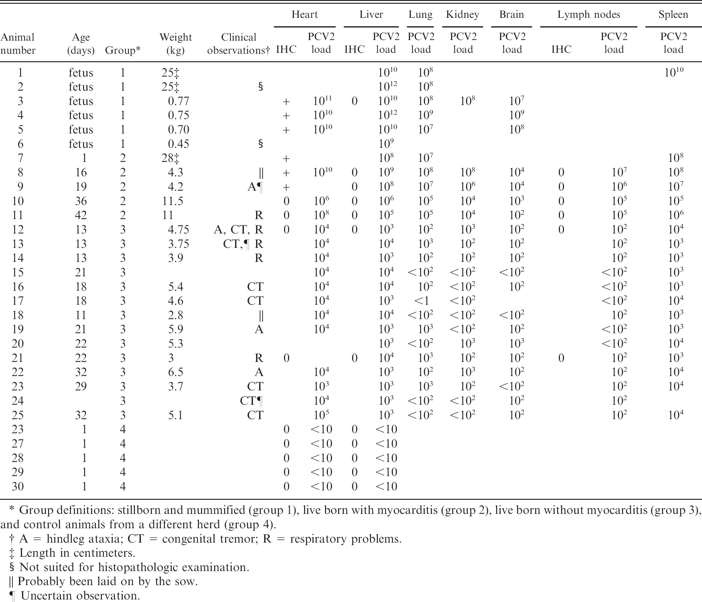
Group definitions: stillborn and mummified (group 1), live born with myocarditis (group 2), live born without myocarditis (group 3), and control animals from a different herd (group 4).
A = hindleg ataxia; CT = congenital tremor; R = respiratory problems.
Length in centimeters.
Not suited for histopathologic examination.
Probably been laid on by the sow.
Uncertain observation.
Results
Clinical observations, immunohistochemical findings, and PCV2 DNA load are presented in Table 1.
Pathological findings. Three of the fetuses were mummified, 2 fetuses showed no changes, and 1 was severely autolytic. Salient findings demonstrated in live born piglets were hydrothorax, ascites, and, in some cases, uni- or bilaterally dilatation of the heart. The myocardium was mottled in 4 piglets. A few piglets showed cerebellar petechial hemorrhages.
Myocardial lesions were found in 9 animals (animals 1, 3–5, and 7–11), and were characterized by multifocal fibronecrosis and infiltrations by mononuclear cells (Fig. 1). In 3 piglets, myocardial fibrosis was the only lesion observed (animals 3–5). In the lungs of the live born piglets, mild to moderate interstitial infiltrations of mononuclear cells were the salient findings (animals 8 and 11–22), with a concurrent purulent bronchopneumonia in 2 animals (animals 13 and 15), and only bronchopneumonia in 2 animals (animals 9 and 24). Liver lesions consisted either of multifocal infiltrates with both mononuclear and polymorphonuclear leukocytes (animals 13–20 and 25), or periacinar necrosis (animals 10–11 and 21), or both (animals 3 and 9). In the lymph nodes, follicular hyperplasia was found in some piglets (animals 8, 11, 16–21, and 23–25). Others showed lymphocytic depletion both in spleen and lymph nodes (animals 12–15) or in spleen alone (animals 8, 12–19, and 21–23). Lymphadenitis was found in the lymph nodes of 2 animals (animals 9 and 15). In the kidneys, the glomerular capillaries displayed micro-thrombi formation and sparse fibrin exudation (animals 12–19). Acute tubulointerstitial inflammation was found in 1 piglet (animal 9). In the central nervous system, fresh hemorrhages were found in the cerebellum in 3 piglets (animals 9, 11, and 21) whereas in 4 piglets acute inflammatory changes with fibrin exudations were revealed in the meninges, the ventricles, and the central canal of the spinal cord (animals 22–25).
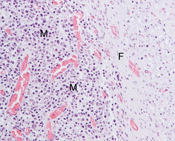
Myocardial inflammatory changes characterized by infiltrating mononuclear cells (M) and incipient fibrosis (F). Hematoxylin and eosin (HE). ×200.
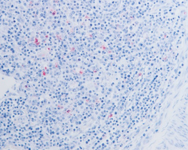
Intracytoplasmatic porcine circovirus type 2 (PCV2) antigen (red) demonstrated by immunohistochemistry in myocardial infiltrating mononuclear cells using polyclonal anti-PCV2-antibody. ×200.
Immunohistochemical staining. Immunohistochemical staining of the myocardium from 6 of the 8 fetuses or piglets with myocarditis and/or myocardial fibrosis revealed positive cytoplasmatic staining of mononuclear inflammatory cells (Fig. 2). There was no positive staining in the livers and lymph nodes that were examined. No positive staining was observed in the livers and myocardium from the negative control animals.
Virological and bacteriological investigations. Only the negative control animals were below the detection limit of the quantitative PCR. The PCV2 DNA load differed between animals and tissues, and the highest loads were observed in the liver, spleen, and myocardium. In the liver, the highest load was observed in mummified and stillborn piglets (defined as group 1) and in live born piglets with myocarditis (defined as group 2) as compared to live born piglets without myocarditis (defined as group 3) and negative control animals (defined as group 4) (P < 0.0001) (Fig. 3). Similar results were observed in myocardium and spleen, although the groups were too small to detect any significant differences.
Encephalomyocarditis virus was not detected in any of the animals from the affected farm, and all 10 animals tested for PPV were negative in the HA test. Streptococcus suis/Streptococcus sp. was isolated from 8 animals (animals 12 and 14–20), Bordetella bronchiseptica was isolated from 2 animals (animals 9 and 21), and nonpathogenic bacteria or no growth was observed from 12 animals (animals 1–8, 10–11, 13, and 25).
Discussion
A link between PCV2 infection and abortion was proposed by several authors, 2,5,10,20,24,26,36 and although PCV2 infection of fetuses during pregnancy has been demonstrated experimentally, little is known about the PCV2 titer in fetuses under field conditions. In the present case study, high amounts of PCV2 DNA were found in all stillborn piglets examined, regardless of the state of autolysis, indicating that the detected DNA was protected within viral capsids. Even though the material was limited to stillborn piglets from only 1 farm, the high amount of PCV2 DNA in the fetal tissues in the absence of both PPV and EMCV, combined with the absence of PCV2 DNA in the myocardium and liver from the control animals, strongly indicate PCV2 as a contributing factor to the elevated number of mummified and stillborn piglets in the affected herd. Myocarditis, characterized by multifocal fibronecrosis and infiltrations by mononuclear cells or myocardial fibrosis alone, was observed in 9 piglets, including all 4 stillborn piglets suited for histological examination. Similar observations of the association between PCV2 and myocarditis are reported by others. 26,36,25 The PCV2 DNA load was highest in the myocardium in all but 2 animals (Table 1). As neither PPV nor EMCV were detected in the material examined, the presence of PCV2 within the lesions in the myocardium, as confirmed by immunohistochemical staining, together with the high PCV2 DNA load in all tissues examined and especially in the myocardium, further corroborates the theory that myocardium is a target tissue for PCV2 in fetuses and young pigs. 31 The negative immunohistochemical results from the liver may be due to the absence of inflammatory cell infiltration, as the myocardium staining was limited to the cytoplasm of leukocytes, indicating an accumulation of antigen in these cells. The animals with high PCV2 DNA load in liver and spleen were not bled before death, and there is a possibility that the observed PCR results were caused by PCV2 in the retained blood. In cases of post-weaning multisystemic wasting syndrome, the PCV2 load in plasma can be very high, 6 and this may contribute to a background level of PCV2 DNA in tissues with a high retention of blood or plasma.
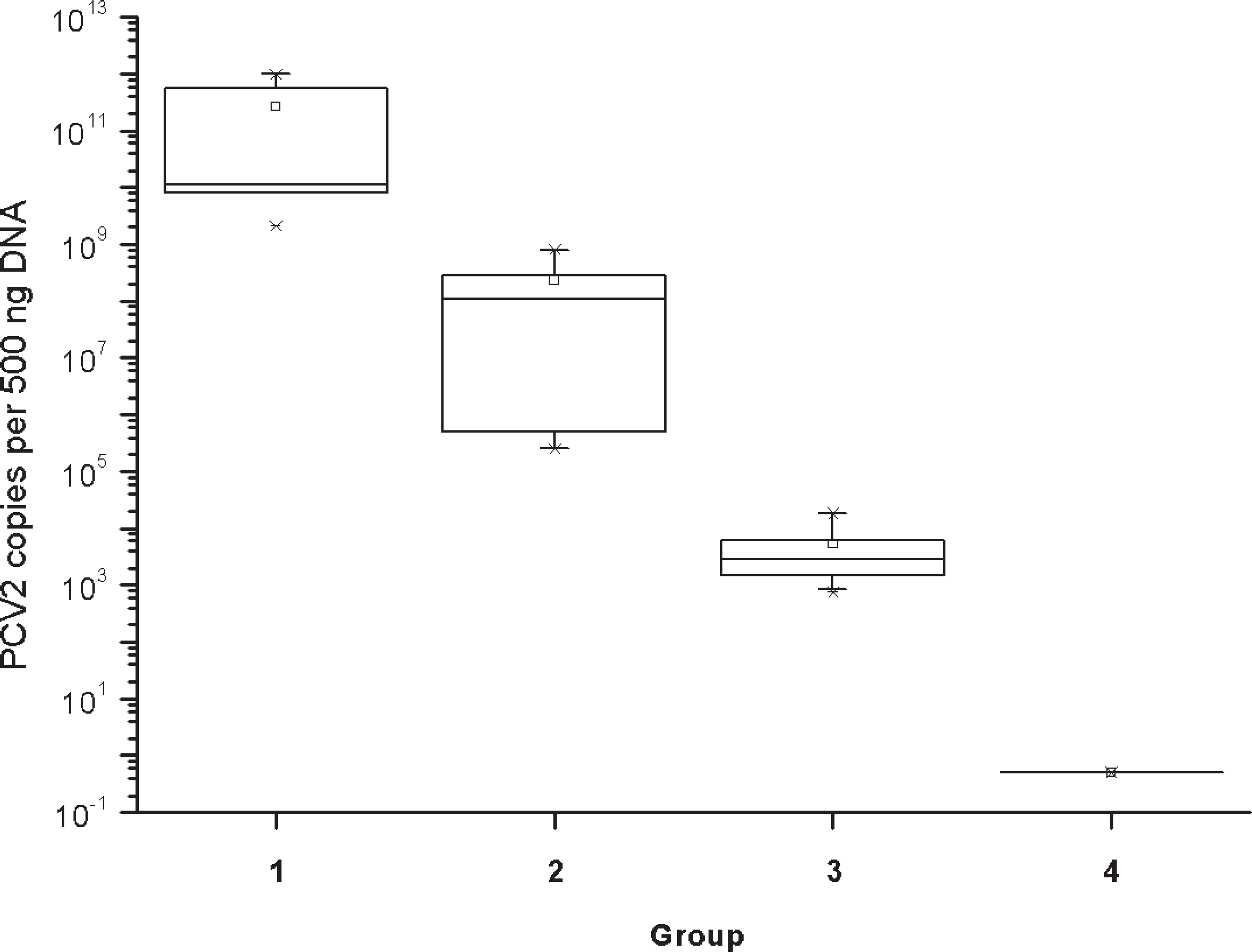
Box plot of estimated porcine circovirus type 2 load (copies per 500 ng DNA) in liver from stillborn fetuses (group 1, N = 6), live born with myocarditis (group 2, N = 5), live born without myocarditis (group 3, N = 14), and negative control animals (group 4, N = 5). The horizontal lines in the box denote the 25th, 50th, and 75th percentile values. The error bars denote the 5th and 95th percentile values. The 2 symbols below the 5th percentile error bar denote the 0th and 1st percentile values. The 2 symbols above the 95th percentile error bar denote the 99th and 100th percentiles. The square symbol denotes the mean of the column of data. Some of the symbols denoting different percentiles coincide because of low numbers of observations within each group.
In a recent study in which pregnant sows were inoculated intranasally with PCV2, Park et al. were unable to detect any virus in the myocardium from fetuses either by immunohistochemistry or in situ hybridization. 27 The main target organs in that study were lymphoid organs. The difference between those results and the present study may be due to different variants of PCV2, differences in immunocompetence at the time of infection, or other unknown factors of the host animal. In the study by Park et al., the sows were inoculated 3 weeks before the expected farrowing date, while natural infection could occur at all stages of pregnancy, and the presence of myocardial lesions may be dependent on infection early in pregnancy. Different co-infections or intoxications may also influence the clinical and pathological findings.
When the animals were grouped as stillborn (group 1) or live born with and without myocardial lesions, (group 2) and (group 3), respectively, as well as negative controls (group 4) there were statistically significant differences in mean liver PCV2 load among the 4 groups (P < 0.0001), and a similar tendency was observed in the myocardial tissue samples, but the groups were too small to detect any significant differences. The pathogenicity of PCV2 in the fetus may be affected by the age of the fetus at the time of infection. 29 In the study by Park et al., gross lesions were observed in fetuses inoculated at 57 days of gestation, whereas no gross lesions were observed in fetuses inoculated at 75 and 92 days of gestation. As in the present study, case reports on stillbirths connected to PCV2 infection have been referring to problems experienced in newly established herds with a high proportion of gilts. 13,23,26,36 Establishing new herds often involves moving and mixing of pregnant gilts with different backgrounds of infection. This may cause an increase in PCV2 load in the blood, either as a result of immune suppression or as a result of a primary or superinfection with PCV2 or a primary infection with another agent. An increase in blood PCV2 load could cause transplacental infection, resulting in an increase in stillbirths. In the present study, the gilts from a single multiplier herd gave rise to litters with 20.8% (40/192) mummified or stillborn piglets, whereas the gilts from the 3 other multiplier herds only gave rise to litters with 0.1 % (2/252) mummified or stillborn. This difference may be due to differing immune and health status in the multiplier herds from which the gilts originated.
Prevalence studies from other countries indicate that the role of PCV2 in stillbirths and abortions is less significant than infections with PPV or PRRS virus. 3,17,20,21 Porcine reproductive and respiratory syndrome has never been detected in Norwegian pigs, and no antibodies against PRRS virus have been detected in the serum samples included in the national surveillance and control program for specific virus infections in swine herds in Norway. 19 Most herds have an effective vaccination program against PPV, resulting in very few reported cases.
The occurrence of PCV2 infection in pigs with CT was investigated earlier, but the significance of PCV2 in this disorder remains dubious. 7,11,14,34 The results from the present study corroborate those of other authors, 14 who found no connection between PCV2 and CT. Although PCV2 was only detected in relatively low numbers in tissues from piglets with tremors, and the brain and spinal cord from the control pigs were not analyzed, piglets with myocarditis had a high viral load, and the viral load was highest in the myocardium, suggesting that myocardium rather than the central nervous system was the target organ. When group 3 was divided in 2 regarding CT, there was no significant difference in the viral load in the liver in animals with CT or not (data not shown). Inflammatory changes in the central nervous system, the lungs, and the kidney were probably because of a concurrent bacterial infection (Table 1). The diverging observations on the association of PCV2 infection with CT and stillbirths may be because of the differing sensitivities of the assays used for PCV2 detection. Although all animals from the affected farm were PCR positive for PCV2 in the present study, the use of a quantitative assay revealed low PCV2 load in animals without myocarditis, which does not support a link between CT and PCV2. However, it is still possible that the CT and ataxia observed postpartum may be a result of an in utero infection with PCV2. Further studies are needed to elucidate this hypothesis.
In conclusion, high PCV2 load was observed in tissues from aborted fetuses and piglets with myocarditis. Viral loads in pigs with tremors but no myocarditis were significantly lower than those in pigs with myocarditis. Samples from myocardium, liver, and spleen were found to be most suited for routine PCV2 testing by quantitative PCR.
Acknowledgements
This study was supported by 14328601 from the Norwegian Research Council. Dr. Gordon M. Allan at the Department of Agriculture for Northern Ireland, Veterinary Science Division, Belfast, UK, kindly provided the antibodies used for immunohistochemistry. We would also like to thank Hilde Lussand Selheim and Lars Selheim for providing the material and Marit Nedkvitne Leinaas, Irene Haugen, and Randi Terland for excellent technical assistance.
Footnotes
a.
Polyclonal rabbit anti-PCV2 antiserum; kindly provided by Gordon M. Allan, Department of Agriculture for Northern Ireland, Veterinary Science Division, Belfast, UK.
b.
DNeasy; Qiagen, Valencia, CA.
c.
Known PPV positive serum, J. No. 1986-08-298/719; National Veterinary Institute, Oslo, Norway.
d.
ATCC CCL10; LGC, Teddington, Middlesex, UK.
e.
JMP5.0.1a, SAS Institute Inc., Cary, NC.