
Abstract
Anti-porcine circovirus type 2 (anti-PCV2) immunostaining was associated with cerebellar lymphohistiocytic vasculitis combined with hemorrhages (50 pigs) or with lymphohistiocytic meningitis (23 pigs) in pigs naturally affected with postweaning multisystemic wasting syndrome (PMWS). The animals originated from 12 farms in Rio Grande do Sul, Brazil. In total, 456 unthrifty 3- to 5-month-old postweaning pigs confirmed as PMWS cases were necropsied. Although most findings mimicked those extensively reported in PMWS-affected pigs, there were distinctive brain lesions that included multiple hemorrhages in the cerebellar leptomeninges associated with lymphohistiocytic vasculitis and fibrinoid degeneration in vessels of the cerebellum and periventricular areas (69 pigs). These vascular lesions were also seen in conjunction with lymphohistiocytic meningitis (38 additional pigs). PCV2 antigen was immunohistochemically demonstrated in the cytoplasm and nuclei from intralesional perivascular macrophages and endothelial-like cells in brain tissues. Together these findings suggest that these lesions were caused by PCV2.
Postweaning multisystemic wasting syndrome (PMWS), porcine dermatitis and nephropathy syndrome (PDNS), reproductive disorders, and porcine respiratory disease complex have all been associated with porcine circovirus type 2 (PCV2) infections. 2,6,10,16 Among these conditions, PMWS has caused the greatest economic losses to the pig industry. The disease is characterized by wasting, dyspnea, and paleness, combined with pathological findings of enlarged lymph nodes, interstitial pneumonia, and nephritis. 1,6 Lymphocyte depletion and histiocytic to granulomatous inflammation in lymphoid tissues and certain organs are the main histological changes. 6,17 The demonstration of a moderate to high amount of PCV2 antigen or nucleic acids in close association with characteristic microscopic lesions confirms the diagnosis. 18 Porcine circovirus type 2 antigens have been detected by immunohistochemistry in a wide range of cells from pigs naturally or experimentally infected with PCV2. 9,13,14
Leptomeningitis, 6 meningoencephalitis, 7,14 and encephalitis 23 have also been linked to PCV2 infection. Moreover, PCV2 antigens have been demonstrated in neurons, cerebral perivascular cellular infiltrates in mononuclear cells, fibroblast-like cells, and endothelial cells in the meninges, choroid plexus, cerebrum, and cerebellum from infected pigs. 3,7,9,20 This report describes PCV2-associated brain lesions in PMWS-affected pigs.
As part of an experimental study, 456 unthrifty 3- to 5-month-old postweaning pigs confirmed as PMWS 18 cases were necropsied. Animals originated from 12 herds in Rio Grande do Sul, Brazil. Pigs were necropsied weekly in groups of 30–40 immediately after euthanasia. Brains were systematically examined and sectioned at the cerebellum and cortex. Brains, in which macroscopic changes could be seen, were sectioned in the medulla oblongata (at obex), cerebellum and cerebellar peduncules, midbrain (at caudal colliculi), parietal cortex and thalamus, and frontal cortex with basal nuclei. In total, 124 pigs out of the 456 had microscopic brain lesions. Formalin-fixed samples of cerebral and cerebellar cortices from these 124 pigs and from 52 additional pigs (also from the group of 456) in which microscopic brain lesions were not detected were examined by PCV2-immunostaining. Sections were processed for immunohistochemical (IHC) staining using a polyclonal antibody against PCV2 a , 19 at 1: 1.000 dilution in the streptavidin-biotin-immunoperoxidase b technique, with diaminobenzidine b as chromogen. Similarly, selected sections were also stained by IHC for porcine respiratory and reproductive syndrome virus c (PRRSV) and bovine viral diarrhea virus d (BVDV) antigens.
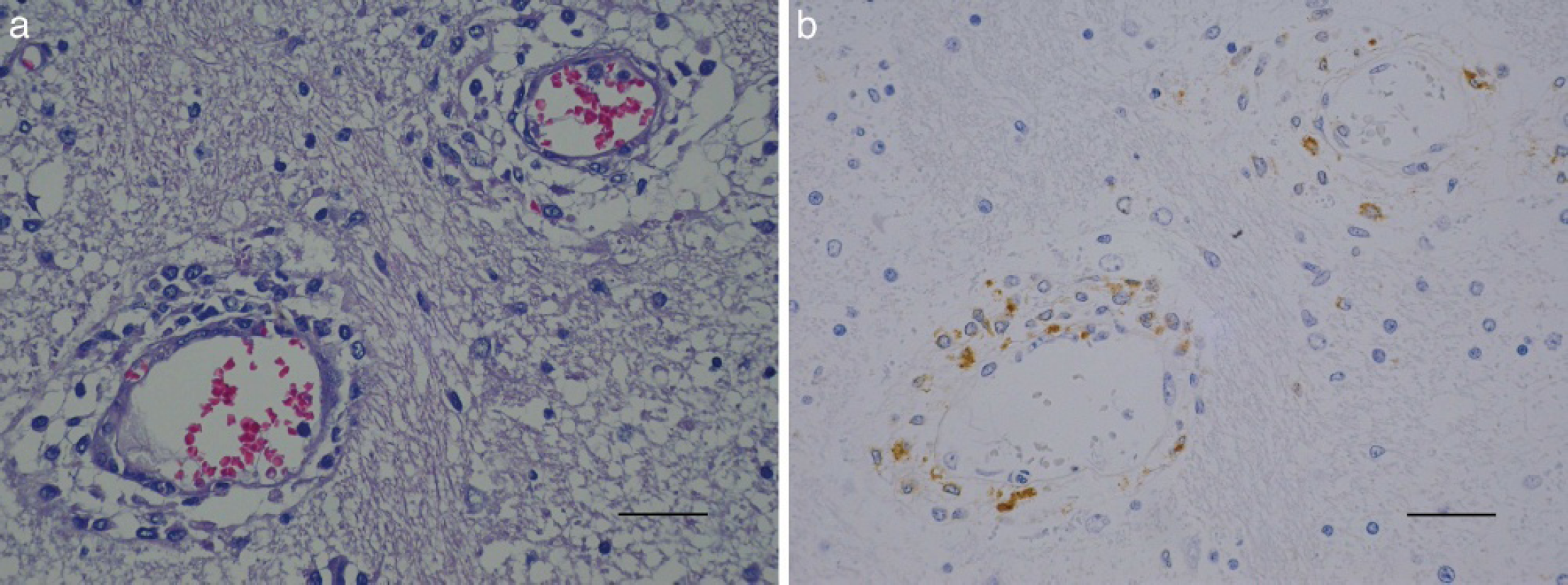
Most clinical and pathological findings mimicked those extensively reported in PMWS-affected pigs. In particular, neurological signs included apathy (94 pigs), ataxia (35 pigs), paddling (12 pigs), and opistotonus (4 pigs). Prominent brain lesions were cerebellar (leptomeninges) multiple petechiae (Fig. 1) (34 pigs), which sporadically were associated with edema (14 pigs) that could be seen on the coronal section at the level of the cerebellum and cerebellar peduncules (Fig. 2). Microscopically, the cerebellar hemorrhages were associated with mononuclear vasculitis in the molecular zone of cerebellum (extending to granular zone), hypertrophied endothelium (Fig. 3), and perivascular lymphohistiocytic infiltrate with deposits of fibrin. Hyaline globules within macrophages (Fig. 3) or extravasated erythrocytes and plasma were often seen in association with degenerated vessels and tumefaction of neuropil in the white substance. The fibrinoid degeneration (Fig. 4A) of the vessels walls was evidenced by phosphotungstic acid-hematoxylin Mallory and periodic acid-Schiff (PAS) staining. In the other sectioned areas (Fig. 5A), these changes were less pronounced and mostly concentrated in the periventricular areas. Lymphohistiocytic infiltrate and vasculitis were also present in the choroid plexus. There was gliosis in medulla oblongata (obex). Multifocal lymphohistiocytic choroiditis with extravasated plasma was also observed. Multifocal mononuclear meningitis associated with perivascular lymphohistiocytic infiltrates and vasculitis was observed in 38 pigs. Finally, whereas gross findings also included 9 cases of purulent meningitis, purulent or fibrin-purulent meningitis was seen microscopically in 17 pigs.
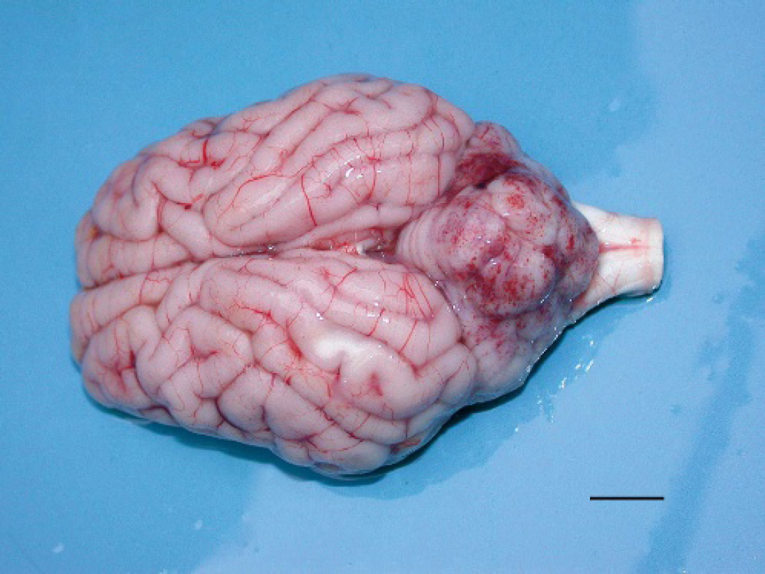
Brain, PMWS-affected pig. Multiple cerebellar petechiae. Bar = 1 cm.
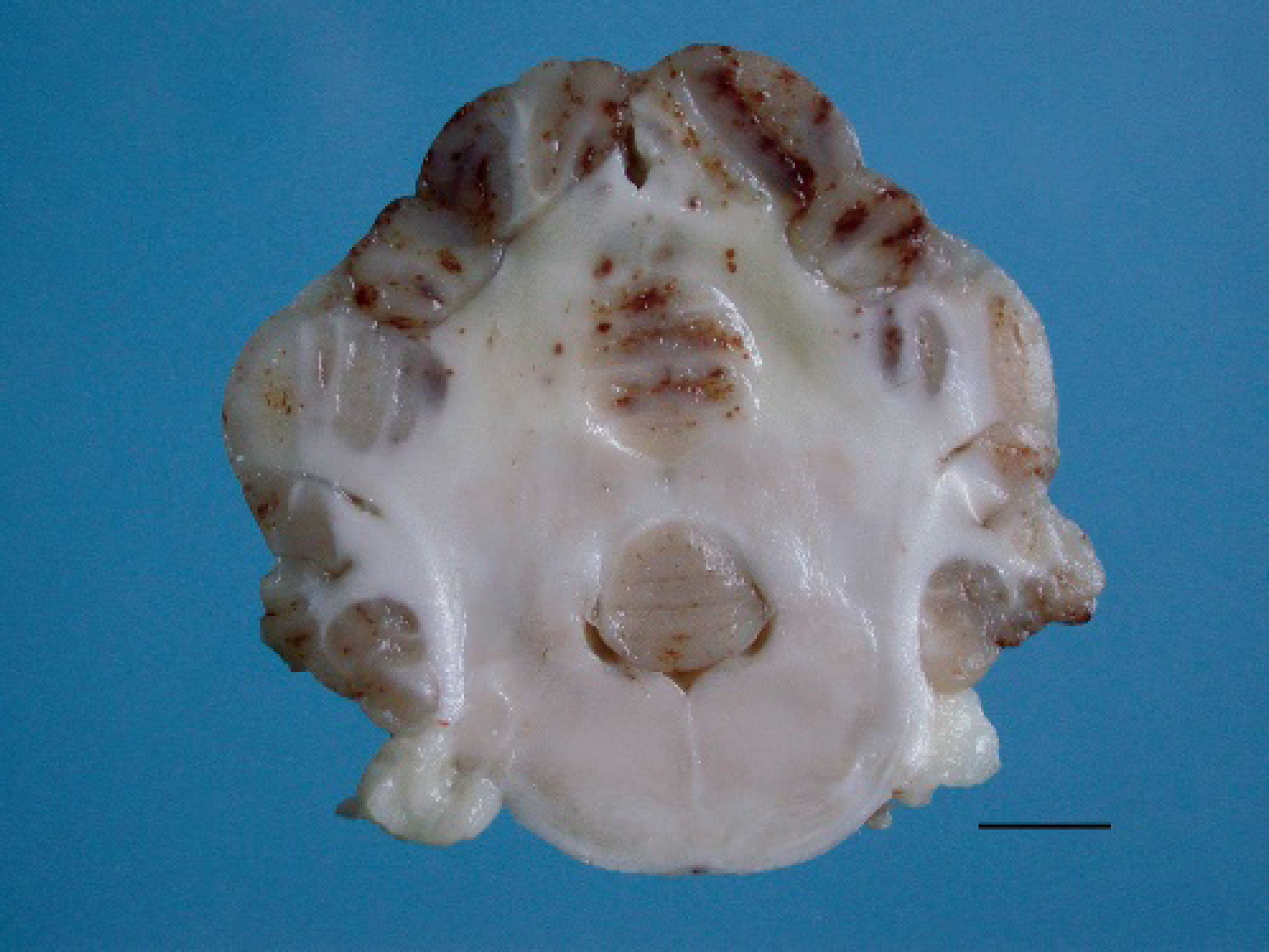
Cerebellum of a PMWS-affected pig, coronal section at the level of cerebellum and cerebellar peduncules. Note the cortical hemorrhages and the edematous white substance. Bar = 0.5 cm.
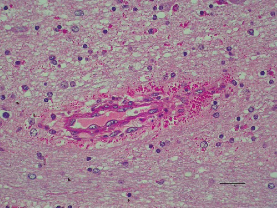
Cerebellum of a PMWS-affected pig. Capillary vessel showing hypertrophied endothelium, extravasated plasma, and hyaline globules within macrophages. PAS. Bar = 30 mm.
Large amounts of PCV2 antigen were observed in the cytoplasm and nuclei from intralesional perivascular macrophages and endothelial-like cells in brain tissues (Figs. 4B, 5B). The most intense PCV2 antigen staining in brain sections were observed in the cerebellar vessels and leptomeninges. Scarce and punctiform areas of PCV2-immunoreactivity were observed in unaffected areas from brains with lesions in 14 pigs. The microscopic findings observed in the brains from PMWS-affected pigs are summarized in Table 1. Escherichia coli and Salmonella sp. were isolated from intestinal samples in 11 and 9 pigs, respectively. Streptococcus sp. and Haemophilus sp. were isolated from brain samples in 3 and 2 pigs, respectively.
Pigs were classified as PMWS cases on the basis of clinical, pathological, and immunohistochemical evidence as previously described. 18 Whereas most findings were similar to those described in previous reports of the disease, 2,6,11,16 this report emphasizes the presence of distinctive PCV2-associated brain lesions in PMWS naturally affected pigs. Brain lesions in pigs affected by PMWS have already been associated with neurological symptoms in 1 study. 23 Clinical signs characteristic of nervous system diseases were observed in some animals within the group with brain lesions (124 pigs) and could be correlated with cerebellar hemorrhages/vasculitis, meningitis/vasculitis, and suppurative meningitis in 21, 16, and 12 pigs, respectively; however, lethargy or apathy may also be linked to the deteriorating conditions of these animals and, therefore, could have been a confusing factor.
Brain lesions have been associated with PCV2 infection, 3,6,7,9,14,22,23 and the role of systemic vasculitis on the pathogenesis of PCV2 has been highlighted in 1 report. 13 In that report, lymphohistiocytic and plasmacytic arteritis was observed in several organs, including the central nervous system. Brain lesions, especially characterized by lymphohistiocytic vasculitis combined or not with lymphohistiocytic meningitis, were observed in 107 PMWS-affected pigs, 73 of which were associated with PCV2 immunostaining. It cannot be ruled out that the presence of PCV2 and brain lesions were completely independent; however, PCV2 antigens were detected in most of the brains with lesions but, except for a few cases, were absent in brains without lesions. A number of diseases have been associated with swine encephalopathies. Pigs in this study originated from herds that had previously tested negative for antibodies against Aujeszky disease virus and classical swine fever. Furthermore, according to official records, these diseases are not present in the state of Rio Grande do Sul, Brazil.
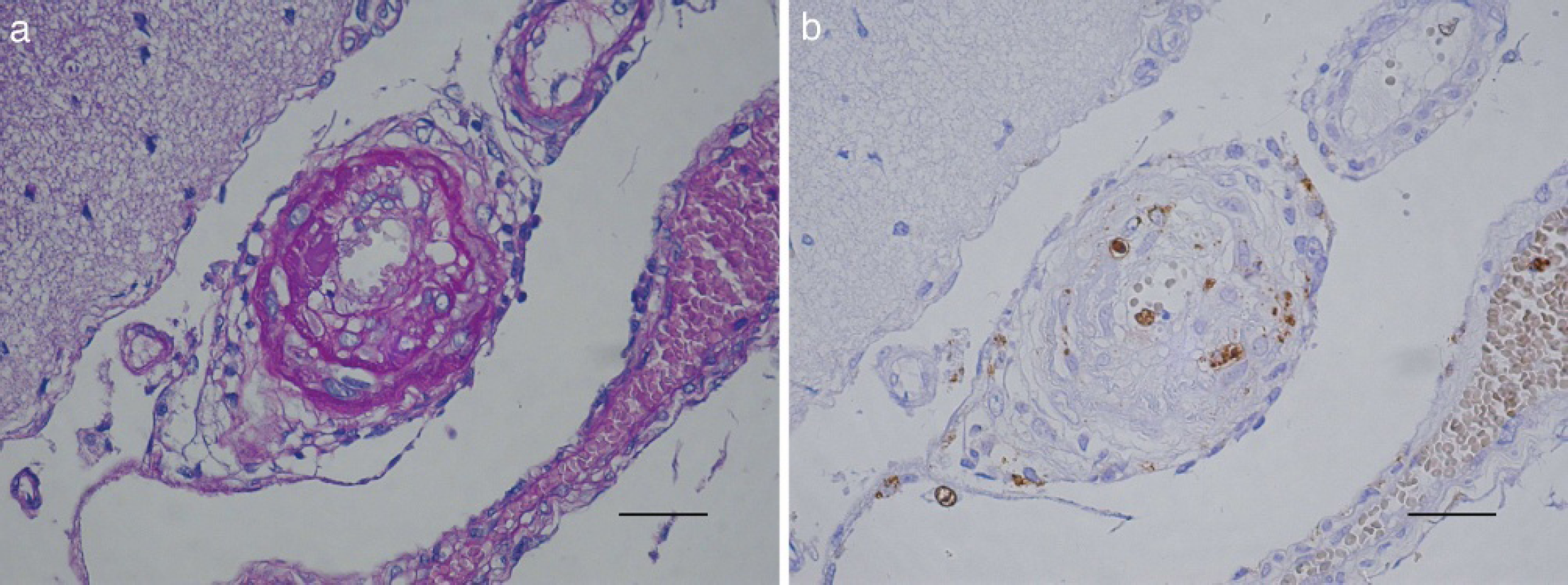
Cerebellum, PMWS-affected pig. Arteriole wall displaying fibrinoid degeneration. PAS. Bar = 60 μm.
Lymphohistiocytic meningoencephalitis has also been linked to PRRSV infection 8,15 ; however, IHC staining for PRRSV antigen was negative in lung and brain sections from 26 pigs in which anti-PCV2 IHC staining was positive in brain sections. In addition, despite the efforts to diagnose PRRSV, a recent survey 5 failed to detect the infection in Brazil. IHC for BVDV was also negative in samples of brains (25 pigs) confirmed as IHC-PCV2 positive. Based on these results, it seems unlikely that these agents were involved in the pathogenic process. However, it is probable that a viral coinfection such as porcine parvovirus could have participated in at least some of these cases. Unfortunately, this could not be tested. Similar vascular lesions have been described in swine cerebrospinal angiopathy, 12 which occurs at similar age, but usually displays characteristic edema of eyelids, snout, stomach, mesenteries, and gall bladder as well as typical bilateral symmetrical foci of encephalomalacia in the brain stem, 4,21 which were not seen. Moreover, although E. coli was detected in intestinal samples from 11 pigs, none of them were in the group with brain lesions. Furthermore, the edematous lesions observed here showed characteristics consistent with a vasogenic origin. 21 Although the positive PCV2 immunostaining is consistent evidence supporting a PCV2-involvement in the genesis of those lesions, it cannot be ruled out that some cases were edema disease in conjunction with PCV2 infection. Typically, suppurative or fibrino-suppurative leptomeningitis processes with abundant effusion of polymorphonuclear cells were associated with Streptococcus suis and Haemophilusparasuis in 3 and 2 pigs, respectively. In 2 of the pigs with suppurative meningitis, there was also positive PCV2 immunostaining, confirming that concurrent infections with bacterial or viral agents may be expected in PMWS-affected pigs. As a systemic disease, PCV2 infection is expected to affect any organ or system. It might be possible that the lesions of lymphohistiocytic meningitis observed here were subsequent to or precipitated by initial vasculitis. Collectively, the results suggest that PCV2 antigens were associated with the brain lesions described here.
Summary of microscopic findings observed in the brains from 176 pigs affected by PMWS. *
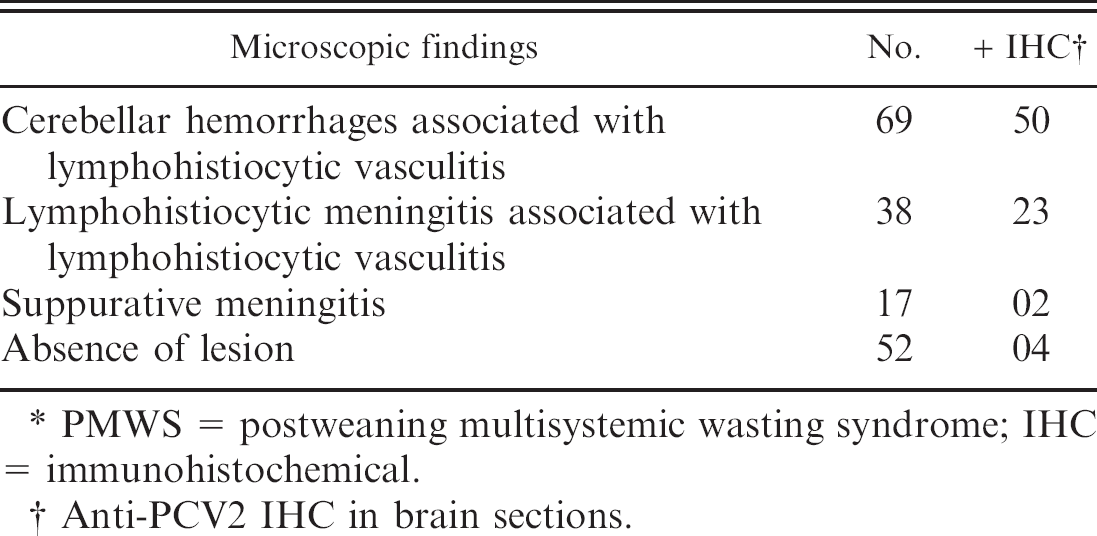
PMWS = postweaning multisystemic wasting syndrome; IHC = immunohistochemical.
Anti-PCV2 IHC in brain sections.
Acknowledgements. This work was supported by grants from the Coordenação de Aperfeiçoamento de Pessoal de Nível Superior (CAPES) and Conselho Nacional de Desenvolvimento Científico e Tecnológico (CNPq), Brazil.
Footnotes
a.
Department of Veterinary Diagnostic and Production Animal Medicine, Iowa State University, Ames, IA.
b.
DAKO LSAB 2 kit, DAKO Corp., Carpinteria, CA.
c.
VMRD Inc., Pullman, WA.
d.
Syracuse BioAnalytical, Syracuse, NY.