
Abstract
A case of unilateral suppurative epididymo-orchitis associated with Salmonella enterica subsp. diarizonae serovar 61:k:1,5,(7) infection is described in a 2-year-old ram. Gross lesions were characterized by severe enlargement of the scrotal contents, fibrous adhesions between testicular layers, coexistence of epididymal abscesses and foci of fibrinous exudate, and testicular atrophy. Microscopically, testicular and epididymal microabscesses and diffuse inflammatory infiltrates with abundant macrophages containing short Gram-negative rods were observed. Superimposed on the chronic lesions were fibrin deposits with clusters of neutrophils, as well as walled-off granulation tissue. Bacterial colonies were also identified in thrombosed spermatic cord vessels, scrotal lymph nodes, lung, and liver. S. enterica subsp. diarizonae serovar 61:k:1,5,(7) was isolated from the affected testis. To the authors' knowledge, this is the first report of infection of the testis and epididymis by Salmonella in rams. This organism must be taken into account in the differential diagnosis of ovine genital infections.
Keywords
Infectious epididymitis and orchitis in rams can be caused by different bacteria 3 and often lead to subnormal fertility or infertility. 9 In rams, Brucella ovis appears to be the most important etiological agent, whereas Actinobacillus seminis and Histophilus ovis are the 2 most common organisms isolated from younger animals. 3
Orchitis associated with Salmonella species has been rarely reported in animals. 5,14,18 In humans, however, Salmonella species is frequently isolated from suppurative testicular lesions, as an extraintestinal complication of enteric salmonellosis 1,21 or in cases of unique testis involvement. 7,11 Arizona group of organisms, designed as subgenus III of genus Salmonella, 15 are divided into 2 subspecies: S. enterica subsp. arizonae (subsp. IIIa) and S. enterica subsp. diarizonae (subsp. IIIb); they have been mostly isolated from reptiles. 2,20 Isolation of Arizona group organisms have been reported from sheep in Canada 8 and the United Kingdom 10 ; they are typically associated with gastroenteritis and abortion. The serotype 61:k:1,5, (7) is one of the most frequently isolated and is considered to be well adapted to sheep. 6,19 Recently, this serovar was isolated from 14% of sheep in a flock in Norway 17 and in 11% of slaughtered sheep in Switzerland, 22 suggesting that sheep are a reservoir for this pathogen.
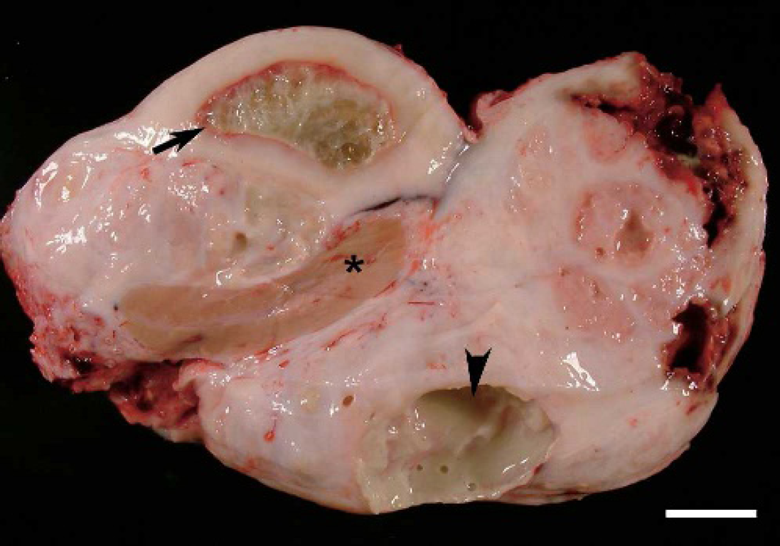
Cut surface of right scrotal contents: areas of intense fibrosis, cavities filled with fibrinous (black arrow) and purulent (arrowhead) exudates; testicular atrophy (black asterisk). Bar = 3 cm.
This report describes the clinical and pathological features of a case of orchitis and epididymitis in a ram, associated with S. enterica subsp. diarizonae serovar 61:k:1,5,(7). A 2-year-old Assaf ram from a flock used for intensive milk production in Zamora province in the Castilla y León region of Spain presented with subnormal fertility and scrotal enlargement, reduced mobility, and induration of the right testis and epididymis, which was painful at palpation. The right testis and epididymis were surgically removed, with the aim of preserving the normal left testis. The animal died during the surgical procedure and was submitted to the Pathology Diagnostic Service of the Veterinary Faculty of León.
After gross examination, samples for histology were obtained from the testes, epididymides, kidneys, liver, lungs, intestines, spleen and several lymph nodes. Tissues were fixed in 10% neutral-buffered formalin and dehydrated through graded alcohols before being embedded in paraffin wax. Sections (4 μm) were cut from each sample and stained with hematoxylin and eosin (HE), Masson Goldner, periodic acid-Schiff (PAS), and Gram staining methods.
For bacterial isolation, 1 g of testicular tissues was homogenized in 9 ml Buffered Peptone Water (BPW) a using a stomacher. b The suspension was incubated at 37°C (±1) for 16–20 hr, and 0.1 ml was transferred onto the surface of a Modified Semi-solid Rappaport Vassiliadis (MSRV) agar plate in 3 equally spaced drops. The MSRV plate was incubated at 41.5°C (±0.5) for 24–48 hr. Additionally, 1 ml of the testicular homogenate in BPW was inoculated into 9 ml of an enrichment broth, Muller-Kauffman Tetrathionate with novobiocine (MKTTn), and incubated at 37°C (±0.1) for 18–24 hr. Each of the 2 enrichment media was inoculated into 2 selective agar plates, XLD and Brilliant Green Agar, a and incubated at 37°C (±1) for 24–48 hr. Colonies resembling Salmonella were selected and identified by conventional biochemical tests (Triple Sugar Iron agar, Lysine agar), the GN card of the VITEK 2 System, a and a polyclonal antiserum. c Serotyping was carried out at the Central Veterinary Laboratory (Algete, Madrid, Spain).
Grossly, the scrotal contents were markedly enlarged (30 X 19 × 9 cm). On cross section, there were extensive fibrous adhesions between the scrotal skin, tunica dartos, and visceral and parietal layers of the tunica vaginalis. The cut surface (Fig. 1) was composed of heterogeneous elements: areas of dense fibrous tissue, abscesses with a creamy, greenish purulent exudate, dark red necrotic lesions, and foci of fibrinous exudate surrounded by a zone of hyperemia and areas of surviving epididymal parenchyma. The right testis was severely compressed and atrophied. Medial iliac and scrotal lymph nodes were enlarged. No macroscopical lesions were seen in the contralateral testis and the tunica vaginalis except for the presence of 3 small cysts adjacent to the head of the epididymis. Hydropericardium with a few fibrin strands, mild ascites, pulmonary congestion and edema, and pale postmortem clots in the atria and ventricles of the heart were other lesions found at necropsy.
Histologically, there was an intense pyogranulomatous inflammatory reaction in the right testis, affecting the seminiferous tubules, with a multifocal distribution (Fig. 2). Neutrophils infiltrated diffusely the tubular epithelium and accumulated in the lumen (Fig. 2). Frequently, tubular epithelium was replaced by microabscesses that disrupted the basement membrane. They were composed of a central cavity filled with necrotic cell debris, neutrophils, and bacterial colonies surrounded by a rim of lymphocytes, large macrophages with a granular basophilic cytoplasm, plasma cells, fibroblasts, and layers of collagen fibers on the outside disposed concentrically (Fig. 2). The remaining atrophic thick-walled seminiferous tubules contained only Sertoli cells lining folded basement membranes (Fig. 2) that occasionally were calcified. The epididymal duct showed epithelial hyperplasia and intraepithelial cysts containing neutrophils and macrophages, with similar morphology to those observed in testis (Fig. 3). This duct was surrounded by a diffuse dense infiltrate of plasma cells, macrophages and lymphocytes (Fig. 3). In some duct sections the epithelium was necrotic and the lumen occluded by aggregates of neutrophils forming microabscesses. Superimposed on these chronic lesions were eosinophilic fibrin deposits with clusters of neutrophils walled of granulation tissue. Numerous fibrin thrombi admixed with bacterial colonies were seen in spermatic cord vessels. In all the affected areas, a characteristic feature was the presence of large macrophages with greatly swollen cytoplasm containing large numbers of short, Gram-negative rods that gave the macrophage cytoplasm a distinctive purple color (Fig. 4). Identical bacterial colonies were observed in the testicular and epididymal abscesses. Salmonella enterica subsp. diarizonae was isolated, by bacteriological culture, from these lesions and the strain was serotyped as serovar 61:k:1, 5, (7).
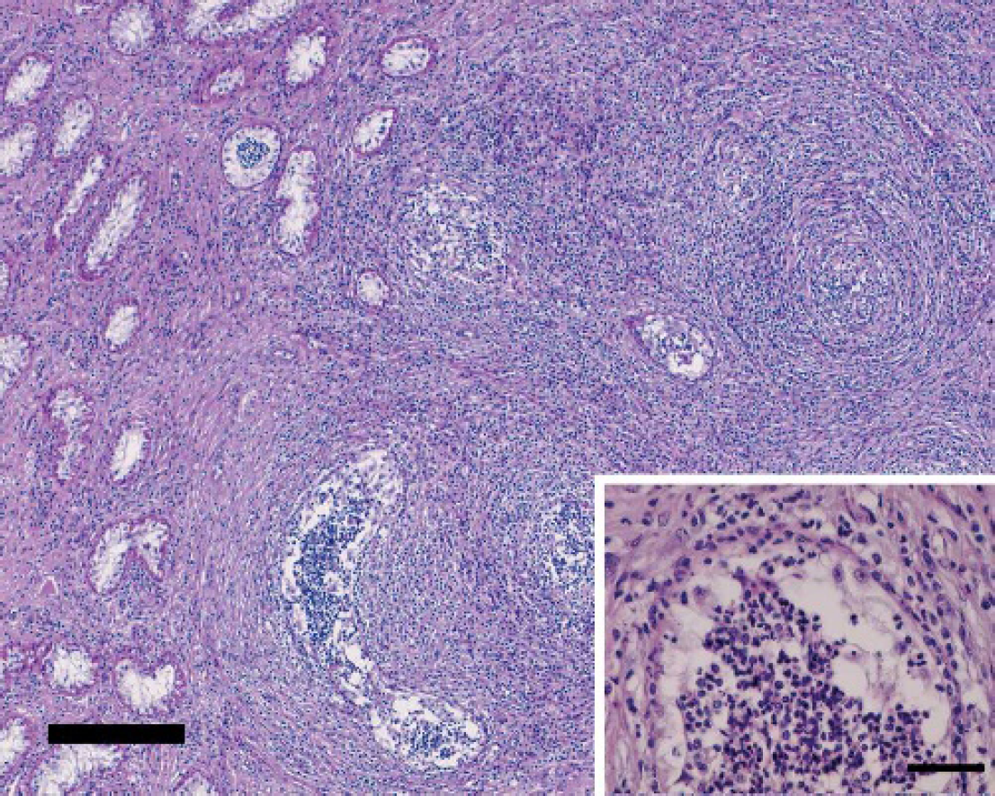
Testis. Pyogranulomatous orchitis characterized by the presence of microabscesses. On the left, atrophic seminiferous tubules are present. PAS stain; bar = 300 μm. The inset shows a seminiferous tubule with only Sertoli cells lining the basement membrane and neutrophils in the lumen. PAS stain; bar = 150 μm.
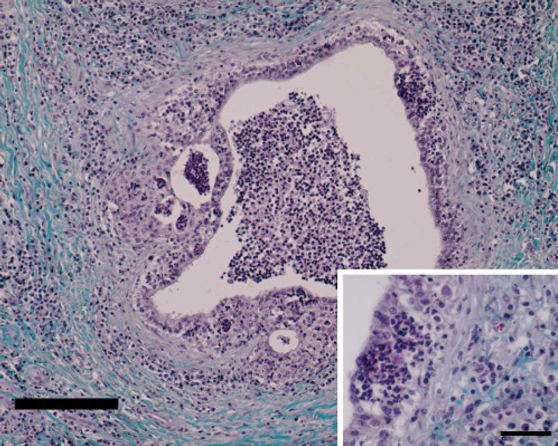
Epididymis. The duct is surrounded by an infiltrate of plasma cells, macrophages, and lymphocytes. Neutrophils are accumulated in the lumen. Masson Goldner stain; bar = 200 μm. The inset shows epithelial hyperplasia and an intraepithelial cyst. Bar = 50 μm.
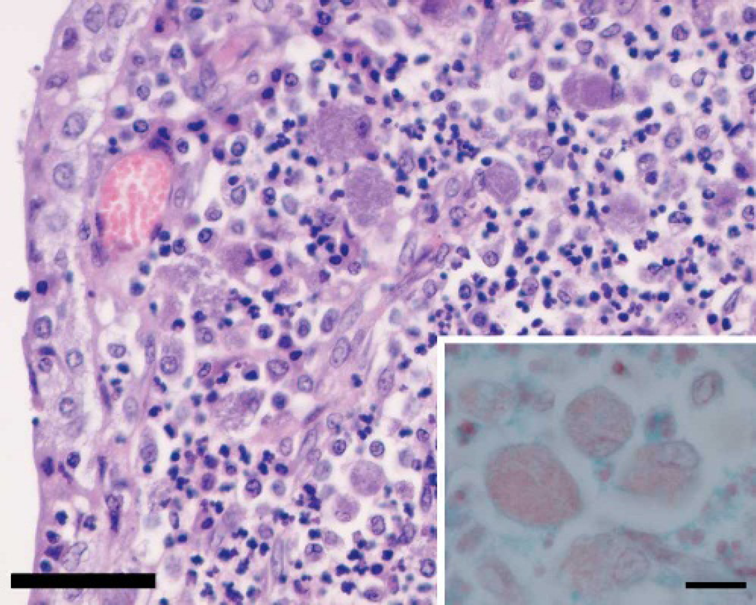
Epididymis. The inflammatory exudate contains large basophilic macrophages. HE, bar = 50 μm.
The seminiferous tubules of the left testis showed a normal architecture, with germinal cells in different stages of the spermatogenic cycle and minimal signs of testicular degeneration, such as occasional presence of multinucleated giant cells in tubular lumina. The epididymis contained abundant mature spermatozoa, and epididymal cysts around the head were lined by cuboidal epithelial cells. Fibrin thrombi, indicative of disseminated intravascular coagulation (DIC), were observed in renal and myocardial vessels. These and other organs such as the lung, liver, and lymph nodes showed edema, congestion, and hemorrhages. Gram-negative bacterial colonies were identified in scrotal lymph nodes, lung, and liver.
This study reports a case of suppurative epididymo-orchitis in a ram, associated with S. enterica subsp. diarizonae serovar 61:k:1,5,(7). To the authors' knowledge, this is the first report in which this Gram-negative organism has been found pathogenic for the ram genitalia. This work also confirms a previous report suggesting that this Salmonella serotype is a pathogen of ovines. 8 In sheep, S. enterica subsp. diarizonae serovar 61:k:1,5,(7) has been isolated from apparently healthy sheep at slaughter, 22 but it has also been implicated in cases of abortion and enteritis. 8,19 Salmonella abortus-equi has been isolated from orchitis cases in a stallion 14 and S. gallinarum in a fowl. 18 In humans several cases of orchitis due to salmonella have been reported. 7,11,21
The gross and microscopic genital lesions observed in this study, mainly epididymo-orchitis followed by abscess formation, are in agreement with those described as characteristic of genital involvement in human salmonellosis. 1,7,11,21 The vascular changes (petechiae, echymoses, thrombosis) observed in several organs at necropsy suggest that Salmonella-filled macrophages may have transported the organism from genitalia to lymph nodes, and further multiplication might have caused septicemia, with location of bacteria in many organs and death by endotoxic shock and DIC. 16 Similar lesions have been described in sheep in association with Arizona infection. 8
Factors that predispose the ram to develop testicular and epididymal lesions associated with salmonella are unknown. It has been demonstrated that stressed, debilitated ewes are predisposed to salmonellosis. 8,12 Although there was no evidence of salmonellosis in the flock, the possibility of the existence of asymptomatic carriers cannot be excluded, since S. enterica subsp. diarizonae serovar 61:k:1,5,(7) has been isolated from healthy sheep 22,17 and even wild animals. 2,20
The presence of lesions in ductus epididymis could support the hypothesis of an ascending route of the infection, as has been suggested previously. 13 However, a hematogenous route of spread of the bacteria from the intestinal tract is another possible route of spread. 6
In conclusion, the testicular and epididymal lesions observed in this study, together with the isolation of S. enterica subsp. diarizonae serovar 61:k:1,5,(7) from genital tissues, in the absence of other pathogens, strongly suggest that this bacteria is pathogenic for the ovine genitalia and should be considered in the differential diagnosis of ram epididymo-orchitis. The presence of S. enterica subsp. diarizonae serovar 61:k:1,5,(7) in sheep in Spain could have public health significance, since human infections through the consumption of uncooked meat or contaminated animal products have been described. 4
Acknowledgements. The authors thank Javier Otaola for providing the ram for this study, and Gloria Belver for technical assistance.
Footnotes
a.
bioMérieux, Lyon, France.
b.
Seward, Worthing, West Sussex, UK.
c.
Becton & Dickinson (DIFCO), MD.