
Abstract
An adult female crested porcupine (Hystrix cristata) was evaluated for acute onset of neurologic signs including head tilt, circling, and ataxia. She was found dead in her holding area 2 days after initially exhibiting clinical signs. Necropsy was unremarkable. Histopathology of brain tissue revealed the presence of protozoal cysts associated with inflammation as the underlying cause of clinical signs and death. Immunohistochemical staining of brain tissue for Toxoplasma gondii was strongly positive. PCR on fresh brain confirmed T. gondii as the causative organism. An adult male in the same enclosure has demonstrated similar neurologic signs over the past 3 years and has failed to respond to various medical treatments. Clinical disease associated with T. gondii has not been previously reported in this porcupine species or any other Old World porcupines, although there are several reports of clinical toxoplasmosis involving New World porcupine species.
Toxoplasmosis is a protozoal disease with the definitive host consisting of all domestic and free-ranging felids, which commonly shed the infective oocysts into the environment in their feces. 7 Toxoplasmosis infections have been reported as a problem in numerous species within zoological institutions. 4,8,9,10,12,13,16 The exact route of transmission is usually unknown, but is suspected to be domestic felines. Frequently the diagnosis of this disease in exotic animals is made at necropsy. The following case report describes the presenting clinical signs, necropsy, histology, and immunopathology of an African crested porcupine (Hystrix cristata) infected with toxoplasmosis.
A captive adult female African crested porcupine, over 6 years old, housed with a male African crested porcupine since 1999, presented with an acute onset of head tilt, circling, and anorexia; she was easily startled and appeared ataxic. She was housed with a male African crested porcupine that had been exhibiting mild neurologic behaviors of circling since 2002. The animals were housed in a moat exhibit and fed a mixture of dry browse biscuits, dog food, and produce. Treatment was attempted with antibiotics and anthelminthics for the female. She did not respond to treatment and was found dead in her holding stall later that day. The male's symptoms remained unchanged despite treatments of antibiotics and anthelminthics in the past. Differential diagnoses included Baylisascaris procyonis, toxoplasmosis, neoplasia, or trauma.
A complete necropsy was performed on the female porcupine; there were no significant findings. Sections of brain, heart, lungs, liver, kidney, spleen, adrenal, stomach, small intestine, colon, pancreas, and urinary bladder were collected in 10% buffered formalin. Samples were forwarded to the Diagnostic Center for Population and Animal Health, Michigan State University, for histopathologic examination. Additional tissue samples were frozen for further analysis pending histological results.
Formalin-fixed samples were paraffin-embedded, sectioned at 5 μm, and routinely stained with hematoxylin and eosin (HE). Histopathologic examination of brain tissue revealed marked lymphoplasmacytic perivascular cuffing admixed with lesser numbers of neutrophils and eosinophils within a large single focus of malacia in the gray matter of the cerebrum (Fig. 1). A similar focal zone of malacia and inflammation was present in the white matter at the base of the cerebellum. Outside the 2 focal lesions was more widespread gliosis. Severe large eosinophilic protozoal cysts measuring 15–40 μm in diameter were associated with the inflammatory foci, and each cyst contained numerous basophilic zoites (Fig. 2). These findings were consistent with encephalitis induced by an apicomplexan protozoal organism. Other histologic findings included moderate patchy interstitial and subendocardial fibrosis in the heart, patchy pulmonary congestion and hemorrhage, mild centrilobular hepatic congestion, and moderate membranous glomerulonephritis and moderate interstitial fibrosis in the kidney. Sections of adrenal gland, stomach, small intestine, colon, pancreas, and urinary bladder were morphologically normal.
The principal microscopic lesion in this porcupine was protozoal encephalitis. Differential diagnoses for protozoal encephalitis included toxoplasmosis, neosporosis, and sarcocystosis. Immunohistochemistry was performed to help in the differentiation of these organisms. The lesions in the heart, lungs, liver, and kidneys were considered nonspecific, aging lesions which were not likely to have contributed to this animal's acute illness and death.
For immunohistochemical examination, paraffin embedded sections of brain were incubated with polyclonal goat antibodies against Toxoplasma gondii. a The primary antibody was diluted 1:100. A streptavidin-immunoperoxidase labeling procedure was used to demonstrate immune reactions. Positive immunohistochemical controls included domestic cat tissues that contained known T. gondii cysts to which the same primary antibody was reacted. For negative controls, the primary antibody was replaced with homologous irrelevant antiserum. Immunohistochemical staining for T. gondii was strongly positive on the encysted protozoal zoites present within sections of the brain. While this technique was helpful in identifying the protozoal organism, additional molecular techniques were used to confirm the protozoal agent involved.
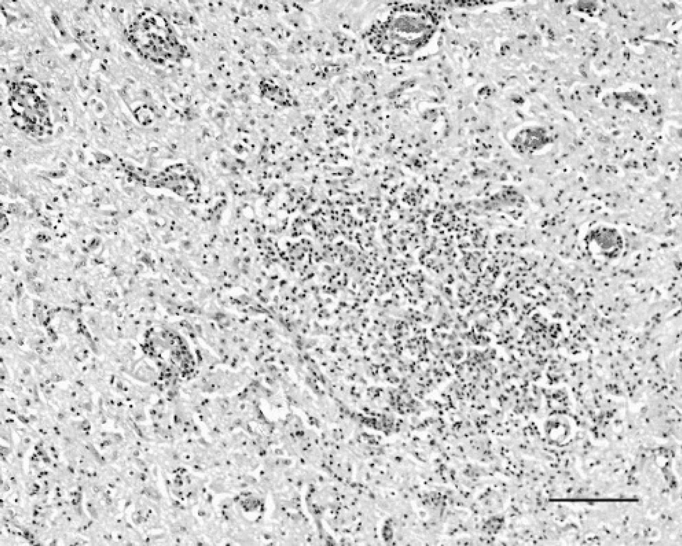
Photomicrograph of a section of brain from an African crested porcupine infected with toxoplasmosis. Note the moderate perivascular infiltrates by predominantly lymphocytes and plasma cells, as well as the central malacic area of gray matter with extensive glial cell and mixed mononuclear leucocyte infiltration. HE stain; bar = 200 μm.
To determine the identity of the parasites, a PCR assay was performed. Briefly, approximately 25 mg of fresh frozen brain was placed in a microcentrifuge tube and digested overnight at 55°C by addition of 200 μl of TL buffer and 20 μl proteinase K solution (DNeasy Tissue Kit). b After digestion, 200 μl of AL buffer was added to the microcentrifuge tube and the manufacturer's instructions for the kit were followed. The PCR assays were designed to detect DNA from the apicomplexan group of parasites or to specifically detect T. gondii or Neospora caninum. The PCR primers used for detection of DNA from apicomplexan parasites were 5′-CTTGTCTTAAAGATTAAGCCATGC-3′ and 5′ -GGATTCCCATCATTCCAATCA-3′. These primers were designed in our laboratory from aligned sequences of the 18S ribosomal RNA gene from apicomplexan organisms. The PCR reaction mixture consisted of 2.5 μl of 10X PCR buffer (10X HotStarTaq Master Mix Kit; b 0.2 mM each dATP, dGTP, dCTP, and dTTP; 1.5 mM MgCl2; 0.4 pmol/μl each primer; 0.625 units HotStar Taq polymerase b 2 μl sample DNA and molecular biology grade water to a final volume of 25 μl. The reaction conditions were 1 cycle of 95°C for 15 minutes; 40 cycles of 95°C for 40 seconds, 55°C for 40 seconds, 72°C for 40 seconds; followed by 1 cycle of 72°C for 5 minutes. The T. gondii-specific PCR primers were derived from the 35-fold repetitive B1 gene 1,3 and were 5′-AACGGGCGAGTAGCACCTGAGGAGA-3′ and 5′-TGGGTCTACGTCGATGGCATGACAAC-3′. The N. caninum-specific PCR primers were 5 ′-CCCAGTGCGTCCAATCCTGTAAC-3′ and 5′-CTCGCCAGTCAACCTACGTCTTCT-3′. 15 The PCR reaction mixture used for the T. gondii or N. caninum-specific primers was as described above except that 0.3 pmol/μl of each primer and 5 μl of DNA were used. The reaction conditions were 1 cycle of 95°C for 15 minutes; 40 cycles of 95°C for 30 seconds, 65°C for 30 seconds, 72°C for 30 seconds; followed by 1 cycle of 72°C for 5 minutes. Amplification products (10 μl) were analyzed in ethidium bromide-stained 1.5% agarose gels after electrophoresis in sodium borate buffer (Fig. 3). 2 The results of the PCR assay supported presence of T. gondii in the brain tissue. To confirm this finding, the amplicon from the apicomplexan PCR assay was excised from the agarose gel, purified with QIAquick Gel Extraction Kit b and eluted in 30 μl of nuclease-free water. An aliquot of the DNA was then sequenced at the Research Technology Support Facility, Michigan State University, using an ABI Prizm 3100 Genetic Analyzer. The 449 bp of sequence that was obtained was a 100% match with published sequences from the T. gondii 18s rRNA gene. Therefore, T. gondii was confirmed as the causative agent responsible for this animal's clinical signs.
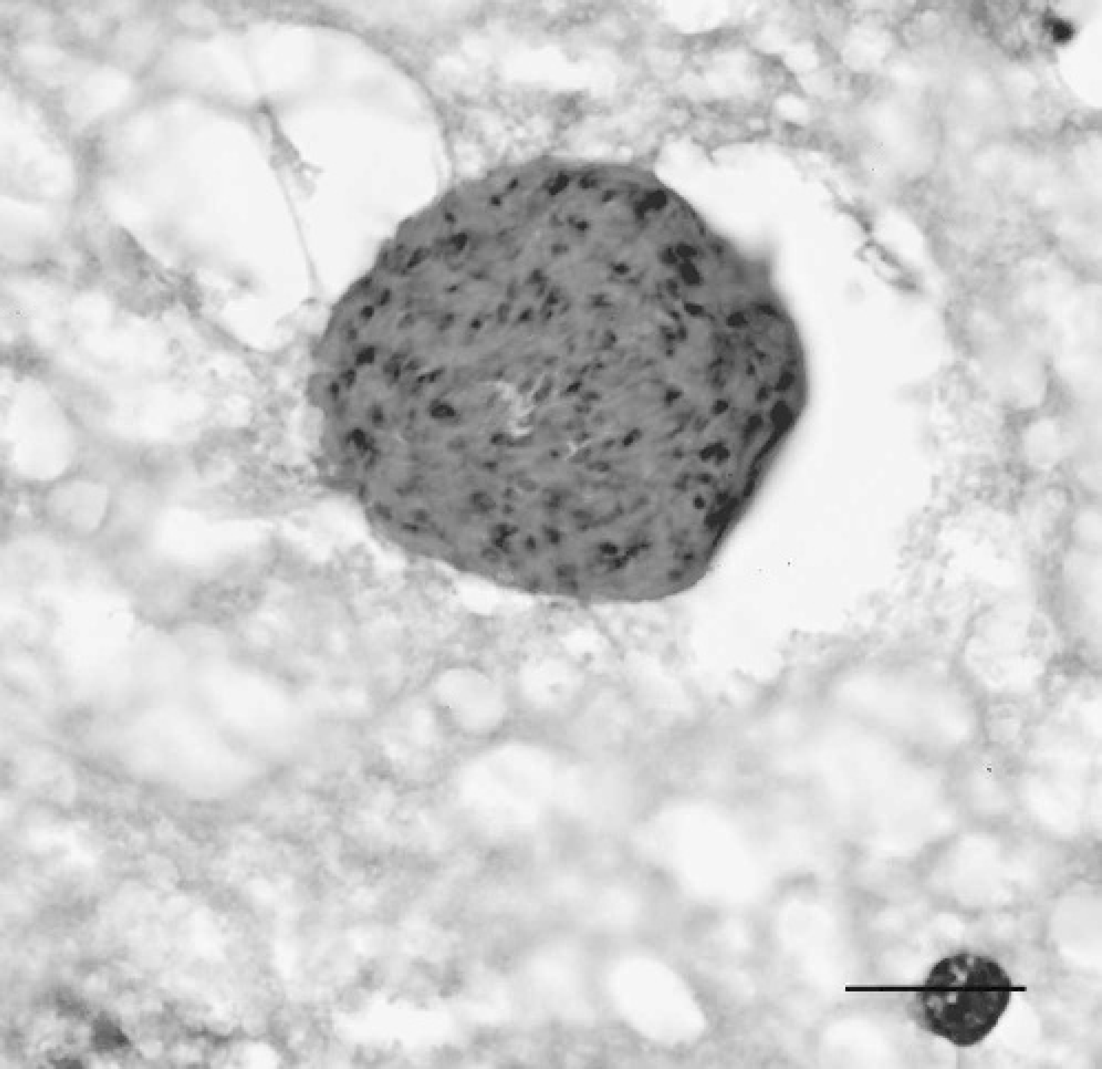
Photomicrograph of T. gondii cyst in brain of an African crested porcupine. HE stain; bar = 20 μm.
The mate of this animal has demonstrated similar clinical signs, including occasional circling, a head tilt to the left, horizontal nystagmus, and occasional ataxia, for over 3 years. However, in contrast to the female, this animal was able to eat and move around the exhibit and holding area with no difficulties. Various medical treatments have been attempted, but none of them have improved his clinical signs. Toxoplasmosis has not been reported in any H. cristata or any other Old World porcupine species, though it has been previously reported in a captive Brazilian prehensile-tailed porcupine (Coendou mexicanus) and 2 North American porcupines (Erethizon dorsatum). 11,14 The prehensile-tailed porcupine was presented for necropsy after sudden death with no associated clinical signs. Both North American porcupines were submitted for necropsy; one was euthanized after demonstrating neurological signs, and the other died suddenly a month later. One of the 2 North American porcupines was concomitantly infected with Baylisascaris.
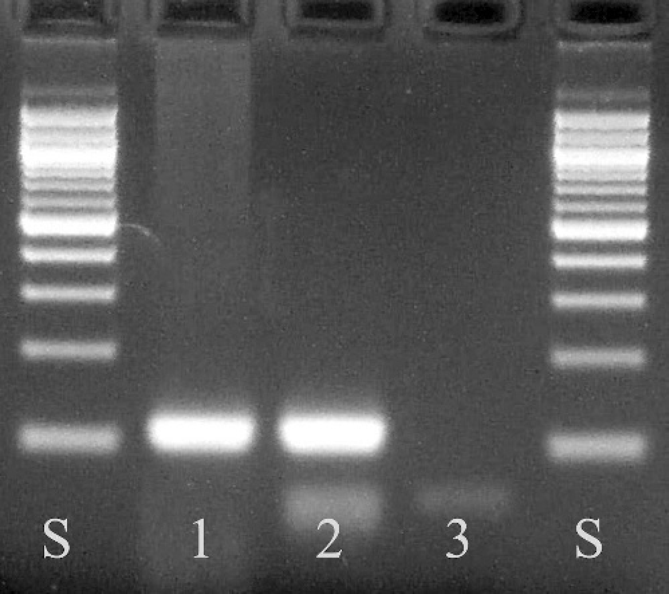
PCR products for T. gondii from a sample of brain from an African crested porcupine. Lanes S = 100-bp ladder scale; lane 1 = porcupine brain sample; lane 2 = positive T. gondii control sample; lane 3 = negative control sample.
Toxoplasmosis has been suspected in several other deaths at this institution. In recent years a red panda (Ailurus fulgens fulgens), and a red-necked wallaby (Macropus rufogriseus) have been diagnosed as having toxoplasmosis histologically. Other macropods and a river otter (Lontra canadensis) have had T. gondii titers. The location of the zoo is near railroad tracks, which attract feral domestic felines. These feral cats have been found occasionally within the property of the zoo. Once they are discovered, they are live-trapped and removed from the premises. The crested porcupine exhibit consists of an animal holding building where the animal was housed overnight and an outdoor exhibit yard, a system that allows free access to roaming cats to the crested porcupine enclosure at night. It is likely that feral cats defecated in the outdoor exhibit area or the wall surrounding the exhibit and shed T. gondii oocysts, resulting in contaminated grounds or feeding stations and leading to exposure of the crested porcupines. It is also possible that the oocysts were carried into the exhibit by the animal caretakers or their cleaning equipment; these same caretakers also cared for several exotic felids housed at the same zoo. The oocysts may have also been brought into the exhibit along with the produce these animals ate, however, the produce fed is fit for human consumption, and this possibility is minimized. Toxoplasmosis is a relatively common disease in zoologic parks, with animal species reported as infected numbering well over 100 and including rodents, carnivores, herbivores, marsupials, primates, marine mammals, and various birds. 7,17 This underscores the importance of feral animal control and biosecurity measures to prevent excessive exposure to infectious diseases and parasites to valuable zoo animal species on exhibit. Further, the importance of toxoplasmosis as a zoonotic pathogen needs to be understood by zoo staff who may be potentially exposed to infective oocysts within zoologic exhibits.
Toxoplasmosis also occurs in free-ranging animal populations. A study performed in the western United States showed that 20% of North American porcupines were seropositive for T. gondii. 10 Another study evaluating seroprevalence of T. gondii in French Guiana showed a seropositive rate of 0% for the Brazilian porcupine (Coendou prehensilis). 5,6 One of the conclusions of the French Guiana survey was that primarily tree-dwelling species, including the Brazilian porcupine, were less likely to come into contact with significant numbers of infectious T. gondii oocysts. 5 African crested porcupines are a predominantly terrestrial and burrowing species, and so are easily exposed to T. gondii oocysts if the ground or feed is contaminated. Despite the possibility of these porcupines having a greater risk of exposure to toxoplasmosis, a survey of other zoological institutions with captive H. cristata did not find any other cases of toxoplasmosis, although some other animal species at these institutions were infected with toxoplasmosis (personal communication). The authors would like to emphasize the importance of feral cat management and exclusion policies in zoological institutions to prevent the exposure of all exotic mammals to diseases transmitted by felines, even species that are not usually associated with felidae infections.
Footnotes
a.
Toxoplasma gondii antiserum, catalog #210-70-TOXO, VMRD, Inc., Pullman, WA.
b.
QIAGEN Inc., Valencia, CA.