
Abstract
Two beef steers accidentally injected into a branch of the auricular artery with an oil-based formulation of ceftiofur died within 5 minutes of injection. Notable pathologic findings included distention and obstruction of cerebral and cerebellar arteries by a whitish tan material and hemorrhages within meningeal spaces, the choroid plexus, cerebrum, and cerebellum. Lipid material was identified within cerebral blood vessels in frozen sections stained with oil red O. This report describes an unusual case of brain ischemia in beef cattle.
Accidental intra-arterial injection of drugs occasionally occurs in equine and bovine veterinary practice. Most notable and more frequently reported has been intracarotid injection of promazine in the horse. In these cases, adverse reactions developed suddenly, occurring within a few seconds of injection, and ranged from excitement, dyspnea, and depression to collapse with seizure activity and death. 2,4 Similar reactions were recorded, although with unpredictable individual variation, in experimental intracarotid promazine injection. 4
Pathologic findings in tissues from either experimental or accidental intracarotid injections were similar. Grossly, brains were swollen and edematous, with congestion of meningeal and cerebral blood vessels and petechial and ecchymotic leptomeningeal and cerebral hemorrhages. Histologic alterations reflected direct severe acute endothelial and vascular injury and included fibrinoid vascular necrosis, ischemic neuronal necrosis, hemorrhage, vacuolation of the white matter, and edema, as suggested by dilation of Virchow-Robin spaces and large astrocytic halos. 2,4
In cattle, a single case of accidental intracarotid artery injection of chloramphenicol was identified in a review of the literature. 6 A 5-year-old Holstein cow collapsed immediately following the injection, developed dyspnea, convulsed, and died within 5 minutes. Gross and microscopic pathologic findings were similar to those described in horses injected with promazine. This report describes the pathologic findings in 2 beef steers dying as a result of accidental injection of an oil-based antibiotic preparation into the auricular artery.
Two dead 12-month-old beef steers of mixed breeding were submitted to the Livestock Disease Diagnostic Center at the University of Kentucky for necropsy. These animals were in a group of 72 recently purchased cattle that were being processed for entry into a stocker operation. Each animal was vaccinated and received a single subcutaneous injection of ceftiofur crystalline-free acid in the right ear. Ear injection of this antibiotic preparation has been shown to be as effective as other routes of administration and has the added benefits of no injection site trimmings at slaughter and, because the ear is an inedible tissue, a short withdrawal time. 5 The 2 presented animals became recumbent 2 to 3 minutes after injection and were dead 5 minutes later as perceived by the owner.
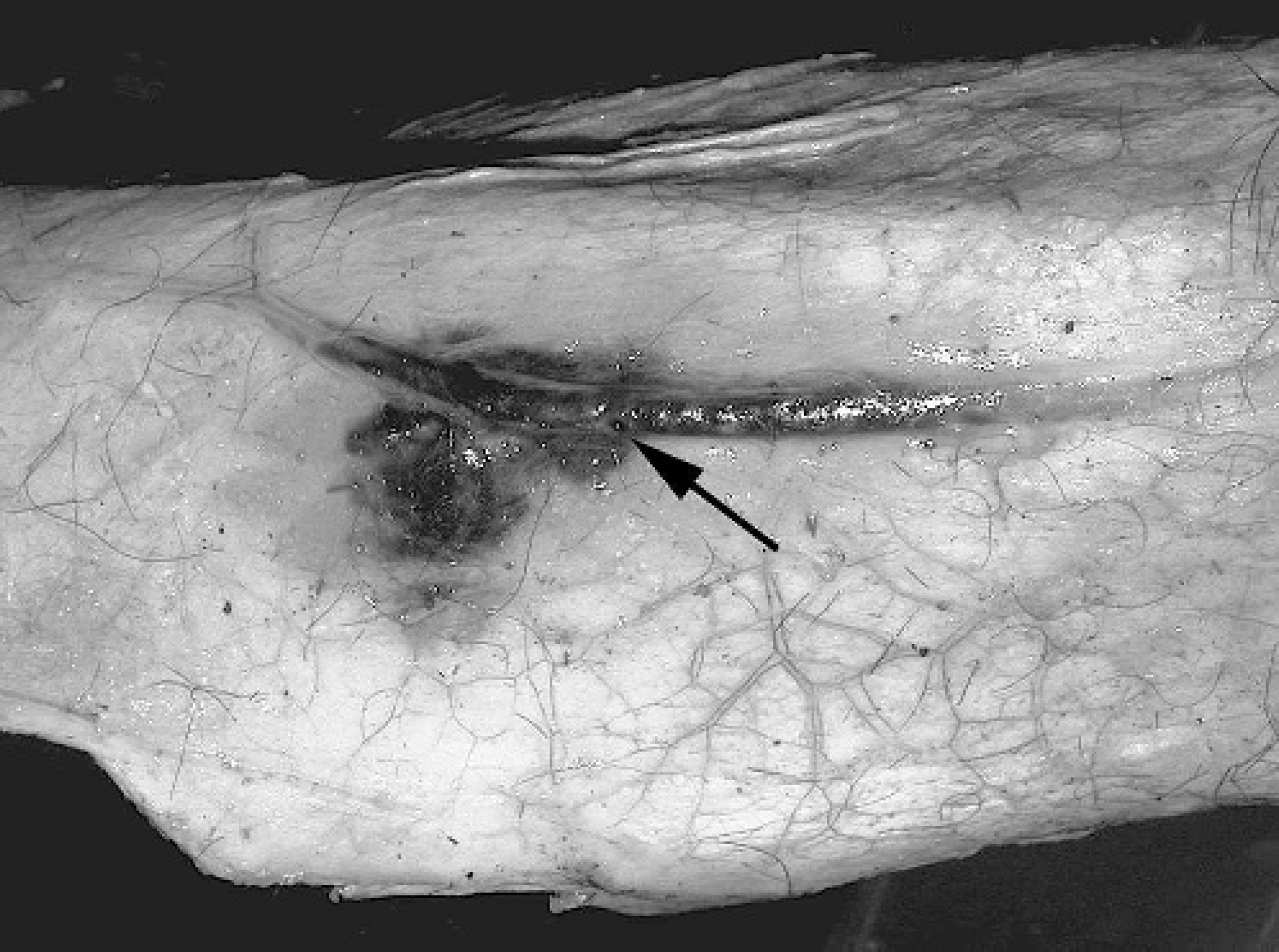
Dilation and thickening of an auricular artery from an affected steer is associated with mild edema and hemorrhage. The puncture site is visible (arrow).
At necropsy, both animals were in good body condition with moderate postmortem autolysis (death occurred the day before necropsy). Because intra-arterial injection of the antibiotic was suspected at the time of submission, the skin covering the right ear of each animal was removed and the injection site examined. In both animals, the auricular artery was punctured, thickened, and surrounded by hemorrhage. There was no visible antibiotic residue in tissues surrounding the artery (Fig. 1). Lesions in the brain of each animal were for the most part confined to the right cerebral hemisphere and right half of the cerebellum. A tan-white material was grossly visible within the distended lumina of the right rostral, middle and caudal cerebral arteries, the common artery of the corpus callosum, and the distal branches of each artery (Fig. 2). The same material was seen less frequently in arteries supplying the left side of the brain. There were foci of cerebral and cerebellar meningeal hemorrhage (both animals) and extensive hemorrhage at the base of the medulla oblongata (1 animal); multiple variably sized randomly distributed hemorrhages in the white matter of the right cerebral hemisphere and choroid plexus of both lateral ventricles (both animals) were also present (Fig. 3). Additional macroscopic alterations included hemorrhages in multiple tissues including the fascia of the neck, tracheal and esophageal mucosa, epicardium, and renal cortex.
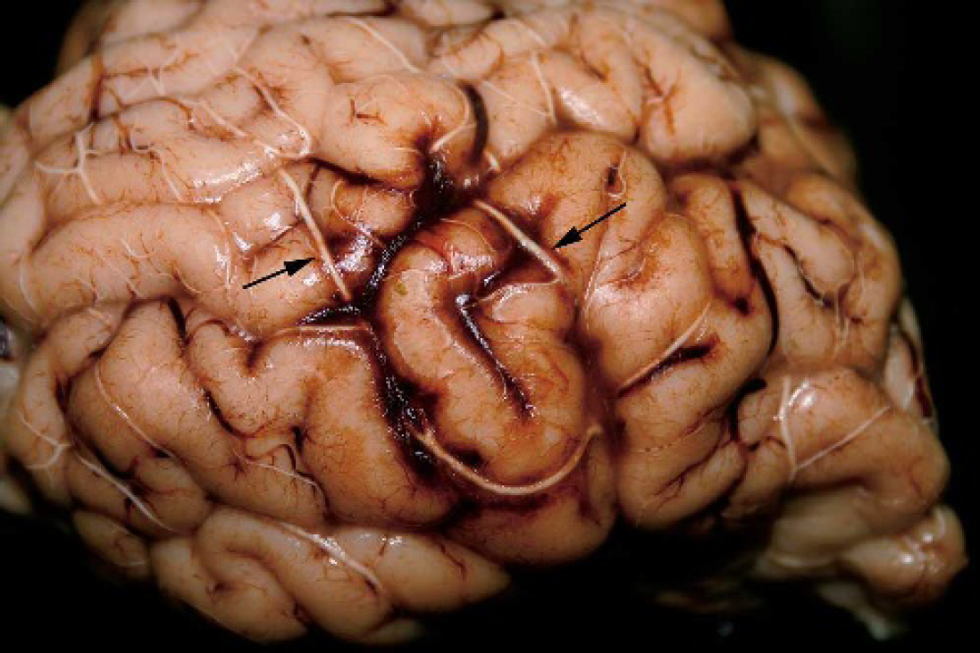
Arteries supplying the right cerebral hemisphere are dilated and filled with a tan-white material (arrows).
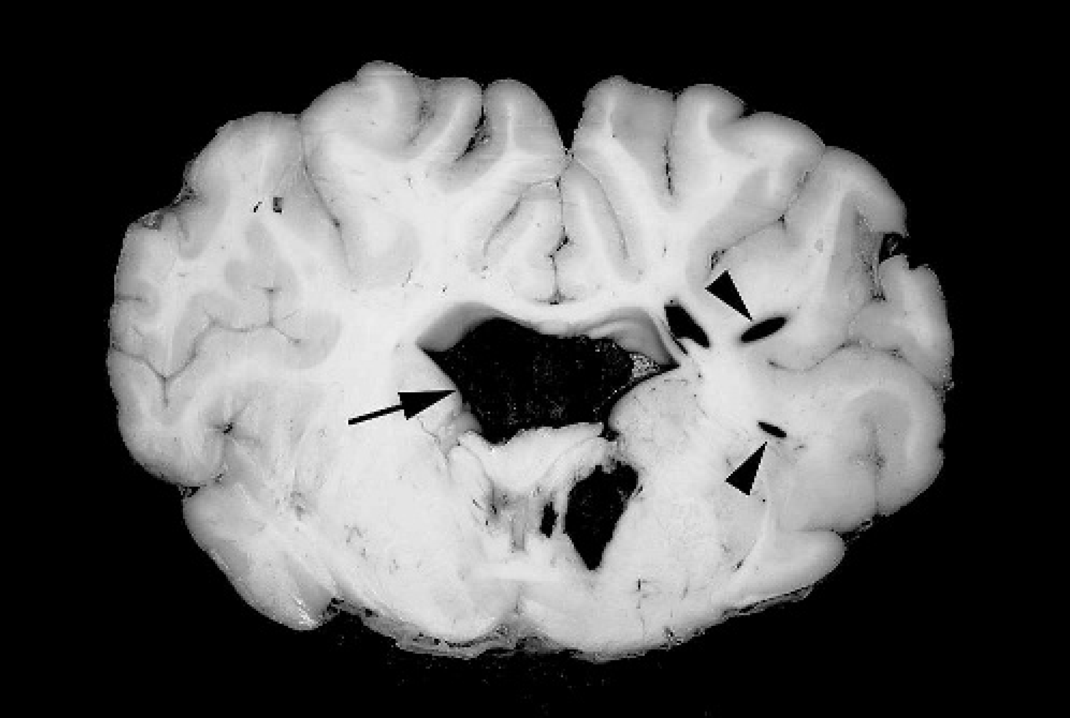
Hemorrhage in the choroid plexuses of the lateral ventricles (arrows) and white matter of the cerebrum (arrowheads).
Samples of lung, liver, spleen, kidney, small and large intestine, and mesenteric and bronchial lymph node were collected for bacterial culture, virus isolation, and fluorescent antibody testing. Brain, lung, liver, kidney, spleen, rumen, omasum, abomasum, and small and large intestine were submerged in 10% neutral buffered formalin and processed for histologic evaluation. Rumen content was collected for toxicologic analysis.
Neither bacteria nor viruses were cultured or isolated, respectively, from submitted tissues. Fluorescent antibody tests for infectious bovine rhinotracheitis virus, bovine viral diarrhea virus, bovine respiratory syncytial virus, and parainfluenza-3 virus were negative. No known toxins were detected in rumen content by gas chromatographic/mass spectrometric analysis.
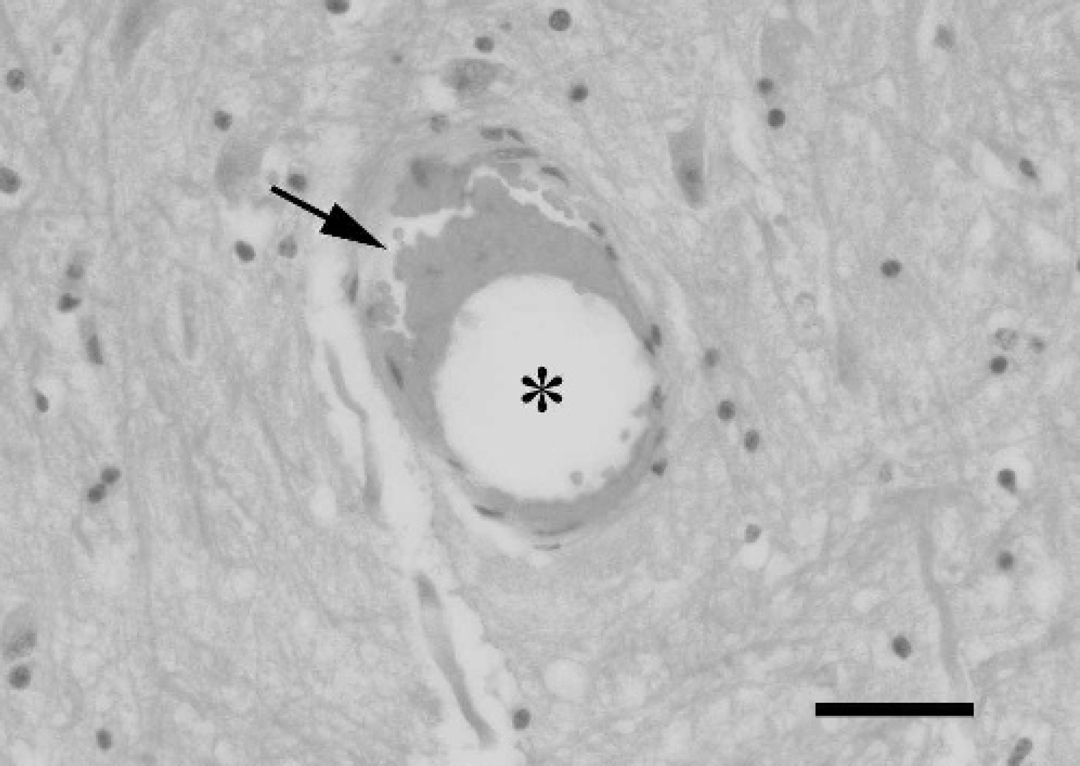
Dilated cerebral blood vessel containing a clear space (*) that represents material removed during processing. Red blood cells (arrow) are along the margin. Hematoxylin and eosin (HE). Bar = 40 μm.
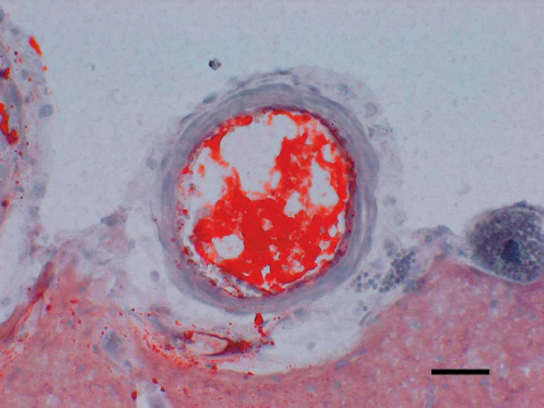
Meningeal artery filled with a homogenous orange lipid material. Frozen section. Oil red O. Bar = 40 μm.
Microscopic findings in these animals were limited to the brain. Even though injections were given into the right ear, many arteries, arterioles, and capillaries in the right and left cerebral hemispheres, thalamus/hypothalamus, hippocampus, midbrain, cerebellum, and medulla oblongata were dilated, had a round to oval profile, and were either empty or had a central clear space with sharp borders and surrounded by red blood cells (Fig. 4). In frozen sections of cerebrum stained with oil red O, a homogenous orange lipid material was within the lumina of many blood vessels (Fig. 5). Multiple variably sized hemorrhages were in meningeal and perivascular spaces and in white matter of the cerebrum and cerebellum. There was no evidence of vasculitis although some blood vessels had mildly swollen endothelial cells. Neuronal necrosis and perivascular edema were not conclusively identified.
This case is unique for several reasons. The pathogenesis is unusual, involving the accidental injection of an oil-based ceftiofur preparation into the auricular artery with subsequent retrograde flushing of the antibiotic into the external carotid artery and subsequently the circulatory system of the brain. Once in the external carotid artery, the material may have either continued moving against a pressure gradient to the opening of the internal carotid artery and hence the brain or flowed with the pressure gradient into the maxillary artery and eventually the branch supplying the rostral epidural rete mirabile (or both). The acute onset of clinical signs in these animals with rapid progression to death supports a direct path of the material to the brain and is similar to the clinical findings and outcome of a cow accidentally receiving intracarotid injection of chloramphenicol. 6 It should be noted that a significant amount of antibiotic should have passed into the maxillary artery with localization in other tissues supplied by this vessel.
From a pathologic perspective, this case is unusual in that focal cerebral ischemia and infarction to this degree are rarely reported in animals. The distribution of occluded arteries in the brains of these steers suggests blood flow to large portions of the right and left cerebral hemispheres, diencephalon, midbrain, cerebellum, and medulla oblongata was permanently interrupted. In general, the ensuing catastrophic physiologic (electrical failure) and metabolic effects (decreased adenosine triphosphate and phosphocreatine concentrations, increased lactate concentration, and fatty acid and protein catabolism) would lead to neuronal necrosis and death. 1 More specifically and in relation to clinical signs, the abrupt transition of the animals from a conscious to a comatose state reflects severe acute injury to the midbrain and medulla oblongata and in particular to components of the ascending reticular activating system, which has projections into the thalamus and from the thalamus to the entire cerebral cortex. 3 Additionally (although not reported by the owner), abnormal breathing patterns and subsequent respiratory arrest most likely occurred as a result of ischemic lesions in respiratory centers in the caudal brain stem.
Although the animals in this report and cow in a previous report died within minutes of accidental injection of the auricular and carotid arteries, respectively, with an antibiotic preparation there were significant differences in histologic alterations in brain and in the intermediate steps leading to cerebral hypoxia/ischemia. In the steers, injection of the antibiotic preparation led to abrupt and complete obstruction of cerebral blood flow without histologic evidence of vasculitis and cerebral edema (increased perivascular spaces containing astrocytic processes, astrocytic halos, and status spongiosis). The lack of conclusive evidence of cerebral edema, however, may have been a result of postmortem decomposition. Additionally, microscopic evidence of neuronal necrosis was not observed because of the extremely short interval between the onset of clinical signs and death of the animal. In the cow, intra-arterial injection of chloramphenicol resulted in extensive vascular necrosis, thrombosis, hemorrhages, and edema. These differences are most likely caused by the composition and irritant properties of the injected materials.
Acknowledgement. This report was published (05-14-050) by permission of the Dean and Director of the Kentucky Agriculture Experiment Station and the College of Agriculture, University of Kentucky, Lexington.