
Abstract
This study aimed to evaluate the antitumor activity of platinum nanoparticles compared with cis-platin both in vitro and in vivo in the treatment of hepatocellular carcinoma induced in rats. The treatment efficacy of platinum nanoparticles was evaluated by measuring antioxidant activities against oxidative stress caused by diethylnitrosamine in liver tissue. The measurements included reduced glutathione content and superoxide dismutase activity, as well as malondialdehyde level. Liver function tests were also determined, in addition to the evaluation of serum alpha-fetoprotein, caspase-3, and cytochrome c in liver tissue. Total RNA extraction from liver tissue samples was also done for the relative quantification of B-cell lymphoma 2, matrix metallopeptidase 9, and tumor protein p53 genes. Histopathological examination was also performed for liver tissue. Results showed that platinum nanoparticles are more potent than cis-platin in treatment of hepatocellular carcinoma induced by diethylnitrosamine in rats as it ameliorated the investigated parameters toward normal control animals. These findings were well appreciated with histopathological studies of diethylnitrosamine group treated with platinum nanoparticles, suggesting that platinum nanoparticles can serve as a good therapeutic agent for the treatment of hepatocellular carcinoma which should attract further studies.
Introduction
The liver is a vital organ that fulfills a wide range of functions including detoxification of various metabolites, protein synthesis, and the regulation of immune responses. 1 Hepatocellular carcinoma (HCC) is a malignant tumor that is the fifth most common type of cancer and the third leading cause of cancer-related death globally. 2 The rate of HCC has been increasing in Egypt with a doubling in the incidence rate in the past years. This has been attributed to several biological and environmental factors. Other factors such as cigarette smoking, occupational exposure to chemicals such as pesticides, and endemic infections in the community, such as schistosomiasis, may have additional roles in the etiology or progression of the disease. 3
Diethylnitrosamine (DEN) is found in a wide variety of foods such as cheese; soybeans; smoked, salted, and dried fish; cured meat; and alcoholic beverages, as well as in ground water having a high level of nitrates. In rats, DEN is a potent hepatocarcinogen influencing the initiation stage of carcinogenesis during a period of enhanced cell proliferation accompanied by hepatocellular necrosis and induces DNA carcinogen adducts, DNA-strand breaks, and in turn HCC without cirrhosis through the development of putative pre-neoplastic focal lesions. 4
It was Barnett Rosenberg in 1965 who accidentally discovered the biological activity of cis-platin, which was recognized as an anticancer drug. The therapeutic activity of cis-platin is achieved by binding with DNA to form crosslinks as major lesions, thus inhibits replication and transcription processes and finally the cell’s repair mechanism and leads to cellular apoptosis.5,6
Despite of good clinical success of cis-platin, it lacks tumor tissue selectivity leading to some severe side effects. Advances in nanotechnology and growing needs in biomedical applications have driven the development of multifunctional nanoparticles. 7
Nanoparticles have the potential to be ideal carriers for delivering anticancer and other therapeutics to diseased sites with minimal collateral damage to normal tissues. 8 Functional platinum nanoparticles (Pt NPs) have evoked keen interest in recent decades owing to their size- and shape-dependent optical, catalytic, and therapeutic properties. Platinum-based nanomaterials have been notable for excellent therapeutic applications. Functional Pt NPs have shown apoptosis-inducing properties through target-specific pathways. Platinum compounds are used as very effective anticancer agents. This property is associated with the inhibition of DNA replication and mitosis by the addition of Pt NPs to DNA strand.9,10 This study aimed to evaluate the antitumor activity of Pt NPs compared with cis-platin both in vitro and in vivo in the treatment of HCC induced in rats.
Materials and methods
Chemical compounds
DEN was purchased from Sigma-Aldrich Corporation (USA). Cis-platin (cis-PtCl2 (NH3)2) was obtained from Oncotec Pharma Produktion GmbH as solution for infusion in a vial of 10 mL Cisplatine Mylan (1 mg/mL).
Hydrogen hexachloroplatinate (IV; H2PtCl6xH2O) was purchased from Loba Chemie (India). Pt NP biosynthesis was done according to Das et al., 11 where the selected bacterial isolate was inoculated into 250-mL Erlenmeyer flask containing 100 mL sterile nutrient broth. The cultured flasks were incubated in a rotating shaker set at 200 r/min for 48 h at room temperature. The culture was then centrifuged at 12,000 r/min for 10 min. The biomass and supernatant were separated and used separately for the synthesis of Pt NPs. The supernatant was used for studying extracellular production of Pt NPs by mixing it with filter-sterilized hexachloroplatinate solution at 1 mM final concentration. All the reaction mixtures were incubated on rotating shaker (200 r/min) at room temperature for a period of 72 h. Visual observation was conducted periodically to check for the nanoparticle formation. Further characterization was conducted for nanoparticle generated through extracellular methods.
Experimental animals
In all, 90 adult male Swiss albino rats (150–200 g) were obtained from the animal farm of the Egyptian Holding Company for Biological Products and Vaccines (VACSERA), Cairo, Egypt. They were maintained on a standard pellet diet and tap water. The animals were housed in suitable cages in conditioned atmosphere (20°C–22°C) and kept on a standard diet.
Methods
In vitro studies
Cytotoxicity assay using crystal violet
HepG2 cells (human cell line of a well-differentiated HCC) obtained from Egyptian National Cancer Institute, Cairo University, were used to determine the cell’s cytotoxic effect of each of the tested treatments according to Vijayan et al. 12
In vivo studies
Determination of LD50 using experimental animals
In screening drugs, determination of LD50 is usually an initial step in the assessment and evaluation of the toxic characteristics of a substance. The LD50 of the studied compounds was determined as described by Akhila et al. 13
Experimental design
Animals were allowed 10 days for adaptation. They were then randomly distributed into six equal groups, 15 rats each. The animal groups were recognized as follows:
Group 1 (Control). Normal healthy control animals.
Group 2 (Pt NPs). Animals were gavaged with 10% of LD50 of Pt NPs (5.75 mg/kg body weight/day) for 6 weeks.
Group 3 (cis-platin): Each animal was injected intra-peritoneally with 10% of LD50 of cis-platin (0.64 mg/kg body weight/day) for 6 weeks.
Group 4 (DEN): Each animal was gavaged with DEN (dissolved in 0.9% normal saline), in a dose of 20 mg/kg, five times a week for 6 weeks according to the modified method of Darwish and El-Boghdady. 14
Group 5 (DEN + Pt NPs): Rats received DEN as in group 4 and then treated with Pt NPs for 6 weeks as in group 2 after induction.
Group 6 (DEN + cis-platin): Rats received DEN as in group 4 and then treated with cis-platin for 6 weeks as in group 3 after induction.
Collection of samples
At the end of the treatment period, animals were fasted overnight prior to dissection under light ether anesthesia. Blood was drawn from the vena cava and centrifuged at 3000g for 10 min. Immediately after blood collection, liver tissue was excised and one portion was used for the histopathological examination. The rest of the tissue was homogenized in 0.25 M ice cold isotonic sucrose to be used for the estimation of the assessed parameters.
Experimental parameters
Lipid peroxidation was measured in liver tissue as described by Ohkawa et al. 15 Reduced glutathione content was assayed colorimetrically as described by Beutler et al. 16 Superoxide dismutase activity was measured according to the method of Nishikimi et al. 17 Total protein level, albumin level, total bilirubin, aminotransferases (alanine aminotransferase (ALT) and aspartate aminotransferase (AST)), and alkaline phosphatase (ALP) activities in serum were determined using colorimetric assays as described by Henry et al., 18 Doumas et al., 19 Malloy and Evelyn, 20 Reitman and Frankel, 21 and Kind and King, 22 respectively, using diagnostic kits. Serum from each group was assayed for alpha-fetoprotein (AFP) using AFP enzyme-linked immunosorbent assay (ELISA) kit purchased from Cloud-Clone Corp. (USA), catalog no. SEA153Ra. Liver tissue homogenates of each group were assayed for caspase-3 (Casp-3) level using rat caspase-3 ELISA kit purchased from MyBioSource, Inc. (USA), catalog no. MBS700575. Cytochrome c (Cyt c) level was determined in liver tissue homogenates using Rat/Mouse Cyt c ELISA kit purchased from R&D Systems Quantikine ELISA (USA; catalog no. MCTC0).
Quantitative real-time polymerase chain reaction
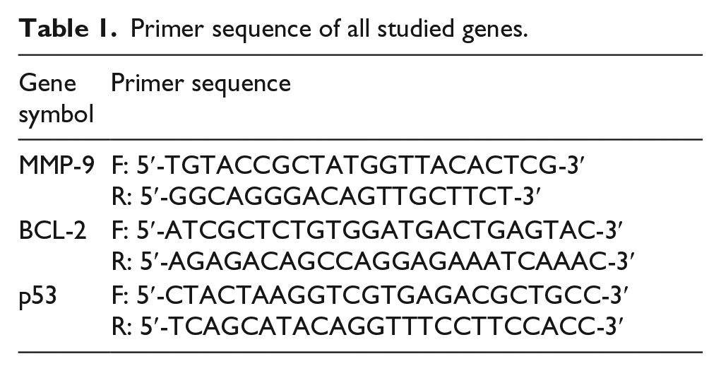
Total RNA was isolated using QIAGEN tissue extraction kit (QIAGEN, USA) according to instructions of manufacturer. Quantitative real-time polymerase chain reaction (qRT-PCR) amplification and analysis were performed using an Applied Biosystems with software version 3.1 (StepOne™, USA). The qRT-PCR assay with the primer sets was optimized at the annealing temperature. All complementary DNAs (cDNAs) were in duplicate and included previously prepared samples for B-cell lymphoma 2 (BCl-2), matrix metallopeptidase 9 (MMP-9), and p53 with glyceraldehyde 3-phosphate dehydrogenase (GAPDH) as an internal control, and water is used as non-template control to confirm the absence of DNA contamination in the reaction mixture (Table 1).
Primer sequence of all studied genes.
Histopathological examination
After sacrificing the rats, liver tissue was rapidly dissected and excised, rinsed in saline solution, and cut into suitable pieces which were fixed in neutral buffered formalin (10%) for 24 h according to the method adopted by Drury and Wallington 23 and examined by light microscope for histopathological investigation.
Statistical analysis
All mean values are reported as the mean ± standard deviation (SD). Data were analyzed using a one-way analysis of variance (ANOVA). The level of significance between mean values was set at p < 0.05 and p < 0.01 (significant and highly significant, respectively). All statistical analyses were performed using SPSS software (version 20.0).
Results
Characterization of Pt NPs
The obtained nanoparticles were characterized by means of transmission electron microscopy (TEM; Figure 1(a)), ultraviolet–visible (UV-vis) spectroscopy (Figure 1(b)), and Fourier transform infrared (FTIR) spectroscopy (Figure 2 (a) and (b)).

Characterization of platinum nanoparticles. (a) Pt NPs using TEM. (b) UV-vis absorption spectrum of Pt NPs. TEM shows that the approximate sizes of the nanoparticles are found to be 20–40 nm. UV-vis spectrum of platinum nanoparticles shows a surface plasmon absorption band with a maximum absorbance at 344 nm which can be taken as indication for Pt NP formation.

Fourier transform infrared (FTIR) spectroscopy. (a) FTIR spectra of extracellular extract of bacterial broth. (b) FTIR spectra of Pt NPs.
FTIR spectrum of extracellular extract of bacterial broth shows a strong intensity peak at 3320.74 cm−1 which represents hydroxyl group, strong peak at 1637.20 cm−1 which represents amide I bonds of proteins, and strong peak at 593.30 cm−1 which represents single bond of C–H or C–N.
The FTIR of Pt NPs showed strong signals at 3318.10 cm−1 which represents hydroxyl group, strong peak at 1636.93 cm−1 which represents amide I bonds of proteins, and strong peak at 602.12 cm−1 which represents single bond of C–H or C–N indicating the interactions of nanoparticles with phytochemicals of Bacillus sp.
Comparing FTIR spectrum of extracellular extract of bacterial broth and Pt NPs, we observed shifting in signal wavenumber per centimeter of the major peaks indicating reduction of platinum and formation of Pt NPs as some R-OH were oxidized and C–H bond was reduced. Overall, it can be concluded that the Bacillus sp. proteins adsorb as a layer over the synthesized Pt NPs, thereby stabilizing the nanoparticles formed through the surface-bound proteins.
In vitro studies
The antitumor activity of Pt NPs and cis-platin was evaluated using HepG2 cell lines. The chemical compounds were applied at different concentrations and results were presented in Figure 3.

Cytotoxic activity of Pt NPs and cis-platin against HepG2 cell line.
In vivo studies
Determination of liver superoxide dismutase, glutathione, and malondialdehyde levels
The data represented in Table 2 indicated that liver superoxide dismutase (SOD) and glutathione (GSH) were significantly decreased in DEN rats than control animals. DEN groups subjected to Pt NPs and cis-platin resulted in significant increase in antioxidants levels compared to DEN group.
Statistical analysis (ANOVA) for liver antioxidant levels in the different groups.

ANOVA: analysis of variance; SOD: superoxide dismutase; DEN: diethylnitrosamine; MDA: malondialdehyde; GSH: glutathione; SD: standard deviation.
Each value is represented as mean ± SD. Data with different superscripts are significantly different at p ≤ 0.05.
Significance versus control group.
Significance versus DEN group.
Significance vs DEN + Pt NPs
DEN animals also showed significant increase in malondialdehyde (MDA) level than normal rats. Treatment with Pt NPs and cis-platin showed significant decrease in MDA level than DEN rats. More pronounced increase in SOD and GSH levels associated with reduction in MDA level was observed in DEN group treated with Pt NPs compared to cis-platin-treated ones.
Liver function tests
A significant decrease was recorded in albumin and protein levels in DEN animals than control group as shown in Table 3.
Statistical analysis (ANOVA) for liver function tests in the different groups.

ANOVA: analysis of variance; ALP: alkaline phosphatase; DEN: diethylnitrosamine; MDA: malondialdehyde; GSH: glutathione; SD: standard deviation; AST: aspartate aminotransferase; ALT: alanine aminotransferase.
Each value is represented as mean ± SD. Data with different superscripts are significantly different at p ≤ 0.05.
Significance versus control group.
Significance versus DEN group.
Significance vs DEN + Pt NPs
Results in Table 3 also indicated significant increase in total bilirubin level, ALT, AST, and ALP activities in DEN rats than normal rats. Amelioration in liver function tests was shown in DEN animals subjected to different treatments toward control animals in comparison to DEN group especially in Pt NPs–treated DEN rats.
Table 4 shows results for serum AFP, liver Casp-3, Cyt c levels, MMP-9, BCL-2, and P53 relative quantification in liver tissue for all groups.
Statistical analysis (ANOVA) for serum AFP, liver Casp-3, Cyt c levels, MMP-9, BCL-2, and p53 relative quantification in the different groups.

ANOVA: analysis of variance; AFP: alpha-fetoprotein; DEN: diethylnitrosamine; MMP-9: matrix metallopeptidase 9; SD: standard deviation; BCL-2: B-cell lymphoma 2.
Each value is represented as mean ± SD. Data with different superscripts are significantly different at p ≤ 0.05.
Significance versus control group.
Significance versus DEN group.
Significance vs DEN + Pt NPs
Serum AFP
Serum AFP level showed significant increase in DEN rats compared to control animals. Treatment of DEN-induced group with Pt NPs and cis-platin showed a significant decrease in AFP level.
Liver caspase-3
Data showed significant decrease in casp-3 level in DEN model than normal rats. Treatment of DEN animals with Pt NPs and cis-platin resulted in significant increase in liver casp-3 level compared to control and DEN groups. Pt NPs’ treatment revealed significant increase in casp-3 level in DEN animals compared to cis-platin-treated group.
Liver Cyt c
Results also revealed significant decrease in liver Cyt c level in DEN rats than normal group. In comparison to control and DEN rats, Pt NPs and cis-platin treatment for DEN animals showed significant increase in Cyt c level. DEN group subjected to Pt NPs revealed also highly significant increase in Cyt c level than DEN animals administered with cis-platin.
Quantitative real-time PCR
MMP-9
Data revealed significant increase in liver MMP-9 gene expression level in DEN rats treated with cis-platin and Pt NPs than normal rats. A highly significant decrease in MMP-9 level was observed in DEN animals administered different treatments than in DEN group. Pt NPs treatment for DEN rats also resulted in a significant decrease in MMP-9 level than in DEN rats administered with cis-platin.
BCL-2
BCL-2 level in liver tissue revealed significant increase in DEN group and DEN animals subjected to Pt NPs and cis-platin than normal rats. Administration of different treatments to DEN animals resulted in significant decrease in BCL-2 level than in DEN group. A highly significant decrease in liver BCL-2 level was seen in Pt NP-treated DEN rats than DEN animals subjected to cis-platin.
P53
Data of liver P53 gene expression level showed significant decrease in DEN animals, while Pt NP-treated DEN showed significant increase in liver P53 gene expression level than normal rats. Pt NP- and cis-platin-treated DEN animals revealed significant increase in P53 level than DEN group. DEN rats subjected to Pt NPs showed significant increase in P53 expression level than DEN group administered with cis-platin.
Histopathological studies
There was no histopathological alteration, and the normal histological structure of the central vein and surrounding hepatocytes in the parenchyma was observed in control, Pt NPs, and cis-platin groups as shown in Figure 4(a)–(c), respectively. Fine fibroblastic cell proliferation dividing the degenerated, necrotic, and dysplastic hepatocytes into nodules was seen in liver of DEN animals (Figure 4(d)). In the liver of DEN rats administered with Pt NPs, dilatation was noticed in the central veins associated with fine fibroblastic cell proliferation dividing the focal vacuolar degenerated hepatocytes into few nodules with no signs of dysplasia (Figure 4(e)). Dilatation in the central veins associated with degeneration and dysplasia in the hepatocytes of the hepatic parenchyma was shown in the liver of DEN group treated with cis-platin (Figure 4(f)).

(a) Liver of rats in normal control group, (b) Pt NP group, and (c) cis-platin group showing normal histological structure of the central vein with surrounding hepatocytes in the parenchyma (H&E 40×). (d) Liver of rats in DEN group showing fine fibrosis dividing the degenerated and necrotic dysplastic hepatocytes into nodules (H&E 16×). (e) Liver of rat in DEN + Pt NPs group showing dilatation of central vein with very fine fibroblastic cell proliferation dividing the vacuolar degenerated focal areas of hepatocytes into few nodules with no signs of dysplasia (H&E 16×). (f) Liver of rats in DEN + cis-platin group showing dilatation of central vein with degeneration and dysplasia of hepatocytes in the parenchyma (H&E 40×).
Discussion
Reports indicate that Pt NPs might be useful as therapeutics in cancer therapy, and Pt NPs in combination with Hadron therapy led to an enhancement of strongly lethal DNA damage caused by double-strand breaks. 24
This study was conducted to evaluate the efficiency of biologically synthesized Pt NPs in the treatment of HCC compared to cis-platin both in vitro and in vivo.
Accordingly, the cytotoxic effects and the biological activity of Pt NPs and cis-platin as antitumor agents were examined in vitro against human liver carcinoma cell line (HepG2) using crystal violet cytotoxicity assay.
Results showed potent effect of Pt NPs more than cis-platin in a dose-dependent manner, where increasing concentration of Pt NPs resulted in increased percentage of dead cells. This result is in harmony with Alshatwi et al. 25 who reported that Pt NPs inhibit cell proliferation via induction of apoptotic cell death. In addition, Pt NPs produce a cytotoxic effect by reducing cell viability and causing inter-nucleosomal DNA fragmentation, G2/M cell-cycle arrest, and hypo-diploid accumulation emphasizing that the Pt NPs have potential anticancer properties and can be applied as cancer therapeutics.
A previous study showed that Pt NPs’ uptake by the cells endocytosis and emphasized intracellular release of Pt+2 ions from Pt NPs (that) blocks cell division by binding to DNA causing DNA damage and contributed to the cytotoxicity and metabolic stress activating cell death via apoptosis. Also, downregulation of proliferating cell nuclear antigen, a factor critical for DNA replication and repair following Pt NP treatment, supports the anti-proliferative effects of Pt NPs. 26
In this study, injection of DEN in male albino rats induced significant deleterious changes in antioxidant status. The results revealed a marked depletion in liver tissue GSH content and SOD activity with a significant increase in MDA level in liver tissue compared to the control group. These findings were in agreement with other studies which reported that DEN confers its hepatocarcinogenicity through the metabolic activation in the hepatic microsomes, resulting in the release of ethylcarbonium ions that bind to the DNA, producing adducts and generating superoxide radicals through lipid peroxidation of phospholipid membrane fatty acids. MDA, a product of lipid peroxidation of polyunsaturated fatty acid metabolism and degradation, has been established as a mutagenic and carcinogenic entity.27,28
Lowering of MDA level and the increase in levels of GSH and SOD in DEN rats treated with Pt NPs and cis-platin indicate their potential as inhibitors of DEN-induced intracellular oxidative stress. Pt NPs has repair effect over cis-platin in remodeling the oxidative stress induced by DEN.
These results are in consonance with others29,30 who showed that post treatment cis-platin in rats injected with DEN suppressed partially the oxidative stress as indicated by significantly decreased hepatic content of MDA and H2O2 and increased content of GSH and SOD in the liver, compared to the untreated HCC group.
Previous studies demonstrated that Pt NPs showed similar activity to that of oxidizing nicotinamide adenine dinucleotide and reducing ubiquinone. Moreover, Pt NPs have unveiled antioxidant properties that scavenge reactive oxygen species (ROS), including superoxide anion (O–2), hydrogen peroxide (H2O2), and free radicals.25,31
The present biochemical results revealed a decrease in total protein and albumin levels, while significant increase was shown in total bilirubin level and ALT, AST, and ALP activities in DEN group than control rats, which agrees with previous studies.32,33 This may be attributed to DEN-induced oxidative stress which induces liver tissue damage and impairment of liver function.
In this study, post treatment of DEN animals with Pt NPs and cis-platin resulted in improvement in liver function tests compared to DEN rats in agreement with others.29,34
The DEN rats also showed significant increase in AFP level compared to control animals. This result is in line with previous studies35,36 that indicated that increase in serum AFP level upon DEN induction is associated with increase in tumor growth.
Decrease in AFP level after treatment with cis-platin conforms with previously published data. 37 Also, administration of Pt NPs resulted in lowering of AFP level compared with DEN rats, which indicates the antitumor activity of Pt NPs and also the decrease in the production rate of tumor as indicated by Yamada et al., 38 who claimed that Pt NPs are thought to serve as a reservoir for platinum ions that can induce DNA damage in cancer cells.
This study showed significant decrease in Casp-3 level in DEN group compared to normal animals as reported before. 39 Treatment with cis-platin and Pt NPs resulted in increase in Casp-3 level compared to DEN rats. These results are in consonance with previous studies, which indicates that both treatments induced apoptosis as reflected by increase in Casp-3 level.30,40
The results also showed a decrease in Cyt c level in DEN rats compared to control group. This result is in line with Moreira et al. 41 who stated that DEN resulted in decrease in Cyt c and increase in apoptotic proteins BCL-2 and B-cell lymphoma-extra-large (BCL-XL) that prevents the release of mitochondrial contents such as Cyt c and thus inhibit apoptosis.
In DEN animals treated with Pt NPs and cis-platin, the level of Cyt c in liver tissue was increased, especially in Pt NP-treated group. Cis-platin induces the death receptors DR4 and DR5 through the upregulation of c-myc and strengthens the activation of caspases via promoting the release of Cyt c, and it also induces apoptosis. 42
This study showed overexpression of MMP-9 in DEN-treated rats which is in concordance with Chang et al. 43 who observed that MMP-9 level was increased as a result of DEN treatment when compared to normal group.
DEN animals treated with Pt NPs and cis-platin showed significant downregulation in MMP-9 gene especially following Pt NPs treatment. These results are in harmony with previous data44,45 where MMP-9 activity was inhibited upon treatment with Pt NPs by acting as scavenger for ROS that acts as an activator for MMP-9.
The upregulation found in liver BCL-2 gene in DEN-treated group is in concordance with others,43,46 where overexpression inhibits apoptosis which is in conjunction with the decrease in Cyt c and Casp-3 levels in DEN animals.
DEN rats administered with Pt NPs and cis-platin showed significant downregulation of BCL-2 gene expression compared to DEN group especially those treated with Pt NPs and also show induction of apoptosis by both treatments.
A previous study showed that cis-platin induced cell death in human liver cancer cell line (HepG2) by increasing Casp-3 and P53 levels to induce apoptosis and downregulation of anti-apoptotic BCL-2 gene and its protein expression. 47 Downregulation in anti-apoptotic BCL-2 gene in A549 lung carcinoma cells and induction of apoptosis upon treatment with Pt NPs were also reported. 48
Results revealed that DEN resulted in downregulation of P53 gene in rats compared to control group. This result correlates with others41,49 who stated that P53 gene was downregulated and its expression was significantly reduced in DEN rats.
Platinum NPs and cis-platin-treated animals showed upregulation in P53 gene expression especially by treatment with Pt NPs which suggest activation of apoptosis. As reported before, p53 protein level increased human lung fibroblasts (IMR-90) and human glioblastoma cells (U251) following treatment with Pt NPs. 26
It was shown that cis-platin-induced apoptosis by ROS generation which significantly caused loss of mitochondrial membrane potential in sensitive cells, induction of p53, apoptosis regulator protein Bax and suppressing anti-apoptotic proteins. 50
Cis-platin caused induction of p53 in both parental and cis-platin-resistant human ovarian cancer 2008 cells, and the increase in p53 correlated with cis-platin-induced apoptosis. 51
In this study, histopathological finding revealed that liver of DEN rats showed fibroblastic cell proliferation dividing the degenerated, necrotic, and dysplastic hepatocytes into nodules. This result correlates with others,36,41,52 and they found that liver tissue of DEN-treated rats showed hydropic degeneration, and focal areas of necrosis, portal inflammation, and hepatocytes showed partial loss of architecture and significant tumor nests.
Platinum NPs and cis-platin treatment showed histopathological improvement. All sections showed more or less normal architecture. Collectively, most of the histological manifestations observed in cancer control were greatly reduced especially when Pt NPs were used in treatment.
Conclusion
From the aforementioned results, it can be concluded that Pt NPs resulted in ameliorating the measured parameters toward normal animals and it is more potent than cis-platin in treatment of HCC induced by DEN in rats. These findings were well appreciated with histopathological studies suggesting that Pt NPs can serve as a good therapeutic agent for the treatment of HCC which should attract further studies.
Footnotes
Declaration of conflicting interests
The author(s) declared no potential conflicts of interest with respect to the research, authorship, and/or publication of this article.
Funding
The author(s) received no financial support for the research, authorship, and/or publication of this article.