
Abstract
STYK1 (Serine/threonine/tyrosine kinase 1), a member of the receptor tyrosine kinase family, exhibits tumorigenicity in many types of cancers. Our study reveals the important role played by STYK1 in nasopharyngeal carcinoma. STYK1 is upregulated in nasopharyngeal carcinoma tissues compared with para-carcinoma. Knockdown of STYK1 inhibits nasopharyngeal carcinoma cell proliferation, migration, and invasion, while ectopic STYK1 expression significantly promoted cell proliferation, migration, and invasion abilities. In addition, we provided lines of evidence supporting the critical role of STYK1 in the regulation of glycolysis via activation of phosphoinositide 3-kinase/AKT pathway. Survival analysis reveals that STYK1 level is an independent prognostic factor for nasopharyngeal carcinoma patients. Our results indicate that STYK1 is a promising therapeutic target in nasopharyngeal carcinoma.
Introduction
Nasopharyngeal carcinoma (NPC), the most common malignancies in South East Asia and southern China, is a type of malignant head and neck cancer originated from the nasopharyngeal epithelium.1,2 Though a decline of incidence and a substantial decrease in mortality due to early diagnosis and advanced radiotherapy and chemotherapy, it remains unsatisfactory for locally advanced and metastatic NPC.3,4 Therefore, an urgent need to identify novel and specific biomarkers for NPC is critically of importance, with a hope to improve the prognosis of NPC.
Serine/threonine/tyrosine kinase 1 (STYK1), a novel oncogene kinase, is a member of the receptor tyrosine kinases family, possibly for activating downstream signaling cascades.5,6 It has a single transmembrane domain and an intracellular domain possessing tyrosine kinase activity but lacks an ectodomain for binding specific ligands.5,6 Notably, the tumorigenicity of STYK1 has been identified in many types of malignancies, such as lung cancer, 7 colorectal cancer, 8 breast cancer, 9 and ovarian cancer. 10 In this article, we investigated the clinicopathological features and prognostic implications of STYK1 expression in patients with NPC and further discussed the biological function of STYK1 and the underlying mechanism.
Methods
Human tissue specimens
In total, 120 tumor tissues and adjacent normal tissue samples were collected at Department of Pathology, Suining Central Hospital, Sichuan province, between January 2002 and December 2015. All the patients were diagnosed and confirmed of NPC by histology, and the patients who had received radiotherapy or chemotherapy were excluded. This study was approved by the Research Ethics Committee of Suining Central Hospital, Sichuan province, China, and informed consents were obtained from all the patients.
Samples were deparaffinized in xylene and rehydrated in a series of graded alcohol. Endogenous peroxidases were blocked by 3% H2O2, and antigen retrieval was done after heating in citrate buffer. The sections were incubated with a rabbit polyclonal antibody against STYK1 (dilution: 1:100; Abcam, Cambridge, MA, USA) at 4°C for overnight and then with horseradish peroxidase (HRP; Gene Tech GTVision III Detection Kit, Shanghai, China) at room temperature for 40 min. Following three washes with phosphate-buffered saline (PBS), the signal was detected with 3,3′-diaminobenzidine (DAB) solution.
Immunohistochemistry scores
A double-blind method was used to analyze immunohistochemistry results, and the evaluation was carried out independently by two pathologists, without access to the patients’ clinical and pathological features. In total, 10 visual fields from different areas of each specimen were chosen at random for the immunohistochemical evaluation. STYK1 expression was scored according to staining intensity and the percentage of positive cells as previously described. 11 The percentage of positive cells was scored as follows—0: no positive cells, 1: ≤10% positive cells, 2: 10%–50% positive cells, 3: >50% positive cells. Staining intensity was scored as follows—0: no staining, 1: faint staining, 2: moderate staining, 3: dark staining. Comprehensive score = staining percentage × intensity. STYK1 expression was classified as follows—≤4: low expression and >4: high expression.
Cell lines and reagents
The nasopharyngeal epithelial cell line NP69 and NPC cell lines CNE1, CNE2, 5-8F, and 6-10B were obtained from the Cell Bank of Chinese Academy of Sciences (Shanghai, China). NP69 cells were maintained in keratinocyte/serum-free medium (Invitrogen, Carlsbad, CA, USA) with epidermal growth factor (Invitrogen). All the NPC cell types were cultured in RPMI-1640 medium (Invitrogen) supplemented with 10% fetal bovine serum (FBS; HyClone, Logan, UT, USA). All cell lines were incubated at 37°C in an atmosphere of 5% CO2. MK2206 was purchased from Selleck Chemicals (Houston, TX, USA).
Plasmids and small interfering RNAs
The plasmids pcDNA3.1-STYK1 (pSTYK1) and control vector pcDNA3.1 were constructed by GeneChem (Shanghai, China). The small interfering RNA (siRNA) targeting STYK1 (siSTYK1) was constructed to knockdown its expression. The target sequence of to STYK1 was 5′-GGTGGTACCTGAACTGTAT-3′, and the control sequence of 5′-GCGCGCTTTGTAGGATTCG-3′ served as a negative control. 12
Transient transfection
Transfection of plasmids and siRNAs into NPC cells was performed using Lipofectamine 2000 and Lipofectamine RNAiMax, respectively (Invitrogen, Grand Island, NY, USA). For transient transfection, the cells were transfected with plasmids or siRNA as indicated for 48 h before performing the functional assays or western blot assays, respectively.
Quantitative real-time polymerase chain reaction
The relative basic expression levels in different tissues and knocked down expression levels of STYK1 messenger RNA (mRNA) in different NPC cells were examined by quantitative real-time polymerase chain reaction (qRT-PCR) as previously described. The expression of glyceraldehyde 3-phosphate dehydrogenase (GAPDH) was also determined and used as an internal control. The relative mRNA expression of each gene was calculated using the comparative threshold cycle (Ct) (2−ΔΔCt) method. The primer sequences of STYK1 and GAPDH were as follows—STYK1: 5′-CAGTGGGAAGGAGGGACTGA-3′ (forward) and 5′-TGCAGCCCAGTGAAATT GGA-3′ (reverse) and GAPDH: 5′-ACAGTCAGCCGCATCTTCTT-3′ (forward) and 5′-GACAAGCTTCCCGTTCTCAG-3′ (reverse).
Western blot analysis
Standard western blotting was carried out using whole-cell protein which was lysed by radioimmunoprecipitation assay (RIPA) buffer and labeled by primary antibodies against STYK1 (dilution 1:1000: Abcam) and GAPDH (dilution: 1:1000; Cell Signaling Technology, Beverly Hills, MA, USA) and a secondary antibody. The procedures were according the manufacturer’s protocol.
Lactate dehydrogenase activity, lactate production, glucose utilization assay, and intracellular adenosine triphosphate level
Tumor cells were transfected with siRNAs and plasmids; 1 × 10⁶ cells were prepared for lactate dehydrogenase (LDH) activity and lactate production assay with the Lactate Dehydrogenase Activity Assay Kit and Lactate Assay Kit, respectively (Sigma, St. Louis, MO, USA). For glucose utilization assay, tumor cells were transfected with plasmids or/and siRNAs or/and treated with MK2206, and the cultures were incubated for 24 h. The media were replaced with phenol-red free RPMI-1640 with 1% FBS to continuous culture for 3 days. Glucose concentrations in media were measured using a Colorimetric Glucose Assay Kit (BioVision, Milpitas, CA, USA) and normalized with cell number. Intracellular adenosine triphosphate (ATP) was measured using a firefly luciferase-based ATP assay kit (Beyotime Biotechnology, Haimen, China) following the manufacturer’s instructions. The protein concentration of each group was also determined using a bicinchoninic acid (BCA) protein assay kit. The relative ATP level is expressed as ATP value/protein value.
Cell Counting Kit-8 cell proliferation assay
The Cell Counting Kit-8 (CCK-8) was used to measure cell proliferation as described previously. 13 After the addition of CCK-8 solution (10 µL/well), an additional incubation of 2 h was carried out before measuring the absorbance at 460 nm.
Cell migration and invasion assay
CNE1 cells were transfected with different reagents respective to different groups. Cells in each group were trypsinized and 1 × 10⁵ cells in a 600 µL of serum-free medium of each group were placed in the upper parts of modified Boyden chambers with a Matrigel-coated/uncoated membrane (Millipore Corporation, MA, USA). RPMI-1640 with 10% FBS was used as a chemoattractant.
Statistical analysis
The results were expressed as mean ± standard error (SE). All statistical analysis was performed using the SPSS 13 software package (SPSS, Chicago, IL, USA). The association between STYK1 and clinicopathological features was analyzed using the χ2 test. Kaplan–Meier curves and the log-rank test were constructed for analysis of survival data. Multivariate analysis was performed by Cox proportional hazards model. The statistical correlation of data between groups was analyzed by one-way analysis of variance (ANOVA) and Student’s t test as appropriate. p < 0.05 was considered to be statistically significant.
Results
STYK1 expression is elevated in NPC tissues
We initially analyzed STYK1 expression in tumor and adjacent normal tissue samples from 10 patients with NPC. The results showed that in eight of the tumor samples, STYK1 expression was increased as compared to matched para-cancerous controls (p < 0.05, Figure 1(a)). STYK1 mRNA and protein levels in the NPC cell lines CNE2, 6-10B, 5-8F, and CNE1 were higher than in the non-tumorigenic human epithelial cell line NP69 (p < 0.05, Figure 1(b) and (c)). Besides, immunohistochemical staining revealed the presence of STYK1 at significant levels in NPC tissues and relatively insignificant levels in paired, non-tumor tissues (Figure 1(d), Table 1). These data indicate that the STYK1 expression is elevated in NPC tissues.

Expression of STYK1 in human nasopharyngeal carcinoma tissues and cell lines. (a) Expression of STYK1 in 10 representative matched samples of NPC tissues and adjacent non-tumorous tissues. (b) Quantitative real-time PCR (c) and western blotting were employed to detect STYK1 expression in NPC cell lines and NP69. (d) Immunohistochemical staining of STYK1 in (d1) adjacent non-tumorous tissues (d2) and NPC tissues. (d3) The number of cases with STYK1 expression between NPC and paired adjacent normal tissues. High STYK1 expression was detected in NPC specimen.
Comparison of STYK1 expression between NPC and paired adjacent normal tissues (N = 120).

STYK1: serine threonine tyrosine kinase 1; NPC: nasopharyngeal carcinoma.
p < 0.05.
STYK1 functions as an independent predictor of overall survival in patients with NPC
We further evaluated the correlation between STYK1 expression and the clinicopathological characteristics. The patients were divided into two groups (low STYK1 and high STYK1) according to the immunohistochemical scores in NPC tissue samples (Figure 2(a)). STYK1 overexpression was significantly correlated with lymph node metastasis, distant metastasis, and advanced tumor–node–metastasis (TNM) stage (p < 0.05, Table 2). Patients with NPC tumors expressing low STYK1 levels had significantly higher overall survival (OS) than those expressing high STYK1 levels (p < 0.05, Figure 2(b)).

Cumulative Kaplan–Meier overall survival curves of 120 nasopharyngeal carcinoma patients segmented by STYK1. (a) Representative images showed STYK1 expression level: (a1) low STYK1 and (a2) high STYK1. (b) Patients with NPC tumors expressing low STYK1 levels had significantly higher OS than those expressing high STYK1 levels. p values were calculated by the log-rank test.
Correlation between the clinicopathological characteristics and STYK1 expression (N = 120).
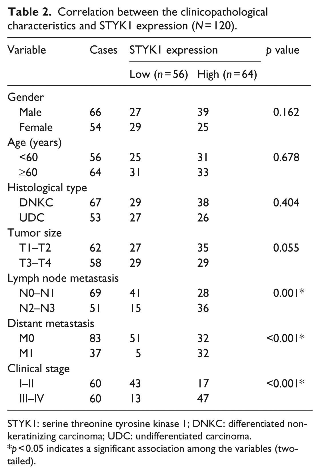
STYK1: serine threonine tyrosine kinase 1; DNKC: differentiated non-keratinizing carcinoma; UDC: undifferentiated carcinoma.
p < 0.05 indicates a significant association among the variables (two-tailed).
Univariate analysis indicated that STYK1 level, lymph node metastasis, distant metastasis, and clinical stage were correlated with patient survival (p < 0.001, p = 0.04, p = 0.003, and p < 0.001, respectively). Multivariate analysis showed that STYK1 level and clinical stage were independent prognostic factors for NPC patients (p < 0.001 and p = 0.004, respectively, Table 3).
Summary of univariate and multivariate Cox regression analysis of overall survival duration in all NPC (N = 120).

NPC: nasopharyngeal carcinoma; HR: hazard ratio; CI: confidence interval; STYK1: serine threonine tyrosine kinase 1; UDC: undifferentiated carcinoma; DNKC: differentiated non-keratinizing carcinoma.
p < 0.05.
STYK1 promotes NPC cell proliferation, migration, and invasion via the phosphoinositide 3-kinase/AKT signaling pathways
To explore the role of STYK1 in the cell proliferation and progression of NPC, we knocked down STYK1 in CNE2 and 6-10B (relatively high STYK1 level) and overexpressed STYK1 in CNE1 and 5-8F cell lines (relatively low STYK1 level). Western blotting was employed to confirm the efficiency of STYK1 knockdown/overexpression (p < 0.05, Figure 3(a)–(d)). CCK-8 and transwell assays were performed to assess cell proliferation, migration, and invasion. The results revealed that knockdown of STYK1 suppressed cell proliferation, migration, and invasion capacities in CNE2 and 6-10B cells (p < 0.05, Figure 4(a)–(f)), while ectopic STYK1 expression significantly promoted cell proliferation, migration, and invasion abilities in CNE1 and 5-8F cells (p < 0.05, Figure 4(g)–(l)). Phosphoinositide 3-kinase (PI3K)/AKT signaling pathway plays important roles in migration, invasion, and metastasis of cancer.14–16 We found that STYK1 knockdown decreased the expression of p-AKT in CNE2 and 6-10B cells (Figure 3(a) and (b)), while overexpression of STYK1 increased p-AKT expression in CNE1 and 5-8F cells (Figure 3(c) and (d)). Furthermore, inhibition of AKT by MK2206 significantly reversed the effects on NPC cell proliferation, migration, and invasion induced by SYTK1 (p < 0.05, Figure 4(g)–(l)).

STYK1 activates AKT signaling pathway. Western blotting was employed to confirm the efficiency of (a and b) STYK1 knockdown and (c and d) overexpression. (a and b) STYK1 knockdown decreased the expression of p-AKT. (c and d) Overexpression of STYK1 increased p-AKT expression.

STYK1 promotes nasopharyngeal carcinoma cell proliferation, migration, and invasion. (a–l) CCK-8 and transwell assays were performed to assess cell proliferation, migration, and invasion. Knockdown of STYK1 suppressed cell proliferation, migration, and invasion capacities in (a–c) CNE2 and (d–f) 6-10B cell lines. Ectopic STYK1 expression significantly promoted cell proliferation, migration, and invasion abilities in (g–i) CNE1 and (j–l) 5-8F cell lines. (g–l) Suppression of AKT by MK2206 significantly reversed the effects on cell proliferation, migration, and invasion induced by SYTK1.
STYK1 promotes Warburg effect through PI3K/AKT signaling in NPC cells
Tumor cells are primarily relied on glycolysis to generate energy needed for cellular growth and progression even in normal oxygen environment. This phenomenon is called aerobic glycolysis or Warburg effect. 17 Therefore, we further explored the impact of STYK1 on the Warburg effect, including LDH activity, glucose utilization, lactate production, and intracellular ATP level. After knockdown of STYK1, we found significant decrease in LDH activity, glucose utilization, and lactate production and an increase in intracellular ATP level in CNE2 and 6-10B cells (p < 0.05, Figure 5(a) and (b)). In comparison, STYK1 overexpression markedly increased LDH activity, glucose utilization, and lactate production and decreased intracellular ATP level in CNE1 and 5-8F cells (p < 0.05, Figure 5(c) and (d)). Interestingly, previous study shows that PI3K/AKT signaling pathway plays important roles in Warburg effect.18,19 We found that specific inhibition of AKT by MK2206 attenuated the increasing effect of STYK1 on glucose utilization, lactate production, and LDH activity but elevated intracellular ATP level in CNE1 and 5-8F cells (p < 0.05, Figure 5(c) and (d)). These data indicated that STYK1 may affect the utilization of glucose and production of lactate, which are the main features of the Warburg effect, via regulation of the activity of PI3K/AKT pathway in NPC cells.

STYK1 promotes Warburg effect through PI3K/AKT signaling in NPC. (a and b) Knockdown of STYK1 significantly decreased LDH activity, glucose utilization, and lactate production and increased intracellular ATP level in (a) CNE2 and (b) 6-10B cell lines. (c and d) STYK1 overexpression markedly increased LDH activity, glucose utilization, and lactate production and decreased intracellular ATP level in (c) CNE1 and (d) 5-8F cell lines. (c and d) These effects could be suppressed by AKT inhibitor MK2206.
Discussion
Our study reveals the important role played by STYK1 in NPC. STYK1 is upregulated in NPC compared with para-carcinoma. STYK1 knockdown inhibits NPC cell proliferation, migration, and invasion, while ectopic STYK1 expression significantly promoted cell proliferation, migration, and invasion abilities. In addition, we provided lines of evidence supporting the critical role of STYK1 in regulation of glycolysis via activation of PI3K/AKT pathway in NPC. Survival analysis reveals that a STYK1 level is an independent prognostic factor for NPC patients. Our results indicate that STYK1 is a promising therapeutic target in NPC.
STYK1, a human putative protein kinase, is approximately 30% similar to the mouse fibroblast growth factor and platelet-derived growth factor receptor superfamily. 5 It is predicted to have a transmembrane domain and protein kinase domain, belonging to a receptor protein tyrosine kinase family.5,6 A recent study suggested that it could be involved in carcinogenesis and metastasis.20,21 We observed that STYK1 levels are increased in NPC tissues as compared to matched non-tumor tissues. STYK1 overexpression was significantly correlated with lymph node metastasis, distant metastasis, and advanced TNM stage. Patients with NPC tumors expressing low STYK1 levels had significantly higher OS than those expressing high STYK1 levels. Furthermore, STYK1 suppression inhibited NPC cell proliferation, migration, and invasion in vitro. On the contrary, STYK1 overexpression promoted NPC cell proliferation, migration, and invasion.
We have demonstrated the critical role of STYK1 in NPC development and progression, but the literature contains little evidence revealing the function of STYK1 in cancer metabolism. The Warburg effect is considered as a hallmark of cancer. 22 The Warburg effect is that most cancer cells predominantly produce energy by a high rate of glycolysis followed by lactic acid fermentation in the cytosol, 23 rather than by a comparatively low rate of glycolysis followed by oxidation of pyruvate in mitochondria as in most normal cells.24,25 We found that STYK1 overexpression markedly increased LDH activity, glucose utilization, and lactate production and decreased intracellular ATP level in NPC. In addition, these effects can be attenuated by AKT inhibitor MK2206. Our results indicate that STYK1 promotes Warburg effect through PI3K/AKT signaling in NPC.
In sum, our findings reveal the oncogenic effects of STYK1 in NPC, which promotes Warburg effect through PI3K/AKT signaling and predicts a poor prognosis. These findings suggest that STYK1 is a potential therapeutic target for the treatment of NPC.
Footnotes
Declaration of conflicting interests
The author(s) declared no potential conflicts of interest with respect to the research, authorship, and/or publication of this article.
Funding
The author(s) received no financial support for the research, authorship, and/or publication of this article.