
Abstract
O-6-methylguanine-DNA methyltransferase, DNA repair gene, has been found to be involved with the pathogenesis of the esophageal cancer. DNA hypermethylation and other factors have been suggested to downregulate O-6-methylguanine-DNA methyltransferase. In this communication, the methylation status of O-6-methylguanine-DNA methyltransferase gene and the corresponding O-6-methylguanine-DNA methyltransferase protein expression in esophageal cancer from North India has been studied. In all, 80 samples of tumor tissue along with adjacent normal tissue as controls were analyzed for messenger RNA level of O-6-methylguanine-DNA methyltransferase gene, protein expression, and subcellular localization. The messenger RNA expression was studied using real-time quantitative polymerase chain reaction, protein expression, and its subcellular localization by Western blotting and immunohistochemistry. DNA methylation was assessed through methylation-specific polymerase chain reaction. Clinicopathological parameters were recorded and correlated with the O-6-methylguanine-DNA methyltransferase expression. O-6-methylguanine-DNA methyltransferase messenger RNA expression was found to be downregulated in 65% cases (52/80). The expression of O-6-methylguanine-DNA methyltransferase at the protein level was also found to be absent in 65% (52/80) cases. In all, 52 cases had low or no expression of the protein, whereas out of those 28 remaining cases, 11.25% (09/80) cases had high O-6-methylguanine-DNA methyltransferase protein expression. The absence of O-6-methylguanine-DNA methyltransferase protein coincided with the methylated cases in 84% (38/45), whereas in 07 cases, out of the 45 methylated, O-6-methylguanine-DNA methyltransferase protein was present. The aggressive esophageal cancer patients having methylated O-6-methylguanine-DNA methyltransferase had more than 50% cases with no/mild expression of the O-6-methylguanine-DNA methyltransferase protein (p > 0.001). Loss of O-6-methylguanine-DNA methyltransferase protein was very frequent in the incidence of esophageal cancer from North Indian patients, and methylation of the promoter region of O-6-methylguanine-DNA methyltransferase was significantly associated in its downregulation.
Keywords
Introduction
Esophageal squamous cell carcinoma (ESCC) a major cancer of the gastrointestinal (GI) tract is the eight most common cancer and the sixth most common cause of mortality. 1 The centric reason to this fact is the late detection and thereby poor prognosis ranging from 15% to 50% worse among the different cancers.2,3 India now holds a significant number of people suffering from esophageal cancer. 4
The O-6-methylguanine-DNA methyltransferase (MGMT) found at chromosome 10q26 is active against the alkylating agents in the DNA repair mechanisms in animals. 5 Smoking, tobacco chewing, environmental contaminants, and diet containing polycyclic aromatic hydrocarbon and nitrosamines cause DNA alkylation damage at the O6-position of guanine, forming alkyl adducts that leads to the mispairing with thymine instead of cytosine.6–8 Thereby subsequent replication gives rise to enhanced transition mutation frequency, G:C to A:T. 9 It is this MGMT protein that functions to protect the cells from this damage. 10 Mutations, DNA hypermethylation at the promoter region, and protein mislocalization are some of the backstage actors that play pivotal role in the expression status of the MGMT gene.11–13 Also, it has been suggested in various studies that the methylated form of MGMT protein undergoes conformational changes that lead to its degradation. 14 If the MGMT expression goes down, the DNA repair mechanism is severely affected which leads to enhanced rate of mutations, and if this mutation takes place in the region occupied by an oncogene or a tumor suppressor gene, the chances of cancer become likely. 10 MGMT, a prominent DNA repair gene, has been found to be inactivated or downregulated in different cancers especially GI tract cancers. 15
There is a notion that the epigenetic changes hold significant contribution to the disease progression as the genomic alterations do.16,17 And it is seen that reversing the epigenetic changes by the use of inhibitors is more effective; hence, the potential to more verily understand and bring it to create changes that can suppress the disease initiation and progression is more profound. 18
Although MGMT has been very extensively studied in esophageal cancer, however, its status in esophageal cancer in North India has not been studied to the best of our knowledge. In this communication, we have attempted to explore the status of the MGMT gene in esophageal cancer of the North Indian patients. The relevance of promoter methylation and expression of MGMT messenger RNA (mRNA) and protein level is investigated. Furthermore, association of MGMT with several other clinicopathological factors and prognosis was evaluated.
Materials and methods
Selection of patient material
Esophageal cancer tissue along with adjacent normal tissue was excised from 80 esophageal cancer patients from the North Indian population. The procedure was carried out in the Department of Gastrointestinal Surgery, Govind Ballabh Pant Hospital, between December 2012 and August 2016. Inclusion criteria were as follow: (1) histologically proven primary ESCC with available biopsy specimens; (2) no previous malignant disease or a second primary tumor; (3) no previous treatment or severe complications; (4) no chemotherapy or radiotherapy given; and (5) patient belonged to the North Indian region. Rest of the patients not complying with the above parameters were excluded from the study.
Endoscopy and esophageal surgery were the procedures employed to obtain the specimens. The patients’ record were noted and tracked from the start of the diagnosis till the treatment in October 2016 (Table 1). In compliance with ethical instructions, written permissions were obtained before the tissue sampling was done. This study was approved by the medical ethics committee of Jamia Millia Islamia as well as Govind Ballabh Pant Hospital, New Delhi, India.
Characteristics of study subjects (N = 80).

TNM: tumor–node–metastasis.
Real-time polymerase chain reaction
The excised tissues were stored in the RNAlater (Qiagen), and the RNA was isolated by TRIzol Reagent (Invitrogen) according to the manufacturer’s instructions. Later, the complementary DNA (cDNA) was synthesized using iScript™ Reverse Transcription Reagents (Bio-Rad Laboratories, Inc.) and stored at −20°C. The quantitative polymerase chain reaction (qPCR) amplification was carried with LightCycler® 96 SYBR Green I Master (Roche Diagnostics India Pvt. Ltd.) using primers for MGMT: sense 5′-GCTGAATGCCTATTTCCACCA-3′ reverse 5′-CACAACCTTCAGCAGCTTCCA-3′ which amplified a 123-bp product. As an internal control, β-actin mRNA was also amplified in the same PCR reactions. The primers used were sense 5′-AGATGTGGATCAGCAAGCAG-3′ and antisense 5′-GCGCAGTTAGTTTTGTCA-3′, which amplified a 122-bp product. The experiment was carried out in accordance with the previous literature. 19 Amplification cycles consisted of denaturation at 95°C for 1 min, 35 cycles of denaturation at 94°C for 20 s, annealing at 59°C for 15 s, extension at 72°C for 20 s, and a final elongation at 72°C for 7 min. Measurements were performed in triplicates. The relative amount of mRNA was calculated as the calibrator normalized ratio using LightCycler 96 Software 1.5. The calibrator normalized ratio was measured as the following formula: RQ = 2–∆∆Ct, ∆∆Ct = (Cttargeted gene – Ctβ-actin) targeted sample – (Cttargeted gene – Ctβ-actin) calibration sample.
Genomic DNA extraction
From the esophageal cancer and adjacent normal tissue specimens, high-molecular-weight total genomic DNA was isolated using genomic DNA extraction kit (MDI India) as per the manufacturer’s instructions. Nanodrop ND1000 spectrophotometer was used to assess the quality and quantity of the isolated DNA which was further confirmed by running on the 1% agarose gel stained with ethidium bromide.
Methylation-specific polymerase chain reaction
Genomic DNA isolated from the above procedure was subjected to bisulfite conversion using the EZ DNA Methylation Kit or the EZ DNA Methylation-Lightning Kit (Zymo Research), by following the instruction given by the manufacturer. The bisulfite-converted product was amplified with two different sets of unmethylated and methylated MGMT primers. The primers were designed using MethPrimer tool. 20 The primer pairs used for the methylated detection in the MGMT promoter region were as follows: sense 5′-TATTTTTGTGATAGGAAAAGGTACG-3′ and antisense 5′-TAAAACAATCTACGCATCCTCG-3′, which amplified a 191-bp product; for the unmethylated detection, the primers used were as follows: sense 5′-A TTTTTGTGATAGGAAAAGGTATGG-3′ and antisense 5′-CTAAAACAATCTACACATCCTCACT-3′, which amplified a 191-bp product. Commercially available completely methylated and unmethylated human genomic DNA was taken as positive control, and for negative control, nuclease-free water was used in place of bisulfite-converted DNA. The PCR was performed in 25 µL reaction volume containing 100 ng of bisulfite-converted DNA, 1.5 mM MgCl2, 200 µM of each deoxynucleotide triphosphates (dNTPs), 0.5 µM each of forward and reverse oligonucleotide primers, 1× PCR buffer, and 1 unit of Hot Start Taq Polymerase (Qiagen) hot start master mix and consisted of 35 cycles at 96°C for 20 s, 56°C/53°C for 20 s, and 72°C for 30 s after the initial denaturation step (94°C for 5 min). A final extension was at 72°C for 10 min. Aliquots from PCR products were visualized on 2% agarose gel containing ethidium bromide, analyzed, and photographed using Gel Doc (Bio-Rad Laboratories) under ultraviolet (UV) illumination. The experiment was carried in triplicate and no distortion among the replicates was observed.
Western blotting
For western blot, protein was isolated from the esophageal cancer tissue by Qiagen protein isolation kit, and to measure the isolated protein quantity, Bio-Rad Bradford assay was done according to the manufacturer’s instructions. The isolated protein was then reduced by heating with Laemmli buffer for 10 min. A volume of 60 µg of total protein was loaded in 12% sodium dodecyl sulfate polyacrylamide gel electrophoresis (SDS-PAGE) and later was transferred to nitrocellulose membranes; 5% bovine serum albumin (BSA) containing 0.05% Tween-20 was used for blocking the membrane for 1 h. Overnight incubation at 4°C with primary antibodies against MGMT (1:1000; Abcam) and against β-actin (1:1000 dilution; Santa Cruz Biotechnology) was done. After washing thrice with phosphate-buffered saline with Tween 20 (PBST), the membranes were re-incubated with secondary antibody conjugated with horseradish peroxidase (HRP) anti-rabbit for MGMT, whereas β-actin-treated membrane was incubated with anti-mouse (1:10,000; Santa Cruz Biotechnology) for 1 h at room temperature. The bands were later visualized in the dark room on photographic films using Luminata Forte Western HRP substrate (Merck Millipore) as described elswhere. 21
Immunohistochemistry
Immunohistochemistry (IHC) was performed as reported earlier.
22
Formalin-fixed tissue blocks of the tissue biopsy of the esophageal cancer sample were made. The blocks were sectioned and taken on poly-
Statistical analysis
Comparisons for relevance were generated through the Statistical Package of Social Science (SPSS) version 17.0 for Windows. Here, the data have been expressed as mean ± standard deviation (SD). All the comparisons between the methylation status, mRNA levels, and protein levels with the clinical parameters were performed with Fisher’s exact test. Wilcoxon signed-rank test is a non-parametric test which was applied to evaluate the significance of differences in mRNA expression levels of MGMT/β-actin mRNA. Univariate analyses of time to death as a result of cancer were performed using the Kaplan–Meier method, and survival times were compared using the log-rank test. The p values < 0.05 were considered as significant.
Results
Downregulated MGMT mRNA expression in esophageal tumor tissue
MGMT mRNA expression at the mRNA level was detected in normal and esophageal tumor tissues. The expression was normalized against the β-actin expression. MGMT mRNA expression was found to be downregulated in 65% cases (52/80), and 61.53% cases (32/52) of which belong to the advanced stages III and IV (Table 2). The 52 cases that reported downregulation were found to be 4.79-fold downregulated, and the expression of MGMT at mRNA level in tumor tissue was 4.81 ± 2.94 and expression in normal tissue was 6.92 ± 3.09 (p < 0.005; Figure 1). The mRNA expression was compared with the different clinicopathological parameters of all the patients; however, no significant association was seen.
Correlation analysis of MGMT mRNA expression levels with the clinical parameters of esophageal cancer from North India.

TNM: tumor–node–metastasis; MGMT: O-6-methylguanine-DNA methyltransferase; mRNA: messenger RNA.
Only cases where MGMT mRNA expression was downregulated.

Real-time PCR analysis of MGMT in esophageal cancer patients from North India.
MGMT protein expression is frequently absent in esophageal tumors
The expression of MGMT at the protein level as analyzed by the western blotting and the IHC revealed MGMT protein to be absent in 65% (52/80) cases (Figures 2 and 3; Table 3); 52 cases had low or no expression of the protein, whereas out of 28 remaining cases, 11.25% (09/80) cases had high MGMT protein. In normal esophageal tissue, moderate to high expression of MGMT protein was seen. The IHC and western blotting results very well corroborated with the mRNA level of MGMT. The MGMT protein localization was also studied and predominant nuclear expression was observed (Figure 3).

MGMT and β-actin expression status in esophageal cancer patients was detected by western blotting (no. signifies case number; N: normal tissue T: tumor tissue).

Expression of MGMT as detected by IHC. (a) MGMT expression in normal esophageal tissue. (b) Absence of MGMT expression in esophageal tumor tissue. (c) MGMT mild expression in esophageal tumor tissue. (d) MGMT moderate expression in esophageal tumor tissue. (e) MGMT high expression in esophageal tumor tissue.
Correlation analysis of MGMT protein expression levels with clinical parameters of esophageal cancer patients from North India.

TNM: tumor–node–metastasis; MGMT: O-6-methylguanine-DNA methyltransferase.
Association between MGMT promoter methylation and MGMT protein expression in esophageal cancer
Various studies have shown that promoter hypermethylation of MGMT plays a role in the downregulation of MGMT expression. 23 Therefore, methylation status of the MGMT gene was studied through methylation-specific polymerase chain reaction (MS-PCR). The results in the North Indian population showed that MGMT promoter methylation was found to play an important role in subsiding the expression of the MGMT. The absence of MGMT protein coincided with the methylated cases in 84.4% of the methylated cases (38/45), whereas in 07 cases out of the 45 methylated, MGMT protein was present (Table 4). Also, in esophageal cancer tissue where no methylation was seen, only 40% (14/35) cases showed the absence of MGMT. Therefore, a very strong correlation among the MGMT promoter methylation and the MGMT protein expression was found (p = 0.0001; Figure 4).
Correlation analysis of MGMT promoter methylation with protein expression in esophageal cancer patients from North India.
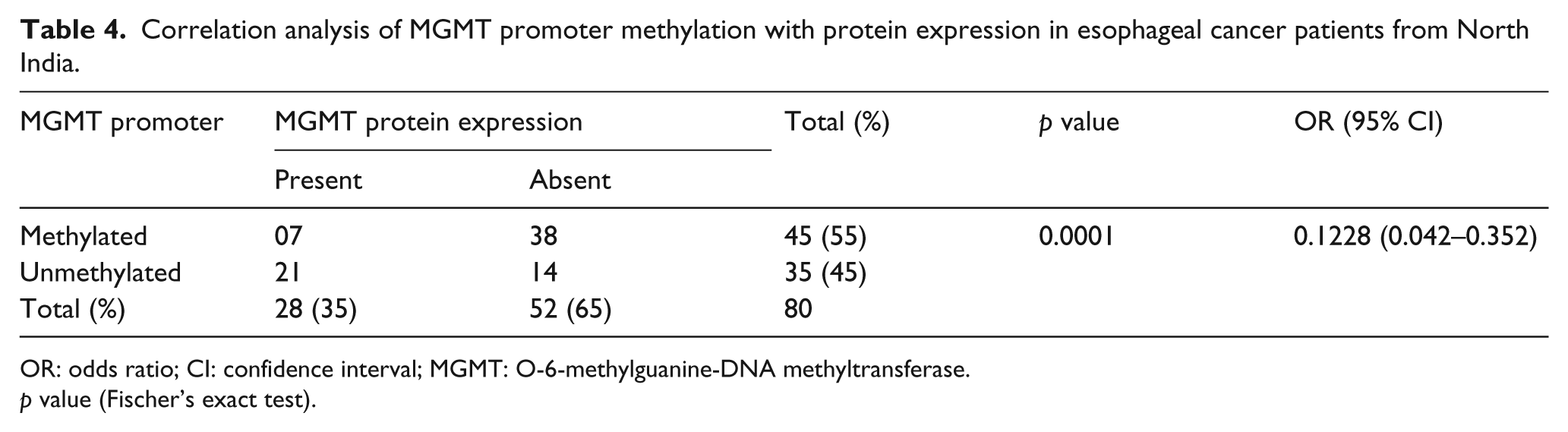
OR: odds ratio; CI: confidence interval; MGMT: O-6-methylguanine-DNA methyltransferase.
p value (Fischer’s exact test).

Methylation-specific PCR analysis of MGMT gene in esophageal cancer patients: DNA methylation was assessed using two specifically designed primers to amplify either methylated DNA (M) 191 bp product or unmethylated DNA (UM) 191 bp product (L: 1 kb DNA ladder; number indicates the case number; N: normal tissue; T: tumor tissue).
Association between MGMT promoter methylation and clinicopathological parameters in esophageal cancer
No significant association was seen when the clinical parameters and the promoter methylation were correlated. However, at certain instances, some linear trends were seen, but as the number of cases increased they diminished. In an aggressive dysphagia grades III and IV, around 55% cases were methylated (Table 5). And when location of the tumor was studied, a slight significance was seen with 71% cases from lower one-third region were methylated (p < 0.02).
Correlation analysis of MGMT promoter methylation with clinicopathological parameters in esophageal cancer patients from North India.

TNM: tumor–node–metastasis; MGMT: O-6-methylguanine-DNA methyltransferase.
Association between MGMT protein expression and clinicopathological parameters in esophageal cancer
The protein expression and clinicopathological parameters again were left with no significant associations; however, more aggressive stages III and IV had subsequently more cases around 71% (32/45) of MGMT protein deficiency as compared to stages I and II which were 57% (20/35; Table 3). Also, 92% (23/25) of grade-III and -IV patients who were methylated for the MGMT promoter region had no MGMT expression (Table 6). A similar scenario was observed with the tumor growth level with 84% (21/25) cases of the lower one-third region had MGMT loss along with methylation.
Correlation analysis of methylation and protein expression in samples having methylated MGMT promoter or MGMT expression loss with clinical parameters of esophageal cancer patients from North Indian population.

Overall, the methylation and protein loss were very much evident in the each clinicopathological parameter when studied at an intra level. The more aggressive esophageal cancer patients had MGMT protein loss significantly correlating the hypermethylation at the promoter region with stages III and IV (p ⩽ 0.001; Table 7). Of the patients with lower growth level of tumors, 60% showed MGMT loss and hypermethylation as compared to the 35% of the upper two-third region cases showing MGMT loss and hypermethylation.
Correlation analysis between methylation and protein expression of MGMT in stratification by various clinical characteristics of esophageal cancer patients from North India.

TNM: tumor–node–metastasis; MGMT: O-6-methylguanine-DNA methyltransferase.
Survival analysis
In this study, 51 esophageal cancer patients received treatment with curative intent, whereas the remaining 29 patients received different palliative treatment or refused the treatment or did not turn up. The Kaplan–Meier survival curve for the esophageal cancer patients was drawn with different factors to assess their role and dynamics in the treatment modality. Some definitive trend was seen with factor studied; however, only some tend to be significant. The mean survival time for stages I and II was 23 months, whereas in the stages III and IV it was 19 months (p = 0.187). Similarly, dysphagia grade II had better survival among all the grades with approximately 24 months as compared to 20 and 15 months in case of grades III and IV, respectively. Female patients were found to be having a better significant survival than the male patients (p = 0.033). However, no significant association could be seen between the survival time of patients with high and low MGMT expression (Table 8).
Survival analysis of esophageal cancer patients from North India.

TNM: tumor–node–metastasis; MGMT: O-6-methylguanine-DNA methyltransferase.
This study also included the negative effects of the smoking and alcohol abuse. It was evident that non-smoker patients had better survival time compared to the smokers (p = 0.008), and also, the same lies with the patients who had history of alcohol abuse with only 15-month survival time against the non-alcoholics who had 24-month survival (p < 0.02).
Discussion
Various studies unraveled the intra tumor and inter tumor heterogeneity among the different individuals.24,25 This led to disparity in the pathogenesis of the cancer and augmented the differences in susceptibility toward cancer. Therefore, studies are required to evaluate the genetic, epigenetic, and environmental disturbances at an individual level and also to figure out the interplay among these factors which might behold the role of a particular gene. Lifestyle and the dietary habits of the North India population further adds to the complexity. Various reports suggested the association of the smoked food/bar-be-cue, common in North India, with incidence of GI cancers to be high.7,26,27 Therefore, North Indian population is faced with an increased risk toward the incidence of the GI cancers.
MGMT is a gene involved in DNA repair whose loss of function has been widely associated with different cancers especially the GI cancers.10,15 The MGMT loss can be attributed to the hypermethylation of the promoter region, different mutations, and other factors.13,28 Researchers have earlier showed that the tobacco abuse and alcohol consumption may alter the epigenetic landscape, which increases the risk of developing cancer.29–31 This study attempts to unravel the status of methylation and its subsequent effect on the MGMT expression level in the esophageal cancer patients.
Here, the data demonstrated that 65% (52/80) cases had downregulated MGMT mRNA expression, and this downregulation was more often in stage-III and -IV cancer. MGMT protein expression was found to be consistent with the studies done on other populations.32,33 Overall, the loss of MGMT protein was evident in 52/80 studied, and these cases had either mild expression or the MGMT protein was absent. Stages III and IV had around 71% cases with low MGMT, whereas stages I and II had 57% patients with downregulated MGMT expression. Based on ours and the data from other groups, MGMT appears to have an important role in the occurrence and the progression of esophageal cancer; however, further functional studies required to characterize its role in esophageal carcinogenesis.
An association between the methylation pattern and the different clinical parameters of the esophageal cancer patients from North India was also studied. Some of the findings are in conjunction with the previous available literatures. In our study, the extend of methylation was higher (56.25%), which is, however, less when compared to a previous study from North East India where the frequency of aberrant methylation was 70%. 33 In 15.5% (07/45) methylated cases, no protein loss was seen probably due to incomplete methylation. 34 For methylation to silence a gene, a particular threshold is required, failing which the silencing might not occur. On the contrary, in 14 cases we found, unmethylation and still MGMT loss were observed. A possible explanation to these results can be mutations which can render MGMT functionless, homozygous deletions, or some unusual post-transcriptional silencing or post-translational modifications.
We further correlated the MGMT promoter methylation and subsequent MGMT protein expression with the various clinical parameters. In dysphagia grade III, 54.5%(20/37) cases were methylated and MGMT was absent, whereas in grade II only 39% cases were methylated for MGMT promoter region along with loss of the MGMT protein. However, in grades IV and V, no trend could be seen, maybe due to the less numbers of cases; therefore, it looks that maybe some more conclusive studies where good numbers of patients falling from each dysphagia grade can be complied on similar lines which can prove to be fruitful in giving some local staging an advantage.
A combined significant association of promoter methylation and MGMT expression with the staging of cancer was found in the esophageal cancer patients. The stages I and II had 40% (14/35) cases methylated and not expressing MGMT, whereas the stages III and IV had 51% (23/45) cases falling in the same line.
In this study, poor prognosis for the patients with history of either tobacco or alcohol abuse was found. The smokers had survival time of 17 months, whereas non-smokers survived for good 26 months (p = 0.008). Similarly, alcoholic patients survived for 15 months and non-alcoholics had survival time of 23 months (p < 0.02). This can be attributed to smoking and alcohol causing damage to the cellular entity and thereby making the treatment regimen less effective. Similar to some previous studies, it was found that nitrosamine present in tobacco can alter genomics and activate signaling pathway that cause cancer progression, invasion, migration, and angiogenesis.
Apart from the MGMT promoter hypermethylation that downregulates the expression, some studies have revealed that Wnt signaling also alters the expression profile of the MGMT. Therefore, during treatment with alkylating drugs, Wnt inhibitors are used to modulate the expression of the MGMT to increase the efficacy of the drugs. 35
In summary, loss of MGMT protein was found to be frequent in the esophageal cancer from North Indian patients, and hypermethylation of the promoter region of MGMT was significantly associated with its downregulation. Future large-scale and preferably, multi-centered studies are required to characterize the MGMT role in the esophageal cancer and to study the factors that may downregulate/upregulate the MGMT expression and also figure out more risk factors to esophageal cancer.
Footnotes
Acknowledgements
A.U.R. and S.S. contributed equally.
Declaration of conflicting interests
The author(s) declared no potential conflicts of interest with respect to the research, authorship, and/or publication of this article.
Funding
The author(s) disclosed receipt of the following financial support for the research, authorship, and/or publication of this article: This study received financial support from Department of Biotechnology (NER-BPMC), Government of India (No. BT/365/NE/TBP/2012; 26 March 2013).