
Abstract
Osteosarcoma is a common primary malignant bone tumor that occurs mainly in children and adolescents. Recent evidence has demonstrated that miR-34a is involved in the invasion and metastasis of osteosarcoma. This study aims to explore the effect of biological behavior of miR-34a on osteosarcoma. First, we collect osteosarcoma and adjacent specimens, and the relative expression of miR-34a and C-IAP2 messenger RNA was quantitated by real-time polymerase chain reaction. Furthermore, miR-34a stimulant is synthesized and transfected onto osteosarcoma MG-63 cells. The effect of overexpression of miR-34a on osteosarcoma was detected by colony-forming assay, Annexin V-fluorescein isothiocyanate Apoptosis Detection Kit I, Transwell assay, and animal experiment in vivo. Finally, the relative levels of C-IAP2 and Bcl-2 protein were checked by western blot, and the activity of caspase-3 and caspase-9 was tested by spectrophotometry assay. In conclusion, miR-34a was downregulated in osteosarcoma cells. And the expression of C-IAP2 and Bcl-2 protein was drastically inhibited, and the activities of caspase-3 and caspase-9 were significantly increased after transfecting miR-34a onto osteosarcoma MG-63 cells. And the overexpression of miR-34a can inhibit cell invasion and metastasis, promote cell apoptosis, and arrest cells in G0/G1 period. And the animal experiment in vivo demonstrated that the overexpression of miR-34a could significantly inhibit the growth of osteosarcoma in animal skin. Taken together, we indicated that miR-34a can inhibit tumor invasion and metastasis in osteosarcoma, and its mechanism may be partly related to downregulating the expression of C-IAP2 and Bcl-2 protein directly or indirectly.
Introduction
Osteosarcoma is the most common primary malignancy of bone, which occurs mostly in children and adolescents and threatens the patients’ lives because of its recurrence easily after surgical excision, early hematogenous metastasis, and then a low 5-year survival rate. 1
MicroRNA-34a (miR-34a) is an important member of the non-coding small RNA molecules that acts as an anti-oncogene in the 1p36.23 and correlates with the regulation of p53 signal pathway. In various cancers, its expression may become abnormal owing to a series of causes, such as promoter methylation, gene deletion, and p53 gene mutation as well as inactivation. 2 The cancer inhibition function of miR-34a has been validated in a number of malignant tumors (including colorectal cancer, lung cancer, glioblastoma, and pancreatic carcinoma). 3 Recent studies 4 have suggested that miR-34a may regulate apoptosis-related genes to promote apoptosis and induce the function of tumor suppression. MiR-34a is closely associated with the genesis and development of cancer.
Cell inhibitor of apoptosis protein 2 (C-IAP2) and B-cell lymphoma-2 (Bcl-2) are the major proto-oncogenes for cell apoptosis, which can suppress the apoptosis by inhibiting the functions of apoptotic pathways of cysteine-containing aspartate-specific proteases (caspase). 5 These genes are overly expressed in a wide variety of cancer cells, which is closely related to tumor progression, prognosis, and tumor sensitivity to radiotherapy and chemotherapy. 6
We assume that miR-34a may affect the biological behavior of osteosarcoma partly by effecting the expression of C-IAP2 and Bcl-2 protein. In this article, quantitative real-time polymerase chain reaction (qRT-PCR) is utilized to analyze the levels of miR-34a and C-IAP2 protein in human osteosarcoma tissues as well as paracarcinoma tissues. Furthermore, miR-34a stimulant is synthesized and transfected onto osteosarcoma MG-63 cells, in order to explore the invasion and metastasis effect of miR-34a on osteosarcoma MG-63 cells and to explore the possible mechanism.
Materials and methods
Materials
The osteosarcoma MG-63 cells were purchased from Shanghai Institute of Biochemistry and Cell Biology, Chinese Academy of Sciences. RPMI 1640, fetal calf serum, and trypsase were all purchased from Gibco (USA). Lipofectamine™ 2000 and TRIzol were purchased from Invitrogen (USA), which also supplied the total RNA isolation nut TRIzol. Reserve-transcription kit Moloney Murine Leukemia Virus (M-MLV) was purchased from Promega (USA). SYBR Premix Ex Taq Kit was sourced from Axygen (USA). The target genes along with the upstream and downstream primers of reference genes were supplied by RiboBio (China). The has-mir-34a as well as the expression carriers and plasmids of chronic viruses for negative control (NC) were synthesized by GeneChem (China). The thiazolyl blue kit (3-(4,5-dimethylthiazol-2-yl)-2,5-diphenyltetrazolium bromide (MTT)) was purchased from Dingguo Biochemistry Ltd. (China). The propidium iodide (PI) was bought from Sigma (USA), whereas RNase A was purchased from Fermentas (Canada). Finally, the cell apoptosis kit was supplied by eBioscience (USA). All animal experiments were approved by the Ethics Committee of the Second Xiangya Hospital of Central South University.
Methods
The expression of miR-34a and C-IAP2 messenger RNA in osteosarcoma and juxta cancerous tissues were detected by RT-PCR
A total of 31 fresh samples of osteosarcoma were collected from the Second Xiangya Hospital of Central South University. Patients had not undergone radiotherapy, chemotherapy, or other related treatments prior to surgery. All these osteosarcoma diagnoses were pathologically validated. Also, the juxta cancerous tissues were 1 cm from osteosarcoma tissues, and they ought to be cut into a size of 0.5 cm × 0.5 cm × 0.5 cm. The total RNA of samples was extracted by TRIzol. Then, the total RNA was transcribed into complementary DNA (cDNA) at 16°C for 30 min, 42°C for 30 min, and 85°C for 30 min and stored at 4°C. The reverse transcription for glyceraldehyde 3-phosphate dehydrogenase (GAPDH) was conducted at 45°C for 1 h and then at 70°C for 10 min and stored at 4°C. At the meantime, the quality and concentration of the total RNA extracted were analyzed as well as measured by NanoDrop 2000 Spectrophotometer. Following that, M-MLV Kit was utilized to reverse transfect such total RNA and synthesize cDNA. In addition, random primers were used in the reverse transfection of C-IAP2 (primer sequence: F: 5′-TCCTAGCTGCAGATTCGTTC-3′; R: 5′-GGTAACTGGCTTGAACTTGAC-3′), whereas stem-loop primer was utilized for miR-34a (primer sequence: F: 5′-CGGTATCATTTGGCAGTGTCT-3′; R: 5′-GTGCAGGGTCCGAGG-3′). The PCR reaction for miR-34a and C-IAP2 was carried out at 95°C for 5 min, 95°C for 15 s, and 60°C for 15 s, and a total of 40 cycles were performed. The reverse transcription for GAPDH was conducted at 95°C for 5 min, 95°C for 15 s, and 60°C for 1 min, and a total of 50 cycles were performed. Fluorescence signals were collected at 85°C, and GAPDH was used as an internal reference. After PCR was performed, the messenger RNA (mRNA) level was calculated by a comparative threshold cycle (Ct) method using the formula 2−ΔΔCt, and the average value of the complex holes was obtained for later statistical analysis. ΔCT computational method: ΔCT = CTmiR-34a or C-IAP2 − CTU6 or GAPDH. ΔΔCT = ΔCTosteosarcoma tissues − ΔCTjuxta cancerous tissues.
Establishing stably infected osteosarcoma cell lines and the level of miR-34a expression were tested by RT-PCR
The transfection of miR-34a: the mature miR-34a mimics for miR-34a transfection were synthesized by GeneChem. The sequence of the mimics was 5′-UGGCAGUGUCUUAGCUGGUUGU-3′/5′-AACCAGCUAAGACACUGCCAUU-3′, whereas the NC sequence was 5′-CAGUACUUUUGUGUAGUACAA-3′/5′-UUGUACUACAAAAGUACUG-3′. They were then transfected using Lipofectamine 2000 Transfection Kit (Invitrogen). The samples were divided into miR-34a mimic group, NC group, and normal group; 24 h prior to transfection, the cells grew for passage and were cultured in non-antibiotic culture medium. As soon as the cells achieved 80% growth, transfection took place. Transfection reagent Lipofectamine 2000 and pending transfection sequence (20 µmol/L) were dissolved in MEMI, following by a 5-min incubation. Subsequently, they were then mixed together and incubated for another 25 min. After that, the compound was added into a six-pore plate drop by drop. The following step was to gently shake the plate to make the mixture incorporate and carry on culturing. The culture medium was changed for fresh doses after 6 h, with normal cultures following. After 48 h of cell transfection, the RNA of each group was extracted utilizing TaqMan miRNA separation kit, while the changes in the expression of miR-34a in the osteosarcoma cells of each group were tested by qRT-PCR.
The cell proliferations were checked by colony formation assay
The cells in the logarithmic growth phase were digested and counted. A total of 1 mL (1 × 10³ cells/mL) of osteosarcoma MG-63 cells stably expressing the miR-34a mimic were seeded in 6-well plates. After 2 weeks, cells in each well were fixed with 100% methanol for 30 min and stained with 0.1% crystal violet for 30 min. Cell colony-forming units were counted. Assays were conducted three times independently.
The cell apoptosis was detected by flow cytometry
This step took place on the 6th day of miR-34a cell transfection. Following centrifugation for 5 min at 800×g, the cells in the logarithmic growth phase in each group were collected through sedimentation, while the supernatant was discarded and washed twice with pre-cooled phosphate-buffered saline (PBS). Subsequently, the cells were gathered in 5 mL centrifugal tubes with three complex pores assigned per group. After centrifugal action, the supernatant was discarded, whereas the cell precipitation was washed with PBS once and centrifuged again before resuspended. Next, the cell suspension was supplemented with Annexin V-allophycocyanin (APC) for staining and properly hidden away from light at room temperature for 10–15 min. Finally, flow cytometry was used to detect the cell apoptosis in each group, and the treatment results were analyzed with ModFit (Verity Software House, USA), the DNA analysis software.
The change of apoptosis cells were observed by transmission electron microscope
The cells in the logarithmic growth phase in each group were digested and counted, and after centrifugation at 1500 r/min for 10 min, supernatant is discarded, and after washing with PBS twice, cell density is adjusted to 1 × 10⁶ cells/group. After repeating centrifugation, cell precipitates are collected; the mixed liquor containing 2.5% glutaraldehyde and 1% paraformaldehyde after 4°C pre-cooling has been adopted as a fixative, and 1% osmic acid is also taken as the fixing agent, and epoxy resin is used for embedding ultrathin section, and uranyl acetate and lemon lead are used for dyeing. Then, observation and photograph recording are achieved under transmission electron microscopy (TEM).
The cell cycle was detected by flow cytometry
The cells in the logarithmic growth phase in each group were digested and counted. The cell concentration was adjusted to 1 × 10⁵ mL and seeded in six-well plates. Each well was provided with 2 mL cell solution, and the cells were cultured in a 5% CO2 incubator at 37°C for 48 h; the fluid culture was collected in a centrifuge tube where the cells were digested and collected at 4°C. The cells were centrifuged at a rate of 1000×g for 5 min. The supernatant was discarded and the cells were washed once with ice-cold PBS. The cells were collected in a centrifuge tube at 4°C and centrifuged at 1000×g for 5 min. The supernatant was discarded, while the ice-cold PBS cells were resuspended, transferred to the Eppendorf tube at 4°C, and centrifuged at 1000×g for 5 min. The supernatant was discarded again and 1 mL PBS was added. The resuspended cells were pre-cooled and fixed with 70% ethanol at 4°C. The cells were centrifuged at 1000×g for 5 min, and the ethanol solution was discarded. Next, the cells were washed twice with PBS and filtered through a 400-mesh sieve prior to PI dye being added at 4°C in darkness for staining for 30 min. The proportion of cells in the G0/G1 phase, S phase, and G2 phase was then detected by flow cytometry.
The cell migration and invasion were examined by Transwell chamber invasion assay
The cells in the logarithmic growth phase in each group were digested and counted after 48 h of miR-34a cell transfection. Osteosarcoma cells were seeded onto the membrane of the upper chamber of the Transwell at a concentration of 3–5 × 10⁵ mL in 2 mL of Dulbecco’s Modified Eagle’s Medium (DMEM) medium. The medium in the upper chamber was serum free. The medium in the lower chamber contained 5% fetal calf serum as a source of chemoattractants. Cells that passed through the Matrigel-coated membrane were stained with Diff-Quik (Sysmex, Japan) and photographed. Eventually, pictures were taken in eight random views under microscope for records.
Animal experiment in vivo
All animal experiments were approved by the Ethics Committee of the Second Xiangya Hospital of Central South University. Research on animals follows internationally recognized guidelines on animal welfare and local and national regulations. The cells in the logarithmic growth phase in each group were injected subcutaneously into the left scapulae of 6-week-old nude mice (2 × 10⁶ cells per mouse). The tumor volume was monitored weekly and was calculated using the following formula: V = 1/2 × width2 × length. After 6 weeks, the mice were killed and the tumor tissues were removed from the left scapulae of nude mice.
The expressions of C-IAP2, Bcl-2, and Bax were detected by western blot
The cells in the logarithmic growth phase in each group were digested and counted after 48 h of miR-34a cell transfection. The total protein (TP) was extracted by TRIzol method. After electrophoresis, wet method is applied to transfer TP onto nitrocellulose membrane. After 1 h of blocking with Tris-buffered saline (TBS), primary antibodies of C-IAP2, Bcl-2, and Bax are used for overnight incubation under 4°C (with a dilution ratio of 1:200). After washing with TBS, secondary antibody of horseradish peroxidase (HRP) is applied for overnight blocking under 37°C. Then, after washing with TBS and enhanced chemiluminescence (ECL) color developing are completed, film processing is finally done. As β-actin is taken as the internal reference, dilution with a ratio of 1:400 is achieved. After the film is shot, Image-Pro Plus 6.0 software is adopted for semi-quantitative analysis.
The activity of caspase-3 and caspase-9 was tested by spectrophotometry assay
The cells in the logarithmic growth phase in each group were collected, and after trypsinization and cell disintegration, centrifugation is realized. When the supernatant is transferred into the pre-cooled centrifuge tube, spectrophotometry is applied for testing the activities of caspase-3 and caspase-9 by Multiscan microplate reader. Through fluorescence released after shearing of the detection substrate by microplate reader (405 nm), the relative fluorescence values from cells of both treatment group and control group are taken as the indexes of the activities of caspase-3 and caspase-9.
Statistical analyses
All statistical analyses were performed with Student’s t-test and represented as mean ± standard deviation (SD) unless noted otherwise. No animal or sample was excluded from the analysis. The p values were designated as *p < 0.05, **p < 0.01, and n.s.—non-significant (p > 0.05).
Results
The expression of miR-34a and C-IAP2 mRNA
The RT-PCR results demonstrated that the miR-34a expression in osteosarcoma tissue was obviously lower than adjacent tissue (Figure 1(a)). However, the C-IAP2 mRNA level in osteosarcoma tissue was significantly higher than adjacent tissue (Figure 1(b)). The results were validated by the non-parametric sum of ranks of paired samples, and the difference was significant (p < 0.05).

Real-time PCR for the relative expression level of miR-34a and C-IAP2 mRNA in osteosarcoma tissues and the adjacent non-tumor tissues. (a) The relative expression level of miR-34a. (b) The relative expression level of C-IAP2 mRNA (*p < 0.05 vs adjacent non-tumor tissues).
The miR-34a mimics were successfully transfected into osteosarcoma MG-63 cells
After 48 h of miR-34a mimic transfection, the outcome indicated that the expression level of the miR-34a mimic group was obviously higher than the NC group and normal group (p < 0.05). However, the difference between the NC group and the mock group was not found with statistical significance (p > 0.05). In short, the miR-34a had successfully transfected into osteosarcoma MG-63 cells (Figure 2).

Effects on miR-34a expression after transfecting the osteosarcoma MG-63 cells with a miR-34a mimic (**p < 0.01 vs the normal or NC group).
MiR-34a inhibits cell proliferation
The colony-forming assay results showed that the number of colony-forming units in the miR-34a mimic group was obviously lower than the NC group and normal group (p < 0.05; Figure 3). It was suggested that the overexpression of miR-34a inhibited colony formation of osteosarcoma MG-63 cells.

The number of colony-forming units in the miR-34a mimic group was obviously lower than the normal group and NC group. (a) Colony formation assay. (b) The number of colony-forming units (**p < 0.01 vs the normal or NC group).
MiR-34a promotes cell apoptosis
The results of flow cytometry demonstrated that the apoptosis percentage of cells in the miR-34a mimic group was significantly higher than the normal group (p < 0.01) and NC group (p < 0.01; Figure 4). It was suggested that the overexpression of miR-34a could promote osteosarcoma MG-63 cell apoptosis.

The effects of miR-34a on apoptosis using flow cytometry (**p < 0.01 vs the normal or NC group).
Morphological changes of apoptosis cells
Under TEM, the morphology of normal and NC group cells line was expressed with large nucleus and obvious euchromatin as well as visible heterochromatin. The cell nucleus is hypertrophic and the edge accumulation is seen. Both nucleosome and rough endoplasmic reticulum are evidently detected. After the action of gemcitabine (GEM), the morphology of apoptotic PANC-1 cells is expressed with increased electron densities of cytoplasm and cell nucleus, smaller volume, concentrated nuclear chromatin, shrunken nucleiform as well as visible apoptotic body, which is also expressed as small patches of chromatin accompanied by a small amount of cytoplasm and is embedded by outer cytomembrane. The apoptotic body is dissociated from cells and drifts in extracellular matrix (Figure 5).

Morphological changes of apoptosis cells under the transmission electron microscopy (TEM 5000×). (Left) Normal group cells. (Middle) NC group cells. (Right) MiR-34a mimic group cells.
MiR-34a arrests tumor cells in G0/G1 phase
The flow cytometry results showed that the cells of G0/G1 phase in the miR-34a mimic group were (69.25% ± 2.58%). The G0/G1 phase cells of the normal group and NC group were (40.12% ± 1.23%) and (41.37% ± 1.97%), respectively, and with statistically significant difference (p < 0.05). It was suggested that the overexpression of miR-34a arrests osteosarcoma MG-63 cells at G0/G1 phase (Figure 6).

The effects of miR-34a on cell cycle using flow cytometry (*p < 0.05 vs the normal or NC group).
MiR-34a inhibits cell migration and invasion
The Transwell chamber invasion results showed that after the transfection of miR-34a mimic, the number of osteosarcoma MG-63 cells that passed through Transwell chamber was significantly decreased (p < 0.01). It was suggested that the overexpression of miR-34a had inhibited the osteosarcoma MG-63 cell migration and invasion (Figure 7).

The effects of miR-34a on cellular migration detected using Transwell invasion chambers (**p < 0.01 vs the normal or NC group).
MiR-34a inhibits tumor growth in animal experiment in vivo
The cells in the logarithmic growth phase in each group were subcutaneously injected into the left scapulae of nude mice, and the animals were closely monitored for tumor growth for 6 weeks. The results demonstrated that miR-34a-overexpressing tumors were significantly larger in size and volume compared with the normal group and NC group (p < 0.01; Figure 8).

Animal experiment in vivo; miR-34a mimic groups were significantly larger in size and volume compared with the normal group and NC group. (a) Photographs of tumors. (b) Growth curve drawn by measuring tumor volumes on the indicated days (*p < 0.05 vs the normal or NC group).
MiR-34a downregulates the expressions of C-IAP2 and Bcl-2 proteins
The western blot results showed that the expression of C-IAP2 and Bcl-2 protein in the miR-34a mimic group was significantly lower than the normal group and NC group (p < 0.01; Figure 9). In addition, the Bax protein expression was also lower than the normal group and NC group, but no significant difference was shown (p > 0.01; Figure 10). It was suggested that the overexpression of miR-34a may downregulate the expressions of C-IAP2 and Bcl-2 proteins directly or indirectly.
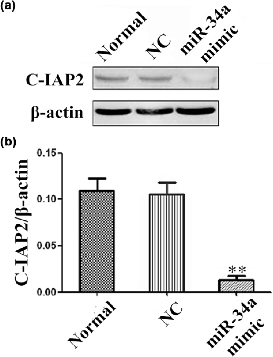
The expression of C-IAP2 protein in osteosarcoma MG-63 cells detected by western blot. (a) Protein blotting stripe. (b) The relative content of C-IAP2 protein (**p < 0.01 vs the normal or NC group).

The expression of Bcl-2 and Bax protein in osteosarcoma MG-63 cells detected by western blot. (a) Protein blotting stripe. (b) The relative content of Bcl-2 and Bax protein (*p < 0.05 vs the normal or NC group; **p > 0.05 vs the normal or NC group).
MiR-34a can activate the caspase-3 and caspase-9
The spectrophotometry assay results showed that the activities of caspase-3 and caspase-9 in the miR-34a mimic group were significantly increased than the normal group and NC group (p < 0.01; Table 1). It was suggested that the overexpression of miR-34a can activate the caspase-3 and caspase-9.
Changes of the caspase-3 and caspase-9 activity in osteosarcoma MG-63 cells (Χ ± s).

Discussion
Osteosarcoma is the most common primary malignancy of bone, accounted for 20% of all bone tumors. The occurrence and development of osteosarcoma are not only the result of uncontrolled cell proliferation and abnormal differentiation but also related to the imbalance of cell apoptosis. 1
MiR-34a is an important member of the non-coding small RNA molecules that acts as an anti-oncogene in the 1p36.23 miR-34a extraordinarily expressed in various tumors, such as promoter methylation and gene deletion. 7 More and more evidence showed that that miR-34a can inhibit tumor cell proliferation. Welch found that mir-34a can inhibit cell proliferation and promotes apoptosis in medulloblastoma cells. 8 This phenomenon also exists in pancreatic cancer cells, 6 in prostate cancer cells,9,10 in head and neck squamous cell carcinoma, 11 in laryngeal squamous cell carcinoma, 12 in uveal melanoma cell, 13 in gastric cancer cell, 14 in human hepatocellular carcinoma cells, 15 in lung cancer cell, 16 and in colon cancer cell, 17 and it is also found by Li et al. 18 that MiR-34a inhibits proliferation and migration of breast cancer through downregulation of Bcl-2 and SIRT1. Wang et al. 19 also found that overexpression of miR-34a reduces ErbB2 expression and suppresses breast cancer cell invasion and growth. But also some studies 20 show that systemic miR-34a-CH delivery affected neither tumor growth nor metastasis to other organs such as lung in the human breast cancer model and the mouse melanoma model. But, in this study, it is found that in both cases, bone metastases were diminished by miR-34a-CH. This raises the intriguing possibility that its anti-cancer effects may reside in other cells that constitute the tumor microenvironment such as the osteoclasts in the bone metastatic niche. Also, in the field of osteosarcoma research, there are much more recent research confirmed that mir-34a can inhibit cell proliferation and promotes apoptosis in human osteosarcoma cells in vitro and in vivo.21–27 So, we are valid to consider that mir-34a could inhibit cell proliferation and promotes apoptosis in osteosarcoma possibly. And our results strongly suggest that miR-34a was downregulated in osteosarcoma cells. In addition, the overexpression of miR-34a can inhibit cell invasion and metastasis, promote cell apoptosis, and arrest cells in G0/G1 period and also inhibit cell migration and invasion. And the further animal experiment in vivo demonstrated that the overexpression of miR-34a could significantly inhibit the growth of osteosarcoma in animal skin. These were consistent with existing studies.
C-IAP2 as the strongest apoptosis-inhibited gene discovered so far has a powerful function in suppressing cell apoptosis. In intrinsic pathway mediated by mitochondrion under the action of cytochrome C, C-IAP2 can both directly combine with procaspase-9 or caspase-3 and inhibit their activities as well and block off the activation of their cutting in turn as well as the feedback activation of caspase-3 to procaspase-9, so as to inhibit cell apoptosis. C-IAP2 is highly expressed in a wide variety of cancer cells, which is closely related to tumor progression, prognosis, and tumor sensitivity to radiotherapy and chemotherapy. Recent research shows that C-IAP2 was closely related with the grade, Enneking stage, and cell types of osteosarcoma. 28 C-IAP2 was not a direct target for miR-34a through targets can software, Miranda software and Pictar software (three miRNA target gene prediction software). In addition, miR-34a regulates target gene through a variety of signaling pathways, and nuclear factor-kB (NF-κB) pathway 29 is the most important signaling pathway.30–35 And C-IAP2 in the downstream of the NF-κB signaling pathway plays an important role in pathway. And our results show that the expression of C-IAP2 protein was drastically inhibited. Therefore, we hypothesized that miR-34a might downregulate the expression of C-IAP2 by NF-κB pathway, which is not its direct effect. But, further experimental investigation is required to confirm this hypothesis.
Bcl-2 family is the proto-oncogene correlated to cell apoptosis, whose expression products are divided into two categories: a kind of inhibitor of apoptosis protein, such as Bcl-2, Bcl-xl, and Bcl-w, and a kind of promoter of apoptosis protein, such as Bax, Bak, and Box. Recent studies suggested that the Bcl-2 protein in G1 period of osteosarcoma of low grade was significantly higher than in G2 period of high grade (p < 0.01). Bcl-2 was a direct target gene for miR-34a, and it can be predicted in miRanda software. 36 And many research showed that miR-34a can directly suppress Bcl-2 gene.17,18,37,38 Many luciferase reporter assays indicated the direct interaction of miR-34a with the Bcl-2 3′-UTR. 38 The western blot results also confirmed that the expression of Bcl-2 protein in the miR-34a mimic group was significantly lower than the normal group and NC group.
Therefore, taken together, we indicated that miR-34a was downregulated in osteosarcoma cells, and the overexpression of miR-34a in osteosarcoma can inhibit cell proliferation and invasion and promote cell apoptosis. And its mechanism may be partly related to downregulating the expression of C-IAP2 and Bcl-2 protein directly or indirectly. MiR-34a may be the potential target and effective molecular marker, and it deserves further studying.
Footnotes
Acknowledgements
The osteosarcoma MG-63 cells were purchased from Shanghai Institute of Biochemistry and Cell Biology, Chinese Academy of Sciences.
Declaration of conflicting interests
The author(s) declared no potential conflicts of interest with respect to the research, authorship, and/or publication of this article.
Funding
The author(s) disclosed receipt of the following financial support for the research, authorship, and/or publication of this article: This work was supported by The Central Laboratory of Xiangya and Nation Natural Science Foundation of China (no. 812721993).