
Abstract
Association of oral diseases and disorders with altered microRNA profiles is firmly recognized. These evidences support the potential use of microRNAs as therapeutic tools for diagnosis, prognosis, and treatment of various diseases. In this review, we highlight the association of altered microRNA signatures in oral cancers and oral inflammatory diseases. Advances in our ability to detect microRNAs in human sera and saliva further highlight their clinical value as potential biomarkers. We have discussed key mechanisms underlying microRNA dysregulation in pathological conditions. The use of microRNAs in diagnostics and their potential therapeutic value in the treatment of oral diseases are reviewed.
Introduction
It has been recognized that oral health and genetics are closely interlinked. 1 Studying the multitude of disorders affecting craniofacial tissues can provide scientists with insight on the role of inflammation in infection and pain, the consequences of depressed immunity, and the changes that can arise from a mutated gene. 2 Recently, there has been a focus on regulation of these inflammatory and cancer pathways by microRNA (miRNA) in innate and adaptive immunity.3–6 In humans, miRNA expression has been shown to affect the pathobiology of oral diseases such as oral squamous cell carcinoma (OSCC), 7 Sjögren’s syndrome (SS), 8 and periodontitis. 9 Recent studies have highlighted the differences in miRNA expression patterns between healthy and inflamed periodontal tissues, which suggest the possible involvement of miRNAs in the regulation of periodontal disease.10,11 This review highlights a largely obscure and unexplored zone: expression of miRNAs in the hosts’ immuno-inflammatory responses and their potential role in the pathophysiology of oral cancer and other oral diseases. Emerging lines of evidence indicate a correlation between oral inflammation and cancer progression. Therefore, identification of biomolecules that can serve as biomarkers for the diagnosis/prognosis of these oral diseases can be of high therapeutic value. Given the capability of miRNA to simultaneously modulate expression of hundreds of target genes, an in-depth study of the role of miRNAs in oral disease can facilitate the understanding of the immune system and efforts taken to achieve a state of homeostasis. We will discuss the probable mechanisms of altered miRNA expression leading to pathology. Finally, the therapeutic potential of miRNAs will be highlighted.
MicroRNA biogenesis
MiRNAs are small, non-coding, single-stranded, ~22-base nucleotide sequences, which bind approximately 60% of all genes.12,13 It has been suggested that the human genome may encode over 2500 miRNAs, which have emerged as one of the key negative regulators of gene expression. 14 MiRNAs bind to the complementary sequences on the 3′ untranslated regions (3′UTRs) of target messenger RNA (mRNA). This interaction is guided by 2–8 nucleotides of the miRNA sequence and results in target cleavage or translational suppression. A single miRNA can have hundreds of target genes, and multiple miRNAs can regulate the same target. Hence, they can influence the expression of a large proportion of proteins.12,15 MiRNAs are differentially expressed at a high dynamic range in developmental stages, cell types, tissues, and disease states16,17 and are also involved in biological processes, such as embryogenesis, differentiation, and carcinogenesis. 18
MiRNA biogenesis commences in the nucleus and terminates in the cytoplasm. The first step being the generation of primary miRNA transcripts (pri-miRNA) usually transcribed by RNA polymerase II with the characteristic 5′ cap and 3′ poly-A tail. 19 Pri-miRNAs are of varied sizes and can be tens of thousands of nucleotides long. 20 The localized secondary structures on the pri-miRNAs are recognized by the RNase III endoribonuclease and Drosha generating 70- to 100-nucleotide-long stem-loop structures termed pre-miRNAs 21 which are transported to the cytoplasm by exportin-5 22 for further processing. In the cytoplasm, pre-miRNAs are acted upon by another RNase III enzyme termed Dicer that crops the stem-loop structure to yield 19- to 25-nucleotide-long double-stranded RNAs. Thereafter, one strand of the duplex is degraded and the other strand accumulates as a mature miRNA inside miRNA-RNA-induced silencing complex (mi-RISC). 23 MiRNAs bind to their respective mRNA targets and mediate gene silencing via the RNA interference mechanism by degradation of mRNA by deadenylation and destabilization 24 and post-transcriptional regulation by repressing translation initiation and protein synthesis.15,16
Altered miRNA profiles in oral diseases
The oral cavity is constantly challenged by a barrage of microbial (i.e. bacteria, viruses, and fungi) and environmental stressors. These stresses contribute to the oral–gene interactions which are likely involved in the onset and/or progression of many oral diseases including those with autoimmune, infectious disease, and carcinogenic characteristics. 25 Reduced protein expression, with/without reduced mRNA expression, has always been the hallmark of gene dysregulation associated with miRNA which can lead to derangements in the immune system, autoimmune disorders, lymphoproliferation, and cancer. 26 It is observed that miRNAs play a discrete role in tooth development and oral epithelial stem cell differentiation. 27 In the past decade, it has been unequivocally demonstrated that miRNAs are important in regulating a healthy immune system, which in turn is the essence of a salubrious existence of oral tissues. 28 Figure 1 illustrates a pathway whereby the identification and validation of 3′UTR targets can be correlated with disease physiology.

Flow chart illustrating the causes of miRNA dysregulation, identification, and validation of miRNA-regulated genes/pathways and association of aberrant miRNA expression in diseases.
Oral cancer
The oral cavity is one of the 10 most frequent sites of cancer globally, with three quarters of cases affecting people in the developing world and is the third most common cancer after stomach and cervical cancers. Worldwide, an estimated 378,500 new cases of intraoral cancer are diagnosed annually. 29 OSCC is seen typically on the lip or lateral part of the tongue usually as a lump or ulcer that is white, red, or mixed white and red. 30 The prevalence of tongue cancer is consistently found to be higher (by approximately 50%) in Blacks compared with Whites within the same regions of the United States. 31 The greatest risk factors for OSCC include tobacco consumption, cigarette smoking, and alcohol, along with an impaired ability to repair DNA damaged by mutagens and genetic aberrations causing immune defects (e.g. miRNA defects). 32 The tumor initiation properties of cancer stem cells such as self-renewal, tumorigenicity, and drug-resistance are regulated by miRNAs. 33 Table 1 summarizes published reports of miRNAs associated with oral cancer and oral premalignant lesions.
Table demonstrating miRNA deregulations in various OCCs and premalignant lesions.

MiRNA: microRNA; OCCs: oral cavity cancers.
Tongue squamous cell carcinoma
Profiling of squamous cell carcinoma of the tongue demonstrated upregulation of miR-184 and miR-21 and downregulation of miR-100, miR-125, miR-133a, and miR-133b signifying the importance of dysregulated expression of specific miRNAs in tongue squamous cell carcinoma (TSCC). 34 Other studies have also identified upregulated plasma levels of miR-184, likely associated with tumor load, 35 along with high miR-21 expression, which inhibited apoptosis and was found to be associated with poor prognosis in TSCC. 36 Likewise, upregulation of miR-24 in TSCC is associated with enhanced proliferation and reduced apoptosis. 37 In contrast, reduced expression of miR-138, 38 miR-195, 39 and miR-7 40 was shown to regulate cancer cell migration, invasion, and progression. These miRNAs may have potential as novel therapeutic targets for TSCC patients at enhanced risk of metastasis. Moreover, miR-222 is reported to play an important role in TSCC invasion and may also serve as a novel therapeutic target. 41
OSCC
Multiple profiling studies on OSCC tissues identified aberrant expression of miRNA indicating a strong association with the etiology of the disease. In a study employing tumor samples from the tongue and floor of the mouth, the most significant miRNAs that were upregulated included miR-21, miR-7, miR-34b, miR-155, miR-182, miR-15b, miR-185, and let-7, while miRNAs that were downregulated included miR-23b, miR-125a, and miR-125b. 42 It has been reported that miR-125b 42 and miR-145 43 have a dramatic effect in controlling cell proliferation. Furthermore, it has been reported that modulation of miR-125b expression helps overcoming radioresistance in OSCC. 44
MiR-155, miR-146a, let-7i, and miR-99a are known to be associated with oral tumor progression.45,46 MiR-155 and miR-146a are well-known miRNAs with immunomodulatory functions.47–52 Identification of miRNAs that shape immune responses supports the notion that immune cells are critical components of cancer development. Interestingly, these miRNAs play antagonistic roles in inflammation. For example, expression of miR-155 is pro-inflammatory and downregulates multiple genes involved in suppression of inflammation including SOCS1, while miR-146a expression is anti-inflammatory and regulates overt immune responses.52–54 Major targets of miR-146a include genes that activate toll-like receptor (TLR) signaling and cytokines.52,55,56 Importantly, in a tumor microenvironment, these miRNAs can gain access to other cells through exosomal pathways. Indeed, it is known that exosomes contain functional biomolecules including miRNAs, mRNAs, and proteins that can be released into recipient cells upon fusion thereby modulating recipient cell function.57,58 For instance, exosomal transfer of miR-155 and miR-146a modulated immune responses of target cells in a mice model of inflammation. 59 Thus, exosomes render a biological system more interconnected and may serve as a mechanism that allows spread of miRNAs that can contribute to cancer pathogenesis.
A very interesting study linked the deregulation of miR-127-3p and miR-363 with human papillomavirus (HPV) infection. 60 Also, let-7d and miR-205 were correlated with poor prognosis in 31 OSCC cavity tumor specimens. 61 It was noted that low miR-126 expression is correlated with tumor progression through the activation of angiogenesis and lymphangiogenesis. 62 Literature reveals that DNA methylation and deregulation in the expression of miR-375, miR-127, miR-137, the miR-200 family, and miR-205 are common, 63 and reduced expression of miR-15a in the blood stream may be associated with OSCC staging. 64 Similarly, a recently published study has demonstrated that the expression of miR-1293, miR-31, and miR-7 was significantly upregulated, while the expression of miR-206, miR-204, and miR-133a was dramatically downregulated in the samples of gingivobuccal cancer. 65
Salivary adenoid cystic carcinoma
Salivary adenoid cystic carcinoma (SACC) is a common type of salivary gland cancer. The poor long-term prognosis of patients with SACC is primarily due to local recurrence, distant metastasis, and perineural invasion. In one study, cell lines from highly metastatic SACC exhibited significant upregulation of miR-4487, miR-4430, miR-5096, miR-1285-3p, miR-1273g-3p, miR-3150b-3p, miR-1273f, miR-1273a, and miR-1273e and downregulation of miR-5191, miR-3131, miR-4278, miR-4498, miR-211-3p, miR-4450, miR-373-5p, and miR-7-5p. 66 In another study, marked downregulation of miR-375, miR-142-3p, miR-142-5p, miR-148, miR-155, miR-33b, and miR-29 family members was noted as was upregulation of miR-455-3p, miR-455-5p, miR-181, and miR-183 with overexpression of miR-17-92 cluster associated with the aggressive behavior of SACC tumors. 67 It was observed that miR-132, miR-15b, miR-140, and miR-223 were differentially expressed in saliva samples derived from patients with malignant parotid gland tumors and benign parotid gland tumors. 68 This study may form a basis for future clinical studies aimed at early detection of salivary gland tumors.
Premalignant lesions
Leukoplakia is one of the major forms of oral precancerous lesions clinically and is characterized as a non-scratchable white lesion of the oral mucosa. It is associated with risk factors including tobacco carcinogen, alcohol, HPV infection, and genetic predisposition. 69 Differential expression of miR-29a, miR-34b, and miR-423 was observed in leukoplakia compared to “control” tissues. 70 While comparing leukoplakia with or without progression, upregulation of miR-21, miR-181b, and miR-345 was associated with an increased severity of oral progressive leukoplakia. 71 It was also reported that upregulation of miR-31 is negatively associated with leukoplakia progression. 72
Oral inflammatory disorders
Chronic inflammation is regarded as the seventh hallmark of cancer and can promote tumor initiation as well as its malignant conversion, thus contributing to an imbalance in genome surveillance by the generation of a cytokine- and chemokine-rich microenvironment. 73 Intrinsic (driven by genetic events that cause neoplasia) and extrinsic (driven by inflammatory conditions which predispose to cancer) pathways link inflammation and cancer. These include leukocytes, cytokines, complement components and are orchestrated by transcription factors, such as nuclear factor-κB (NF-κB) and hypoxia-inducible factor 1-alpha (HIF1-α) pathways which contribute to genetic instability. 74 This leads to uncontrolled expression of pro-survival genes, cell proliferation, subversion of adaptive immunity, angiogenesis, resistance to apoptosis, reduced response to hormones and chemotherapeutic agents, epithelial to mesenchymal transition, and survival of malignant cells.73,74 Similar to other infectious chronic inflammatory diseases, periodontal disease is primarily caused by chronic exposure to pathogenic oral bacteria and associated toxins inducing host-derived inflammatory responses leading to the release of pro-inflammatory cytokines (tumor necrosis factor alpha (TNF-α), interleukin (IL)-1β, IL-6, etc.), growth factors (fibroblast growth factor (FGF), epidermal growth factor (EGF), transforming growth factor beta (TGF-β), etc.), free radicals (reactive oxygen species (ROS), reactive nitrogen species (RNS), etc.) and metalloproteinases (MMP-9). 75 All of the above could represent a pathogenic link between periodontal disease and cancer. 75 Immature myeloid cells such as macrophages and dendritic cells involved in the periodontal immunosuppressive activity activate major transcriptional factors such as NF-κB and STAT3 that could contribute to tumor progression via the release of iNOS, Arginase1, and ROS; induce DNA damage; and also inhibit DNA repair pathway. 76 Cancer-related inflammation represents a target for innovative diagnostic and therapeutic strategies.
Periodontal and endodontic diseases
Chronic inflammatory diseases such as periodontitis are one of the most common causes of bone tissue destruction. 77 Oral bacteria and inflammatory mediators associated with periodontal disease may be co-factors in the initiation and progression of OSCC.78,79 Our data search implicates a definitive relationship between miRNAs and periodontal inflammation owing to the difference in miRNA profiles between periodontally diseased and healthy gingiva. Table 2 encapsulates the various miRNAs that are deregulated in periodontal and endodontic diseases.
Table demonstrating miRNA deregulations in periodontal and endodontic diseases.

MiRNA: microRNA.
Downregulation of miR-29b and let-7f 12 and upregulation of miR-203, 80 miR-146, 81 and miR-584 82 upon exposure of oral pathogens to human gingival epithelial cells or fibroblasts suggest a key role of miRNAs in fine-tuning the host response to periodontal pathogens, by targeting IRAK1, SOCS3, or TRAF6 pathways. A study conducted comparing gingival (gum) biopsy samples derived from chronic periodontitis subjects, peri-implantitis (dental implants) subjects, and healthy subjects showed significant differential expression of miR-146a and miR-499 among these groups. 83 Various studies comparing healthy versus diseased gingiva have demonstrated specific miRNA deregulations in periodontal tissue homeostasis and pathology targeting TLR and TNF-α pathways.10,11,84 Furthermore, our pilot investigation sought to determine whether miRNA expression differed in gingival biopsies obtained from obese/non-obese subjects with or without periodontal disease. We identified upregulation of miR-15a, miR-18a, miR-22, miR-30d, miR-30e, miR-103, miR-106b, miR-130a, miR-142-3p, miR-185, and miR-210, thus providing a breadth of view into possible mechanisms of how risk factors can modulate periodontal inflammation. 85
Members of miR-30 family and miR-142-3p have been demonstrated to regulate multiple functional aspects in various cells. While miR-30 is ubiquitously expressed, miR-142-3p expression is limited to the cells of hematopoietic, myeloid, and lymphoid origin.86–90 Numerous direct targets of these miRNAs have been validated and the list continues to expand. Our studies and that of others have demonstrated that these miRNAs regulate cell migration, myeloid cell differentiation, phagocytosis of various bacteria and opsonized antigens, TLR signaling, autophagy, regulation of actin cytoskeleton network, Wnt and phosphoinositide 3-kinase (PI3K) signaling, and so on.91–97 With such diverse functional roles, dysregulation of these miRNA can have adverse effects on cellular functions and can contribute to disease development and severity. Thus, pursuing such multifunctional miRNAs can help identify potential drug targets. 98 Furthermore, we identified miR-584 and miR-766 to be upregulated in diseased human dental pulps when compared with normal pulps highlighting the role of miRNA in inflammation and immunity of pulpal pathology. 99 Using microarrays, a second study performed by our group identified differential expression of miR-29a, miR-30b, miR-181a-2, miR-181d, miR-455-5p, miR-199-5p, and miR-664 in both periapical and pulp diseased tissues as compared with healthy pulpal tissues. 100
SS
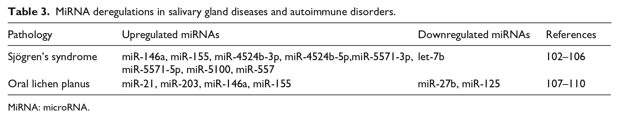
SS is a chronic, autoimmune disorder characterized by reduced secretions of the salivary and lacrimal glands and associated endocrine disturbances. 101 It was noted that miR-146a and miR-155 102 are upregulated and let-7b 103 was downregulated in response to the adaptive immune response in SS. Another study described the deregulation of miR-4524b-3p, miR-4524b-5p, miR-5571-3p, miR-5571-5p, miR-5100, and miR-5572 in patient samples derived from SS patients. 104 The specificity of miRNAs such as miR-203, miR-768-3p, and miR-574-3p within the exosomes of parotid saliva samples from patients with SS holds promise for future development of biomarkers for the diagnosis and prognosis of various salivary gland pathologies. 105
Other oral autoimmune disorders
Oral lichen planus (OLP) is a chronic inflammatory disease of the oral mucosa with an unknown etiology mediated by T helper 1 (Th1) lymphocytes within the basal cell layer of the oral epithelium. Upregulation of mediators including TGF-β, TNF-α, IL-6, COX-2, and MMP-7 has been reported.106–108 Increased expression of miR-21, miR-203, 106 miR-146a, and miR-155 107 and decreased expression of miR-125 108 were observed in OLP compared to normal oral mucosa. MiR-27b was significantly downregulated in OLP tissue, and its expression was more suppressed in atrophic-erosive OLP than in reticular OLP. 109 The unique profile of the cytokines and chemokines that initiate OLP has been suggested to lead to transformation toward OSCC. 110 Table 3 illustrates the various miRNAs that were found to be differentially expressed in salivary gland diseases and autoimmune disorders.
MiRNA deregulations in salivary gland diseases and autoimmune disorders.
MiRNA: microRNA.
Possible causes of altered miRNA expression
The deregulation of miRNA expression is associated with various oral cancers and diseases. There are multiple pathways that can impact miRNA profiles, and these can be broadly categorized as genetic, epigenetic, and processing defects. MiRNA transcription can be effected by genetic changes (i.e. deletions, amplifications, or mutations involving miRNA loci, epigenetic silencing, dysregulation of transcription factors that target specific miRNAs), while defective enzymatic activity associated with miRNA precursors can impact mature miRNA accumulation. 111
Genetic changes
Multiple transcription factors work cooperatively to ignite gene expression, while multiple miRNAs bind to cognate sites in the 3′ UTR of the target mRNAs to modulate expression. Although the binding within the seed region is in perfect Watson–Crick complementarity, base pairing outside the seed region adds a secondary layer of specificity. The complexity of translation is further extended through heterotypic miRNA–mRNA interactions, as genes can harbor binding sites for several miRNAs. 112 A key mechanism behind cancer cell progression is abnormal levels of expression for mature and/or precursor miRNA compared with corresponding normal tissues. Depending on the genes it targets, miRNA can act either as a tumor suppressor gene (TSG) or as an oncogene. MiRNAs located in genomic regions amplified in cancers (such as the miR-17–92 cluster) function as oncogenes, whereas miRNAs located in portions of chromosomes deleted in cancers (such as the miR-15a–miR-16-1 cluster) function as tumor suppressors. 113
Epigenetics
Single-nucleotide polymorphisms (SNPs) in miRNA genes (miRSNPs), whether in a CpG island or not, can be predicted to affect function by modulating the transcription of the primary transcript, pri-miRNA, and pre-miRNA processing and maturation or miRNA–mRNA interactions. 114 Similar to other protein coding genes, the miRNA promoter is susceptible to changes in methylation pattern, and aberrant methylation will impact the expression of miRNA. For instance, hypermethylation will diminish the transcription of the miRNA encoding gene, while hypomethylation will increase miRNA levels. Several studies have reported aberrant methylation in diverse forms of cancers. The fact that statistically significant differences of miR-137 promoter methylation were seen between healthy controls and patients of OLP raises a possibility that miR-137 methylation may serve as a biomarker for malignant prediction in patients with OLP. 115
MiRNA processing defects
Deletion or over-activation of processing genes, even in a single miRNA, may give rise to severe defects in organogenesis. 63 For instance, the defective processing of miRNA pathway effector genes such as Drosha and Dicer-1 can alter the flux of miRNAs. Deletion of Dicer-1 from the dental epithelium (DcrK14−/−) has shown to have aberrations in tooth morphogenesis, terminal cell differentiation, and tissue homeostasis in rodents 116 (multiple, branched, enamel-free incisors and cuspless molars). MiR-140, miR-31, miR-875-5p, miR-141 miR-689, miR-720, miR-711, miR-203, and miR-455 are known to be expressed during various tooth formation stages. Sirtuin 1 (SIRT1) along with tumor suppressor p53 modulates miR-34 processing, thus interfering with effective biogenesis of keratinocyte cell lines. 117 It was also reported that p53 can influence miRNA processing of several miRNAs including miR-16-1/15, miR-145, and miR-107 resulting in failure to produce mature miRNA. 117 X-linked miRNAs examined in one of the studies suggested that SNPs could alternate Drosha or Dicer excision sites. The resultant mutant alleles, namely, miR-502-C/G, miR-510-T/C, miR-890-C/G, and miR-892b-T/C (less-detectable mature miRNAs) could be the etiological determinants of diseases such as schizophrenia and autism. 113
MiRNA alterations as diagnostic biomarkers
MiRNAs aberrantly expressed during different stages of oral diseases unveil their potential utility as biomarkers in understanding the pathogenesis of disease and in monitoring disease activity and effects of therapy. Given the abundance of miRNA alterations in oral cancer and their stability in a wide range of tissues, miRNA detection may prove to be a valuable diagnostic tool. 63 Plasma levels of miR-31 (under-expressed), 118 miR-184 (over-expressed), 35 and miR-10b (over-expressed) 119 were suggested to serve as tumor biomarkers for early detection of OSCC recurrence, which significantly dropped in patients following surgical removal of the tumor. There is accumulating evidence implying the feasibility and potential superiority of salivary-based miRNA biomarkers for the earlier diagnosis and prognosis of patients with oral cancers due to their slower degradation and shuttling via exosomes. MiRNA-125a and miR-200a were significantly decreased, and miR-31 over-expressed 120 with aberrant methylation of miR-200c, miR-141, and atypical expression of miR-375, miR-200a 120 in saliva samples of OSCC patients. When circulating miRNAs in sera of the patients with oral cancer were evaluated, miR-16, let-7b, miR-338-3p, miR-223, and miR-29a were downregulated in high-risk oral lesions, suggesting their utility as novel biomarkers for detection of oral cancer at the early stages. 121 Furthermore, methylation of miR-9-1 and miR-9-3 122 and inhibition of integrin beta-1 (fibronectin receptor beta) by miR-124, which is found to suppress tumor invasion and migration, 123 have great potential to serve as highly specific prognostic indicators for oral and oropharyngeal carcinomas. The differential expression patterns of miR-768-3p and miR-574 124 associated with salivary gland inflammation and miR-5100 104 in SS represent an innovative group of promising biomarkers.
MiRNA signatures pertaining to oral diseases based on their tissue of origin, disease progression, and so on could be useful for diagnostic, therapeutic, and preventive measures. A study employing quantitative reverse transcription polymerase chain reaction (qRT-PCR) and clustering analysis methods successfully identified miRNA signature profiles based on specific tissue of origin and their link to physiologic and disease processes in head and neck/oral cavity cancer cell lines. 125 A signature of 114 miRNAs have been noted to be differentially expressed between OSCC and normal oral epithelium, with the downregulation of miR-375 and upregulation of miR-31 as the most significant aberrations. 47 MiRNA signatures aberrantly expressed in low-grade dysplastic/progressive leukoplakia were reported as follows: miR-10b, miR-660, miR-708, and miR-30e were significantly overexpressed, while miR-145, miR-99b, miR-181c, and miR-197 were under-expressed. 71 In a second study, miR-21, miR-181b, miR-345, and miR-146a were upregulated, and miR-196a and miR-206 were downregulated. 126 A signature of five abundantly expressed miRNAs, miR-223, miR-191, miR-16, miR-203, and miR-24, has been detected in whole human saliva (healthy patients) which can be used in salivary diagnostics as biomarkers for detection of diseased states. 34 In an attempt to identify a miRNA signature specific to OSCC, 148 miRNAs in 18 cancer cell lines were examined. Upregulated miRNAs included miR-374, miR-340, miR-224, miR-31, and miR-9, while downregulated miRNAs were miR-137, miR-139a, miR-133b, miR-138, miR-34b, miR-137, miR-193a, and miR-203. 127 When combined, these studies identify individual miRNAs as potential tools for monitoring cancer precursor lesions.
MiRNA-based therapies: attractive alternate therapeutics
MiRNA—an emerging therapeutic target
MiRNA therapeutics in cancer either target or mimic miRNAs involved in cancer onset, progression, angiogenesis, and metastasis by behaving as tumor-promoting miRNAs (oncomiRNAs and metastamiRNAs) or as tumor suppressor miRNAs.128–130 The authentication and validation of miRNA expression in physiological as well as pathological conditions such as cancer, inflammatory disease, and autoimmune and endocrine disorders depend on detection using high-throughput techniques such as microarrays, reverse transcription polymerase chain reaction (RT-PCR), and RNA sequencing. mRNA degradation serves as one of the major final outcomes for miRNA-targeted transcripts. 131 To therapeutically target miRNA dysregulation, various modalities have proven to be promising including chemically stabilized modifications of RNA, miRNA inhibition therapy (antisense miRNA oligonucleotides, ASOs), and replacement therapy using miRNA mimics in plasmid DNA. MiRNA has become a promising tool owing to its advantages over conventional gene intervention therapies due to its exclusive biogenesis, mechanism of action that minimizes off-target effects, and tissue specificity and toxicity. 132 Combinations of anti-miRNA and miRNA replacement strategies have demonstrated synergistic anticancer activities which has been proven to be an effective strategy for tumor therapy. 133 Indeed, much interest has been given to developing and optimizing miRNA delivery systems including lipid-based delivery systems, oligonucleotides, dendrimers, exosomes, inorganic materials, and magnetic nanoparticles, 134 for example, miRNA antagonists such as miR-122 for Hep C virus—phase 2 of clinical trial (Santaris Pharma, San Diego, CA, USA), miR-208 for chronic heart failure—pre-clinical phase (miRagen Therapeutics, Boulder, CO, USA), miRNA replacement therapy such as miR-34 and let-7 for cancer—pre-clinical phase (MiRna therapeutics, Austin, TX, USA). 134
Clinical implications of miRNA therapy tool
Detection of miRNAs in serum, plasma, and saliva supports the clinical application of miRNAs as potential cancer biomarkers. 135 Rather than intercepting a single target as is the case of other gene therapies, miRNAs modulate entire gene programs. Therapeutically, it may be possible to combine with synthetically derived siRNAs or anti-miRNA oligonucleotides (AMOs) which can be delivered to patients to match the target mRNA and provide translational repression analogous to tumor suppressors. Because tissues are highly heterogeneous cell population, the accurate identification of cell type that predominantly contributes to altered miRNA levels and disease is highly desired. Minimal yet effective doses of miRNA may avoid immune responses, which cause the body to reject foreign RNA. Combining miRNA regulation with gene therapy has targeted the expression of “transgenes” in stem cell therapy. 131 The idea of using miRNAs in therapeutics is appealing owing to the outcomes from manipulating these molecules. For instance, it has been observed that siRNA derivatives of HIV-1 miRNAs could be used to inhibit HIV-1 viral replication in mice. HIV-1 Nef is necessary in the regulation of viral replication, and overexpression of Nef-derived miR-N367 can repress Nef transcription, assuring that miRNAs are highly useful in the treatment of diseases like HIV-1. 135
The development of chemoresistant oral cancers curbs the efficacy of chemotherapeutic drugs. Evaluating miRNA levels in such scenarios may serve as effective biomarkers for monitoring the post-transcriptional regulation of apoptosis, epithelial–mesenchymal transition (EMT), and stemness. For instance, silencing of chemoresistant miR-214 and miR-23a and rescue of chemosensitive miR-21 (oncomiR) could aid in abolishing cisplatin resistance in TSCC. 136 Furthermore, docetaxel-induced multi-chemoresistance observed in the treatment of head and neck SCC showed downregulation of miR-100, miR-130a, and miR-197 and upregulation of miR-101, miR-181b, and miR-195. 137 Thus, investigating the role of miRNAs may provide a deeper understanding of the challenges present in oral cancer chemotherapy.
Conclusion
Application of miRNA genomic and proteomic technologies in early detection of oral diseases may prove beneficial during the course of therapy. This review begins to summarize the significance of miRNAs in various oral disorders. Characterization of miRNA as potential noninvasive biomarkers is important and can prove to be greatly effective for the diagnosis and management of oral diseases. MiRNA target prediction and modulation of their expression can be used for therapeutic purposes. However, the local/systemic delivery options need to be investigated in detail. Targeted delivery of miRNA with antibody fusion protein, encapsulation with liposomes, and intravenous delivery systems represent encouraging areas of investigation. However, challenges remain including elicitation of interferon response and disruption of endogenous miRNA biogenesis. Fortunately, it may be possible to overcome these challenges by techniques such as directed knockdown.
Footnotes
Acknowledgements
Varun Kulkarni, Juhi Raju Uttamani, and Afsar Raza Naqvi equally contributed to this work.
Declaration of conflicting interests
The author(s) declared no potential conflicts of interest with respect to the research, authorship, and/or publication of this article.
Funding
The author(s) disclosed receipt of the following financial support for the research, authorship, and/or publication of this article: This review was supported by the NIH/NIDCR (DE021052) and in part by the University of Illinois at Chicago Edward C. Wach Fund.