
Abstract
Gastric cancer remains a big health problem in China. Gastric cancer cells contain a small subpopulation of cells that exhibit capabilities of differentiation and tumorigenicity. A putative explanation for ineffective therapy is the presence of cancer stem-like cells. Side population cells, which have cancer stem-like cells’ property, are characterized by the high efflux ability of Hoechst 33342 dye. Side population cells have been isolated from gastric cancer cell lines in previous studies. The epithelial–mesenchymal transition is very important in the invasion and metastasis of epithelial-derived cancers. More and more studies showed that gastric cancer stem-like cells possess high invasive ability and epithelial–mesenchymal transition property. A brief overview of the recent advancements in gastric cancer stem-like cells and epithelial–mesenchymal transition will be helpful for providing novel insight into gastric cancer treatment.
Introduction
Gastric cancer is the second most common cancer worldwide, especially in China. 1 In China, the incidence of gastric cancer ranks third among all malignant tumors, with an estimated 380,000 new cases annually. 2 Some cancer cells may have different properties than others, including tumorigenicity and self-renewal. 3 The first modern evidence for cancer stem cells (CSCs) came in 1994 with a study of human acute myeloid leukemia. 4 Although the gold standard for CSCs identification has not yet been reported, two approaches have been used to identify CSCs in published studies.5,6 One is an in vitro method termed “spheroid colony formation,” 5 and another is an vivo method involving implantation of candidate CSCs in immunodeficient mice. 6 Both the two methods were used to demonstrate self-renewal ability and tumorigenicity of CSCs. Due to constant research, the concept of CSCs has been found in many tumors, such as breast, 7 brain, 8 liver, 9 and gastrointestinal tumor.10,11 At the meeting of the American Association for Cancer Research in 2006, Reya et al. 12 proposed the definition of CSCs, which have four characteristics: tumorigenicity, multipotency, self-renewal, and drug resistance. Gastric CSCs are a group of heterogeneous cells that can self-regenerate and have the potential for multiple differentiation, metastasis, and drug resistance.13,14 In this review, we mainly discuss the link between epithelial–mesenchymal transition (EMT) and gastric cancer stem-like cells.
Side population cells of gastric cancer cell lines
Side population (SP) cells in some human cancers and cell lines that possess the characteristics of CSCs have been described as a CSC-rich population. 15 Hence, the characterization of SP cells may be a useful tool for analysis of CSCs, especially when specific CSC surface markers are unknown. SP are cell clusters with strong ability to efflux DNA dye Hoechst 33342 via various types of “ATP-binding cassette (ABC)” transporters. 16 The three ABC transporters, ABCB1 (MDR1/P-glycoprotein), ABCC1, and ABCG2 (BCRP), had the highest expression in SP cells.17–19
SP cells have been reported in gastric cancer cells. 20 In the study of Fukuda et al., 21 they isolated SP cells from five human gastric cancer cell lines (MKN45, KATOIII, MKN74, MKN28, and MKN1) by using flow cytometry, ranging from 0.02% ± 0.001% to 1.93% ± 0.16%. Nishii et al. 22 found that 0.46%, 0.29%, and 5.24% of the SP cells in OCUM-2M, OCUM-2D, and OCUM-2MD3 cells by using flow cytometric analysis, respectively. Another study showed that the ratios of SP cells were 0.5% for SGC-7901 cells and 0.6% for both BGC-823 and MGC-803 cells. 23 Schmuck et al. 24 obtained SP cells from AGS and MKN45 gastric cancer cells by using Hoechst 33342 staining and fluorescence-activated cell sorting, and ranged from 0.1% to 0.8%. Li et al. 25 found a small proportion (2.3% ± 0.78%) of the SGC-7901 cells were SP cells in their study. SP cells from the total population accounted for 0.57% in KATOIII, 1.04% in Hs-746T, and 0.02% in AGS. 26 In the study of Wang et al., 27 the MKN28, AGS, SGC-7901, and HGC-27 cell lines contained a distinct fraction of SP cells ranging from 0.18% ± 0.01% to 0.41% ± 0.03% of the total cell population. According to the studies described above, the SP cells were isolated from gastric cancer cell lines, including MKN45, KATOIII, MKN74, MKN28, MKN1, OCUM-2M, OCUM-2D, OCUM-2MD3, HGC-27, SGC-7901, BGC-823, MGC-803, Hs-746T, and AGS. In addition, they all demonstrated that SP cells have stem cell-like characteristics.
However, Zhang et al. 28 found that SP cells from BGC-823 did not have CSC properties in their study. Their results indicated that not all SP cells contain CSCs in gastric cancer. The main reason for the inconsistent results may be a dynamic equilibrium with some reversibility in transitions between cancer cell populations. 29 In our opinion, we tend to think that SP cell is a phase of cancer cells but not a specific subpopulation. The evidence in favor and against cancer stem-like cells are still debated.
EMT and gastric cancer
EMT is a process that epithelial-derived tumor cells reduced intercellular adhesion and acquired more mesenchymal phenotype.30,31 Epithelia converts between epithelia and mesenchyme through multiple rounds of EMT and the reversible process mesenchymal–epithelial transition (MET) during organ development. 32 An essential EMT step is the E-cadherin to N-cadherin switch which has critical functions in motility and migration of cancer cell 33 (Figure 1). EMT has been linked to the progression of cancer and increased stemness of tumors,34,35 and observed in the formation, invasion, and metastasis of gastric cancer.36–39 E-cadherin is a calcium-dependent cell membrane protein involved in cell–cell adhesion. 40 In various cancer cells, the abnormal expression of N-cadherin also correlates with the cell motility. 41 N-cadherin expression in gastric cancer tissues is significantly higher than that in the matched normal tissues, while the E-cadherin level is lower. 42 In addition, Gabbert et al. 43 found that the gastric cancer patients with E-cadherin-positive tumors had favorable survival rates than the patients with E-cadherin-negative tumors. Non-EMT phenotype is mainly distributed in the patients with early-stage gastric cancer. 44 EMT promotes gastric cancer cell motility and metastasis mostly at advanced-stage gastric cancer. 44 These results indicated that EMT plays an important role in gastric cancer (Figure 2).
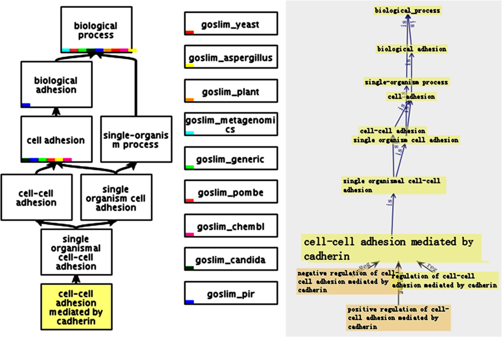
The canonical signaling pathways of EMT were generated by using the Gene Ontology (GO) software.

The proposed model of EMT pathways in cancer was generated by using the Ingenuity Pathway Analysis (IPA).
EMT and Helicobacter pylori
Moreover, Helicobacter pylori infection is the strongest risk factor for gastric cancer development.45,46 The imbalance between proliferation and apoptosis in H. pylori–infected cells is the main cause for gastric cancer. Previous studies have reported that the promoter methylation status of many tumor suppressor genes in gastric cancer is regulated by H. pylori infection, including E-cadherin. 47 Yu et al. 48 reported that cytotoxin-associated gene A (CagA), a H. pylori virulence factor, inhibits E-cadherin by down-regulation of programmed cell death protein 4 (PDCD4). Choi et al. 49 also observed the enhanced expression of four EMT-mesenchymal markers (Twist, Snail, Slug, and vimentin), whereas the expression of the E-cadherin was more suppressed in H. pylori–positive subjects compared with those in H. pylori–negative ones. Bessède et al. 50 demonstrated that H. pylori is responsible for an EMT phenotype associated with CD44 expression in gastric epithelial cell lines via CagA.
EMT and gastric CSC
Gastric CSCs possess high invasive ability and EMT property. In the study of Yang et al., 51 they found that gastric cancer stem-like cells from SGC-7901 cell line showed more invasive than their counterpart and exhibited an elevated vimentin but decreased E-cadherin expression. Aggressive clinical features in gastric cancer are associated with the EMT-related proteins, E-cadherin and vimentin, and stem cell-like phenotypes. 52 Loss of RUNX3 (Runt-related transcription factor 3) in gastric epithelial cells results in spontaneous EMT and induced a remarkably increased expression of the gastric stem cell marker Lgr5 (leucine-rich repeat-containing g-protein coupled receptor 5). 53 Aldehyde dehydrogenase 1 (ALDH1) is a detoxifying enzyme that catalyzes oxidation of intracellular aldehydes 54 and has been used to isolate CSCs from many cancers.55–58 In a recent study, Wu et al. 59 found that aldehyde dehydrogenase (ALDH) bright cells expressed decreased levels of E-cadherin but increased levels of Snail and vimentin.
Hypoxic microenvironment is another pivotal factor in regulating invasion and progression of cancer. 60 Hypoxia facilitates gastric cancer invasion and metastasis through EMT. 61 Hypoxic pretreatment also promoted proliferation, colony formation, migration, and invasion of gastric cancer cells (BGC-823 and SGC-7901). 62 These results indicated that hypoxic microenvironment induces EMT and upgrades stem-like properties of gastric cancer cells.
Conclusion
EMT plays a significant role in the formation and metastasis of gastric cancer. Accumulating evidence indicate that there is a link between CSC and EMT in gastric cancer. EMT could provide a new perspective for CSC theory. However, the potential mechanisms have to be studied in future studies. Specific targeting of stem-like cancer cells could reduce tumor growth, metastasis formation, and drug resistance. Therapeutic agents aimed at the inhibition of stemness via an approach based on EMT could prove beneficial for the treatment of gastric cancer patients.
Footnotes
Declaration of conflicting interests
The author(s) declared no potential conflicts of interest with respect to the research, authorship, and/or publication of this article.
Funding
The author(s) disclosed receipt of the following financial support for the research, authorship, and/or publication of this article: This study was supported by National Natural Scientific Foundation of China (No. 81572777 and No. 81502558).