
Abstract
Background
Colon cancer continues to be the third most commonly occurring cancer around the globe with both lifestyle-related risk factors and genetic dispositions. Owing to the increased incidence of the disease, there is a need to find new treatment options which are easily accessible and less toxic to the cells. Cynaropicrin, a sesquiterpene extract from the artichoke plant exhibits varied properties such as anti-inflammatory, anti-cancer, and antioxidant among others. In the current investigation, the anti-cancer potential of the cynaropicrin extract was observed against colon cancer in HCT-116 cell lines.
Materials and Methods
The drug exhibited a considerable decrease in cell proliferation. Also analyzed was the reactive oxygen species (ROS) generation and apoptosis induction by cynaropicrin on the HCT-116 cell line. The AO/EtBr staining confirmed the apoptotic induction through chromatin condensation by fluorescence microscopic examination followed by DAPI staining of the treated HCT-116 cells which showed nuclear condensation and disintegration.
Results
Furthermore, it was observed that cynaropicrin elevated the levels of ROS in the cells which modified the mitochondrial membrane permeability in the HCT-116 cell lines. The study also evaluated the levels of apoptotic proteins and the expression of inflammatory cytokines concerning cells treated with cynaropicrin and untreated control cells.
Conclusion
It has been observed that cynaropicrin treatment can lead to apoptosis in the HCT-116 cell line via mitochondria-mediated apoptotic pathway and if further evaluated, it can turn out to be a potent anti-cancer drug for effective management of colon cancer.
Introduction
Malignancies of the colon and rectum are known to be the 3rd most frequently occurring cancer worldwide and contribute heavily to the cause of cancer-associated mortalities annually. Roughly more than one million people are affected by colon cancer worldwide and 0.5 million deaths are estimated because of the same (Kamel et al., 2022; Saltz, 2022). Cancer formation is a multi-stage process that happens over a long period. Pedunculated polypoid structures are the first to arise and grow into the lumen of the colon and with time, it develops into highly disordered histology and dysplastic cytology. The condition is recognized to be cancerous only when these cells rupture the basement membrane of the underlying epithelial cells (Venugopal & Carethers, 2022).
The rate of incidence and mortality of colon cancer varies greatly as the process is complex and also the disease progresses due to lifestyle changes as well as old age (Shinji et al., 2022). Several cohort studies support the evidence that there is an increased risk in the incidence of the disease with the intake of red meat, reduced folate consumption as well a sedentary lifestyle (Rawla et al., 2019). Germline mutation in the key genes involved in colon cancer leads to the incidence of hereditary colon cancer that accounts only for 3%−7% of the overall cases every year (Liu et al., 2014). Ideally, colon cancers can be mitigated through early detection along with the elimination of pre-malignant adenomas. Surgical excision of the tumor is the most beneficial treatment outcome of cancer. It has been observed that the disease is mostly presented at an advanced stage while diagnosing, the majority having distal metastases along with unresectable tumors making the treatment options scarce. The overall five-year survival rate of the disease is known to be poor because of the late presentation of the disease making the tumor grading the most important factor in predicting the survival of the disease (Kamel et al., 2022).
Among the different risk factors involved in the progression of the disease, a recognized process for the progression of colorectal carcinoma involves the disproportion between cell regeneration and cell death with proliferation being favored. Apoptosis a highly regulated process is necessary for the maintenance of the genomic balance and several proteins are employed as potential anti-cancer regulators (Jaganathan et al., 2011). The major proteins involved in the process of apoptosis include the Bax/Bcl-2 and the proteins of the caspase family (Hosseini et al., 2020). The levels of Bax and Bcl-2 are regulated by the mitochondria and the stimulation of caspase family proteins plays a crucial role in apoptotic cell death.
It is important to understand the molecular basis of human cancer to provide corrective treatment options. Much work remains in the field of understanding the molecular mechanisms and the genes involved that contribute to the disease. This leads to the scenario where finding new alternative treatments is a necessity and the use of agents that are specific and less harmful becomes an absolute essential shining light on the potential of natural compounds to serve as an anti-cancer agents (Sithara et al., 2017; Zheng et al., 2020).
Cynaropicrin a sesquiterpene lactone obtained from artichoke (Cynara scolymus L.) in 1960 and later was discovered in other species of the same genus (Moujir et al., 2020). Cynaropicrin is a major biologically significant secondary metabolite and is considered to be a chemotaxonomic indicator for artichoke plant species. Similar to other sesquiterpene lactones, the biological activities of cynaropicrin are related to the γ-butyrolactone ring. The compound possesses several pharmacological activities including anti-tumor, anti-inflammatory, anti-trypanosomal, and anti-hepatitis C virus among many other potentials. Owing to these multiple specific potentials, cynaropicrin seems to be more significant and will serve towards the development of pharmaceutical substances (Elsebai et al., 2016). A recent study revealed that cynaropicrin demonstrated strong cytotoxicity on glioblastoma cells (Rotondo et al., 2022), melanoma cells (De Cicco et al., 2021), and revealed antioxidative and anti-neuroinflammatory activity in an ischemic stroke model (Jin & Leng, 2022).
The primary objective of the current investigation is to determine the anticancer potential of the sesquiterpene compound of the artichoke―cynaropicrin against Human Colon Cancer Cells―HCT-116. The effect of cynaropicrin on the cell viability by (3-(4,5-dimethylthiazol-2-yl)-2,3-diphenyltetrazolium Bromide) (MTT) assay, apoptotic induction by AO/EtBr, DAPI staining, mitochondrial membrane potential (MMP) by Rh-123 staining, reactive oxygen species (ROS) generation via 2’,7’-dichloro-dihydro-fluorescin diacetate (DCFH-DA) staining and the expression of the apoptotic proteins―caspase-9, caspase-3, Bax, and Bcl-2 and to analyze the expression levels of TNF-alpha, NF-kB, COX-2 and IL-6 using ELISA technique has been investigated.
Materials and Methods
Chemicals
All the chemicals including cynaropicrin were purchased from Sigma-Aldrich, USA. All the kits for biochemical assays were acquired from Thermo Fisher Scientific, USA.
Culturing of HCT-116 Cell Line
The human colon HCT-116 cancer cell line was procured from the ATCC, USA. At 37℃, the cells were grown in EMEM with FBS (10%) in a humidified environment with a 5% CO2 supply. Every 48 h, the cells were checked for confluency and were replenished with fresh complete media and subcultured using 0.25% trypsin-EDTA solution.
MTT Cytotoxicity Assay
HCT-116 cells were seeded into a 96-well plate at a density of 5000 cells per mL, and they were then incubated for 24 h at 37°C (Mosmann, 1983). After 24 h the cells were checked for adherence and the existing media was discarded and the cells were exposed to fresh media with cynaropicrin drug at different concentrations: 5, 10, 15, 20, 25, 30, 35, 40, and 45 µM and was left for incubation for 24 h at 37℃. Afterwards, the cells were treated with 10 µL of 5mg/mL of MTT and were incubated in the dark at 37°C for 3 h. Later, cells were suspended in 10 µL of DMSO which aids in dissolving the purple color formazan crystals formed after incubation with MTT. The cells were incubated with DMSO for a minimum of 15 min and the optical density was read at 570 nm.
Estimation of Intracellular ROS Level
The amount of ROS produced by cynaropicrin against colon HCT-116 cells was examined using the DCFH-DA staining. The HCT-116 cells were exposed to cynaropicrin at the IC50 concentration (25 µM) for 24 h followed by staining with DCFH-DA. The cells were subjected to staining with DCFH-DA dye (10 mM) and were incubated for 30 min in the dark. The cells were given time to incubate with the dye and then were given two PBS washes before being examined with a fluorescence microscope (Olympus).
Estimation of Mitochondrial Membrane Potential
The onset of the apoptosis process is detected by the change in the cell’s level of MMP. Rhodamine-123 is a cationic dye specific to the mitochondria and is used in the determination of change in the MMP. The cells were treated with cynaropicrin at the IC50 value and were incubated for a period of 24 h at 37°C with a supply of 5% CO2. Before staining with Rh-123 dye, the cells were rinsed using PBS and stained with Rh-123 at a concentration of 10 mg/mL for 30 min. The cells were rinsed using PBS post 30 min staining with Rh-123 dye. With the aid of a fluorescent microscope, the cells were examined.
Apoptosis Estimation by AO/EtBr Staining
To understand and assess the apoptotic induction potential of cynaropicrin on the human colon HCT-116 cells, the AO/EtBr staining approach was used. The seeded cells were treated with cynaropicrin at the IC50 concentration and were incubated for 24 h. Post the drug exposure, the cells were subjected to staining with an equal combination of acridine orange and ethidium bromide and were incubated in the dark for 5 min. The stained cells were washed with PBS post 5 min incubation and the cells were subjected to a fluorescent microscopic examination.
Apoptosis Estimation by DAPI Staining
To examine the morphological changes occurring within the nucleus of the cells, the HCT-116 cells are stained with DAPI. HCT-116 cells (2 × 10⁵ /well) were seeded in a 6-well plate and were grown overnight in DMEM medium with 10% FBS. After 24 h, a new medium containing 0.2% FBS was added to the cells, and cynaropicrin at the IC50 concentration was administered to the cells. The drug treatment was given for 24 h duration after which the media was discarded with PBS two times with cold PBS. The cells were then fixed using 100% ethanol at 37°C for 20 min. Following the fixing step, the cells were again washed with cold PBS twice. The cells were examined using the fluorescence microscope (Olympus).
Involvement of Caspase-3, Caspase-9, Bcl-2, and Bax in HCT-116 Cell Line
The cell lysates of control and treated cells were prepared and centrifuged at 10000 rpm for 15 min at 4°C. The resultant supernatant was kept at −80°C. The concentrations of the caspase-9, caspase-3, Bcl-2, and Bax in the extract were determined with the help of the ELISA kit as per the guidelines provided by the manufacturer (Thermo Fisher, USA).
Involvement of TNF-alpha, NF-kB, COX-2, and IL-6 in HCT-116 Cell Line
The cell lysates of control and treated cells were prepared and centrifuged at 10000 rpm for 15 min at 4°C. The resultant supernatant was utilized to assess the concentrations and expression of TNF-alpha, NF-kB, COX-2, and IL-6 determined with the help of the ELISA kit as per the instructions provided by the manufacturer (Thermo Fisher, USA).
Statistical Analysis
All the experimental analyses done with different drug concentrations were carried out in triplicates and the data were statistically analyzed with GraphPad Prism software. A one-way ANOVA test was utilized to evaluate the data, followed by the Student Newman Keuls test. The data were reported as mean ± SEM, with a statistical significance level of p < 0.05.
Results
Cytotoxic Potential of Cynaropicrin on HCT-116
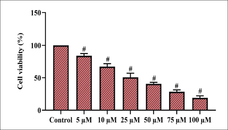
The experimental study was planned and executed to understand and evaluate the cytotoxic potential of cynaropicrin on human colon HCT-116 cancer cells at varying concentrations ranging from 5 to 45 µM. Each concentration was tested in triplicates to ensure the sensitivity and specificity of the test. The findings of the study revealed that cynaropicrin down-regulates cell viability in HCT-116 cells dose-dependently. Statistical investigation of the MTT assay further established that the proliferative functions of the HCT-116 cells were remarkably down-regulated by different concentrations of cynaropicrin (Figure 1). When compared to the control untreated cells, the cell viability was reduced in the treated cells. Among the range of concentrations used to treat the cells, at 24 h 25 µM of the drug showed the maximum effect and was determined to be the IC50, which was further used for the following experiments.
Estimation of Intracellular ROS Level
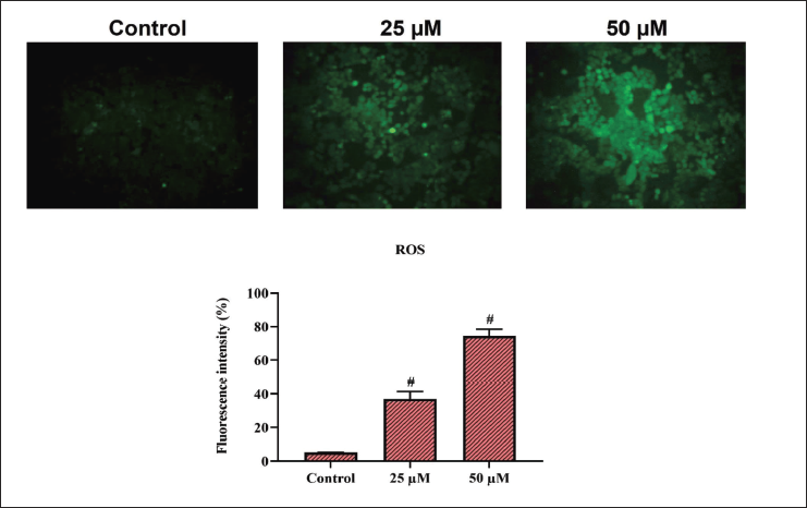
In the HCT-116 cell line, ROS generation after exposure to cynaropicrin was examined using DCFH-DA staining. The concentration of cynaropicrin used was 15 and 25 µM and the incubation period was 24 h. After 24 h, the fluorescence intensity was measured. The mean fluorescent intensity was found to be increased at 25 µM when compared to the untreated control cells (Figure 2). ROS intensity increased nearly 3-fold compared to the untreated control after 24 h.
Estimation of MMP in HCT-116
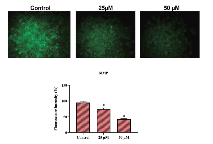
Mitochondria are the major site of ROS formation within the cells. Induction of apoptosis by various agents―both natural and chemotherapeutic agents is complemented with a decrease in the MMP. Rhodamine-123 is used to measure MMP loss. The cells were treated with cynaropicrin at 15 and 25 µM along with untreated control cells. Comparing treated and untreated cells, the mean fluorescence intensity of treated cells was reduced. The MMP levels in the cynaropicrin-treated cells were decreased when compared to the untreated control cells (Figure 3). It was observed that untreated control cells fluoresced brilliant green, but those treated with cynaropicrin at 15 and 25 µM fluoresced dull green, indicating a reduction in MMP in the cells. It has been established from the study that cynaropicrin can diminish MMP levels in HCT-116 cells.
Apoptotic Induction by Cynaropicrin on HCT-116 Cells
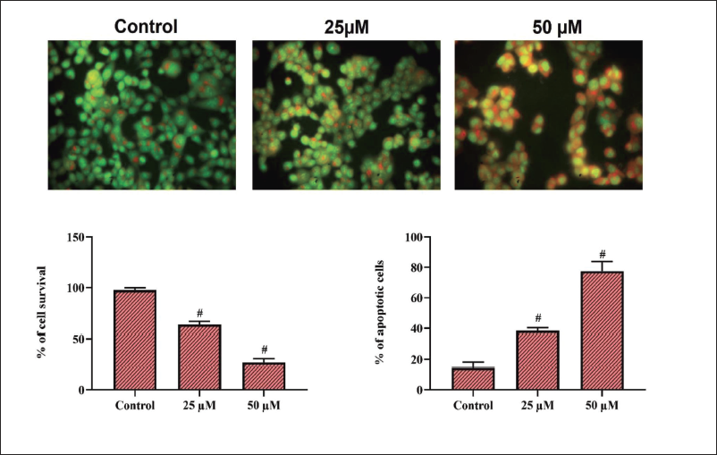
Cell apoptosis is characterized by morphological changes in the cells. AO/EtBr staining is used to assess the morphological changes brought about by the cynaropicrin drug treatment of HCT-116 cells. Staining by AO/EtBr is utilized to differentiate between living and dead cells. EtBr, a red fluorescence dye is capable of selectively staining only the fragmented nuclei of apoptotic cells whereas Acridine Orange, a green, fluorescent dye can enter only the healthy cells. The study indicates that the untreated control cells have a deep green fluorescence whereas the cells treated with 15 µM of cynaropicrin shows a mix of green and orange-stained cells indicating early apoptosis, whereas the cells treated with 25 µM of the cynaropicrin dye―the IC50 of the drug the cells are all stained in red stained fragment representing fragmented nuclei exhibiting late apoptosis (Figure 4). The bar graph depicts the percentage of apoptotic cells in AO/EtBr.
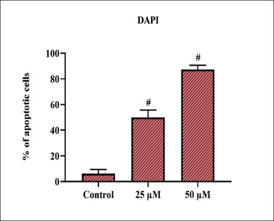
Apoptotic Estimation by DAPI Staining in HCT-116 Cells
To examine whether cynaropicrin treatment induced morphological changes in the treated cells, DAPI staining was used. Phase-contrast microscopy revealed apoptotic bodies. When compared to the untreated control cells, cynaropicrin-treated cells generated nuclear condensation, DNA fragmentation, and perinuclear apoptotic bodies, as shown by DAPI staining (Figure 5).
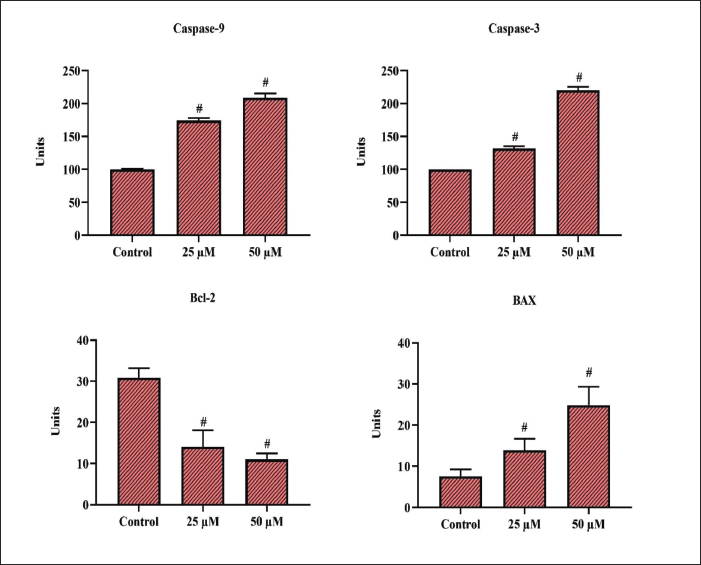
Involvement of Caspase-9, Caspase-3, Bcl-2, and Bax in HCT-116 Cell Line
The levels of caspase-9, caspase-3, Bcl-2, and Bax when HCT-116 cells are treated with cynaropicrin were estimated to understand the contribution of the apoptotic gene in cell death mechanism using the ELISA technique. It was observed that the activity of caspase-9, caspase-3, and Bax were all augmented in the cells treated with 25 µM of cynaropicrin when compared to the levels of the proteins in the untreated control cells (Figure 6). Conversely, the Bcl-2 expression was significantly lowered in the cynaropicrin-treated cells opposite to the effect on control cells.
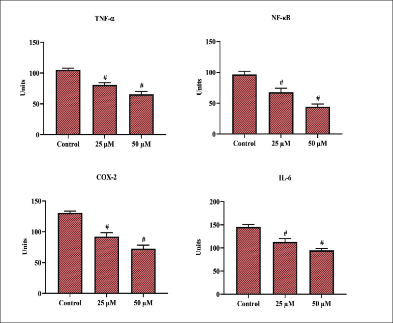
Involvement of TNF-alpha, NF-kB, COX-2, and IL-6 in HCT-116 Cell Line
The contribution of the inflammatory cytokines and their expression levels in HCT-116 cells treated with cynaropicrin and in cells that are untreated control was determined using the ELISA technique. It was observed that the expression of TNF-alpha, NF-kB, COX-2, and IL-6 were all significantly reduced in the cells treated with 25 µM cynaropicrin when compared to the untreated control cells where the expression of these inflammatory cytokines remained high (Figure 7).
Discussion
Colon cancer, which affects the lower digestive system, is one of the tumors with the highest mortality rates worldwide (Benson et al., 2014; Kamel et al., 2022). There has been significant development in the area of colon cancer research including the implementation of several chemotherapeutic drugs to provide proper medical intervention. However, owing to the higher prevalence of colon cancer worldwide and the aggressiveness of the disease, it has become inevitable to find advanced therapies to perfect the imperfect suppression of malignant cells and the subsequent progression of the disease which is a severe clinical challenge in the current scenario. Coming up with novel therapeutic drugs and targets that can combat the disease has become significantly imperative (Marnett & DuBois, 2002). The increased attention on plant compounds is due to their efficiency and also the non-toxic behavior of the compounds which has been identified as an optimal strategy to reduce the incidence of tumors and the progression of the same (Kim et al., 2006; Su et al., 2019). In this study, cynaropicrin was explored for its anti-tumor activity against colon cancer.
The MTT assay performed to assess the cell viability and cytotoxic potential of cynaropicrin showed anti-proliferative activity dose-dependently. Similar dose-dependent reductions in the cell growth of the HCT-116 cell line were observed in a study conducted by Zheng et al. (2020). Colon cancer cells undergo oxidative damage-arbitrated apoptosis as a result of oxidative stress. Overgeneration or elimination of ROS causes severe damage to the intracellular molecules (Choi et al., 2022). It has been observed that plant-based chemo-preventive agents elevate the generation of ROS and disrupt the homeostasis in cancer cells.
This increased level of free radicals leads to diminished antioxidant levels and encourages early-stage apoptosis in cancer. An increase in ROS formation was noticed in HCT-116 cells treated with cynaropicrin when stained with DCFH-DA probes. An important component of oxidative stress-mediated apoptosis is MMP. The mitochondria play a crucial part in the apoptotic process, and the permeabilization of the outer membrane is a crucial step in apoptotic cell death (Park et al., 2008). As a result, cytochrome c is produced in the cytosol by the mitochondria in response to apoptotic stimuli. The current study exploring the potential of cynaropicrin depicted that the drug-induced oxidative stress-mediated the alteration of outer MMP which was observed by diminished fluorescence intensity. The agent tested cynaropicrin also generated high levels of ROS which could result in the induction of MMP alterations.
To understand and explore the morphological changes induced by cynaropicrin on the HCT-116 cell line, AO-EtBr staining was done. The live cells in the control were stained in a deep green color whereas the apoptotic cells were displayed as orange entities owing to the shrinkage of the nucleus. Chemotherapeutic drugs are known to target the DNA directly to induce cancer cell death. The process is related to various characteristics including nuclear disintegration, chromatin condensation, and membrane blebbing. Once the cells go through these phases, the apoptotic bodies are formed which are then engulfed by the macrophages. Excessive ROS production leads to the mentioned changes in the cells leading to apoptosis. This has been observed in the cells treated with cynaropicrin and is confirmed by the AO/EtBr staining which depicts the above-mentioned apoptotic features. The DAPI staining of the cynaropicrin-treated cells depicted the apoptotic bodies when compared to the untreated normal cells. A study conducted by Su et al. (2019) depicted a similar result on colon cancer cells which analyzed the potential of the compound zingerone extracted from Ginger.
A major and critical mechanism in the treatment options of cancer cells is the induction of programed cell death. The major obstacle in apoptosis induction in cancer cells is the intrinsic resistance of the tumor cells to the apoptotic proteins and the process. The process of apoptosis is manifested by means of two pathways―the intrinsic and the extrinsic pathway (Lin et al., 2010). For the intrinsic pathway, death signals are transmitted through the mitochondria, whereas the extrinsic pathway uses cell surface receptors to transmit the signal. Both pathways eventually lead to the activation of caspases ad results in cell death. Caspases are a family of cysteine proteases which modulate the apoptotic response (Rawla et al., 2019).
The major caspases of the family are caspase-3 which is an executioner and caspase-9 which is an initiator in mitochondria-mediated apoptosis (Umesalma & Sudhandiran, 2010). The study evaluated the expression of the major apoptotic proteins including caspase-3 and caspase-9 using an ELISA technique which showed that the HCT-116 cells treated with cynaropicrin showed increased expression of both caspase-3 and caspase-9 which represents the apoptosis induction in the cells. Mitochondria-dependent apoptosis is regulated by the Bcl-2 family of proteins. Bax is a pro-apoptotic protein of the family of Bcl-2 and is triggered by apoptotic signals (Waldner et al., 2012). By lowering the MMP, releasing cytochrome c, and activating caspases, the migration of Bax to the mitochondria triggers the onset of apoptosis. The current study shows that the cells treated with cynaropicrin had upregulated expression of Bax whereas the expression of Bcl-2 was downregulated. This finding is in concordance with a prior study that involved salinomycin’s potential in regulating apoptosis in colorectal cancer cell lines (Zhou et al., 2013).
It is well-recognized that the tumor microenvironment affects the development of cancer. Inflammation is known to exist in the tumor microenvironment and is known to be induced by inflammatory mediators including IL-6 (Beckeret al., 2005, Borrelli et al., 2014), TNF-alpha (Al Obeed et al., 2014), NF-kB (Slattery et al., 2018, Tong et al., 2016), and COX-2 (Sinicrope & Gill, 2004; Zhang & An, 2007). There has been increasing evidence showing that there is a marked increase in inflammatory cytokine expression in the progression of cancer (Plewka et al., 2018). The cells treated with cynaropicrin showed a marked reduction in the expression of the inflammatory cytokines when compared to the normal cells. Overall, cynaropicrin shows potential anti-cancer properties in the HCT-116 cells of human colon cancer. Cynaropicrin is advantageous since it is a natural substance and is soluble in water. It may be used with therapeutic injections to hasten the onset time and lessen the risk of adverse effects. The drug’s inexpensive cost of synthesis and production, together with its straightforward chemical structure, might ease the burden of medical therapy on patients.
Conclusion
Colon cancer serves as a paradigm for our insights into the molecular causes of human cancer. The constant need for finding new treatment options makes it important to find plant-derived compounds that can be efficient in treating cancer. The study explored the anti-cancer properties of cynaropicrin―a secondary metabolite from the artichoke plant. The study looked at all the aspects including cell viability, and the apoptotic-inducing potential of the plant extract and was observed that cynaropicrin can be used as a potential anti-cancer agent against colon cancer as it induces the killing of cancer cells by mitochondria-mediated apoptosis.
Summary
Cynaropicrin is advantageous since it is a natural substance and is soluble in water. It may be used with therapeutic injections to hasten the onset time and lessen the risk of adverse effects.
Cynaropicrin treatment can lead to apoptosis in the HCT-116 cell line via mitochondria-mediated apoptotic pathway and if further evaluated, it can turn out to be a potent anti-cancer drug for effective management of colon cancer.
The drug’s inexpensive cost of synthesis and production, together with its straightforward chemical structure, might ease the burden of medical therapy on patients.
Abbreviations
MTT: (3-(4,5-dimethylthiazol-2-yl)-2,3-diphenyltetrazolium bromide); DCFH-DA: 2′,7′-dichloro-dihydro-fluorescin diacetate; MMP: mitochondrial membrane potential; ROS: reactive oxygen species.
Footnotes
Acknowledgment
Department of Science and Technology of Xinjiang Uygur Autonomous Region, Natural Science Foundation Program No.2021D01C425).
Declaration of Conflicting Interests
The authors declared no potential conflicts of interest with respect to the research, authorship, and/or publication of this article.
Funding
The authors received no financial support for the research, authorship and/or publication of this article.