
Abstract
Background
Excitotoxicity is an early event of cerebral ischemia, which is mainly caused by neuroinflammation, oxidative stress, and dysfunction of Excitatory Amino Acid Transporter-2 (EAAT-2). Generally, tissue plasminogen activators (tPA’s) and anticoagulant therapies are being used as first-line treatment options for cerebral ischemia, but they only restore the cerebral blood flow (CBF) and fail to attenuate the detrimental events associated with ischemic insult in neurons, resulting in neurodegeneration. Based on the earlier studies, we designed a novel combination for targeting neuroinflammatory and excitotoxicity cascades in cerebral ischemia.
Methods
Angiotensin receptor blocker (ARB) Azilsartan (Azi) and a third-generation cephalosporin Ceftriaxone (Cef) were evaluated in in vitro oxygen glucose-deprived (OGD) primary astrocytes and N2a neuronal co-cultures model of cerebral ischemia. Further, the above combination was also investigated in the middle cerebral artery occlusion (MCAo) rat model of cerebral ischemia. Then, neuro-biochemical estimations and molecular techniques like flow cytometry, ELISA and gene expression studies were performed to elucidate the possible mechanism.
Results
The novel combination ameliorated the neurodegeneration by downregulating the ROS, apoptosis, oxidative stress, and excitotoxicity cascades and also enhanced the level of antioxidant enzymes. Moreover, EAAT-2 gene expression was remarkably increased with the treatment with a novel combination of Azi and Cef than with the individual treatment. The above combination significantly reversed the behavioural dysfunction in ischemic rats, which evidences the beneficial effects.
Conclusion
The repurposing of anti-hypertensive, Azi, and antibiotic Cef combination demonstrated an excellent neuroprotective potential, mediating through ameliorating neuroinflammation, excitotoxicity and oxidative stress in in vitro OGD-induced astrocyte-neuron co-culture as well as cerebral ischemic rat model.
Introduction
Cerebral ischemia is a prevalent cerebrovascular disorder that, through prolonged disruption of cerebral blood flow (CBF)—often caused by thrombus formation in the arterial circulation—leads to oxygen and nutrient deprivation and ultimately triggers secondary neurodegenerative processes in affected brain regions. Impairment of oxygen and glucose leads to abruption in the cellular functioning of the neurons, subsequently causing immobility of limbs and death. It has been estimated that more than 80% of stroke cases are ischemic in nature. Co-morbidities such as Diabetes and Hypertension are the relative risk factors for the occurrence of ischemic insults as well as haemorrhagic stroke. The incidence of stroke is increasing day by day due to a sedentary lifestyle, individual habits and preexisting diseases.
The pathophysiology of stroke is a multifaceted, complex phenomenon that mediates through several different pathways in neuronal deterioration, such as oxidative stress, excitotoxicity, and cytokine storm. In general, the normal CBF rate of a healthy adult lies between 50 and 60 mL/100 gm/min, which will accomplish the metabolic needs of normal brain physiology, and also CBF varies in different regions of the brain. When CBF falls to less than 20 mL/100 gm/min, it will commence the activation of the ischemic cascade as a consequence of energy failure. Further, it diminishes the synaptic activity of neurons, and when it falls below 10 mL/100 gm/min, irreversible neuronal damage happens because of oxygen-glucose deprivation (OGD). 1 OGD in brain cells initiates several pathological events, including elevated intracellular cytosolic Ca²+ levels and disruption of ionic gradients. The resulting sustained cellular depolarisation in both neurons and glial cells promotes excessive influx of Na+ and Ca²+ and efflux of K+. This depolarised state overstimulates presynaptic voltage-dependent Ca²+ channels, leading to excessive release of excitatory amino acids such as glutamate and aspartate into the synaptic cleft. Elevated glutamate and aspartate concentrations in the synaptic region during energy-depleted conditions activate Ca2+-dependent N-methyl-D-aspartate (NMDA) and α-amino-3-hydroxy-5-methyl-4-isoxazole propionate acid (AMPA) receptors, which trigger the release of destructive enzymes like protease, lipase, and endonucleases by increasing the intracellular Ca2+ levels. The metabolic byproducts of enzymes liberate free radicals and lead to loss of cellular integrity, oxidative stress further activates downstream signalling pathways such as mitogen-activated protein kinases (MAPK) or nuclear factor-kappa B (NF-κB), which release a variety of pro-inflammatory proteins and cytokines like tumor necrosis factor-α (TNF-α), interleukin-1β (IL-1β) and IL-6.2–6
As of now, intravenous thrombolysis with recombinant tissue plasminogen activators (tPA’s) is being used as a first-line treatment option, but there are some drawbacks associated with tPAs than their benefits. tPAs are expensive drug that may not be quickly available in pharmacy stores, and a delay in hospitalisation often affects the efficacy of thrombolysis. tPA has to be administered within 3–4.5 hours of the onset of the stroke attack. 7 Moreover, treatment with the tPA’s, antiplatelet agents, anticoagulants, and osmotic diuretics will clear the arterial circulation, but the existing therapies could not interfere with neuroinflammatory mechanisms. Since current treatments fail to target the neuroinflammation associated with stroke, recently several researchers have explored the role of the Brain Angiotensin System (BAS) in the central nervous system as a modulator of cerebrovascular and inflammatory processes. Saavedra and his colleague’s research findings stated that Angiotensin-II (Ang-II) of the brain has an important function in the regulation of CBF, hormonal system, innate immune response, drinking behaviour and also reported for its critical role in several neurodegenerative pathological conditions such as cerebral ischemia, malfunctioning of BBB, abnormal stress response and neuroinflammation as a result of (Ang-I) AT1 receptor stimulation. 8 Another experimental evidence has suggested that pressor responses were observed after the cerebral ischemia induction by middle cerebral artery occlusion (MCAo) model in Sprague Dawley (SD) rats. The pressor effect was a result of the activation of the pro-inflammatory mediator chemokine, monocyte chemoattractant protein-1 (MCP-1) by Ang-II in the brain predominantly, which regulates the blood pressure in the rostral ventrolateral medulla of the brain stem, 9 suggesting the role of Ang-II in neuroinflammation. Recent studies in an ischemic mouse model have revealed that the overexpression of human angiotensinogen and renin augmented the neuronal damage due to elevated Ang-II in the OGD mice model, and the expression of Angiotensin converting enzyme-II (ACE-II) induces the conversion of Ang-II into Ang (1-7), thereby ameliorating neuronal damage by reducing the binding of Ang-II on AT1 receptors.7, 8 It has also been noticed that the incidence of ischemic stroke was increased with angiotensinogen promoter and AT1 receptor gene polymorphism. 10 These research findings signify that the BAS and circulatory Ang-II have a crucial role in neuronal damage through neuroinflammation. 11
Earlier studies evidenced that blockade of central AT1 receptors with a non-hypotensive dose of Angiotensin receptor blocker (ARB’s) Telmisartan (5 mg/kg) exhibited neuroprotection via antioxidant and anti-inflammatory mechanisms in a murine model of transient focal ischemia, turndown the expression of cytokine production and reduced the expression of vascular endothelial growth factor (VEGF),12–14 and also administration of AT1 blockers have shown significant improvement in neuronal plasticity and demonstrated functional recovery from ischemic stroke. 15 Another study reported the neuroprotective potential of Azilsartan (Azi) against ischemia-induced secondary brain injury by protecting the enzyme complex activities and thereby preventing mitochondrial dysfunction. Azi treatment further arrested oxidative, apoptotic, inflammatory & histological neuronal damage. 16
A third-generation cephalosporin, Ceftriaxone (Cef), is widely used as a broad-spectrum antibiotic for several bacterial-induced pathological conditions and also evaluated for neuroprotection in in vitro neuronal cell lines and in vivo animal models. 17 Cef is generally administered as a prophylactic antibiotic in patients with stroke and other medical conditions in order to avoid hospital-acquired infections. In a study, it has been shown that Cef upregulated the expression of Excitatory Amino Acid Transporter-2 (EAAT-2) through NF-kB activation in primary human fetal astrocytes (PHFA), where Cef directly binds to the -272 position (EAAT-2 promoter gene) of astrocyte DNA and promotes the transcription of EAAT-2 proteins, which helps in clearing excess glutamate and reducing excitotoxicity. 18 Cef has significantly protected neurons against global ischemia via upregulating the GLT-1 expression in an in vivo animal model, 19 and it also restores the glutamate transporters and prevents chronic intermittent hypoxia-induced excitotoxic neuronal damage in in vitro P16 organotypic hippocampal slices. 20
In the present study, we used the novel combination of Azi and Cef to elucidate the neuroprotective potency in OGD C57BL/6 primary astrocyte—N2a neuron co-culture and transient intraluminal focal ischemic/MCAo rat model, as the combination offers a mechanistically complementary strategy for mitigating ischemic brain injury. Azi exerts neurovascular and anti-inflammatory benefits by reducing AT1R-mediated oxidative stress and improving cerebral perfusion, while Cef enhances astrocytic glutamate clearance and limits excitotoxicity. Given that cerebral ischemia involves simultaneous glutamate accumulation, oxidative imbalance, and inflammatory activation, this combination has the potential to produce synergistic neuroprotection. Therefore, we evaluated the therapeutic efficacy of Azi plus Cef in OGD-induced primary astrocyte–N2a co-cultures and the intraluminal MCAO rat model.
Materials and Methods
In Vitro Neuroprotective Evaluation of Azi and Cef Novel Combination in OGD Neuron-astrocyte Co-culture
Primary Astrocyte Culture
The primary mouse astrocyte culture was prepared using C57BL/6 primary astrocytes (Cellbiologics, USA). Briefly, 10 × 105 cells were plated in a 24-well plate in DMEM/F12 media and supplemented with 10% FBS, 100 U/mL of penicillin and 100 µg/mL of streptomycin. The cells were seeded in a Poly-D-lysine-coated flask in a humidified atmosphere with 5% of CO2 at 37°C, and the medium was changed every two days until the cell population reached 80%–90% of the culture flask. The flasks were shaken vigorously inside of the incubator for 12 hours, the detached cells were eliminated and medium was changed on 11th day, cell debris and residuary cells were digested using 0.25% trypsin, and then cells were seeded in 24-well plates (8 × 10⁵ cells/plate) and maintained with DMEM/F12 medium with an appropriate humidified atmosphere of 95% of oxygen, 5% of CO2 at 37°C. The medium has been refreshed and changed every two days in order to minimise cell debris.
Primary Astrocyte and N2a Neuronal Co-culture
The primary astrocytes and N2a neuronal co-culture was prepared by plating N2a neuronal cells in a 6-well plate (1 × 10⁶ cells/well) and primary astrocytes (8 × 10⁵ cells/well). The astrocyte culture strips were inserted into a neuronal culture medium and incubated in an adequate environment for co-culture development with a humidified atmosphere, 5% of CO2 and 95% of oxygen at 37°C. An inverted phase contrast microscope (Olympus Corporation, Tokyo, Japan) was used to observe the morphology of co-culture and to capture the images of cell density in multilayer surfaces.
OGD and Reperfusion
In vitro ischemic reperfusion stroke was induced using OGD followed by the reperfusion model. The primary astrocyte and N2a neuronal co-culture cells were washed with glucose-free medium, and then the cells were incubated in glucose-free DMEM medium and a humidified oxygen control conditions with 1% of O2, 5% of CO2 and 94% of N2 for 24 hours at 37°C. After 24 hours of OGD, the cells were immediately reperfused by replacing with high glucose DMEM medium and incubated for 72 hours at 37°C with normoxic conditions. 21
Morphological Observations
The astrocyte-neuron co-culture morphology was observed before and after induction of OGD using a confocal microscope.
Cell Viability
The cell viability of neuron-astrocyte co-culture was measured after 24 hours of OGD induction, and treatment with Azi and Cef using the CCK-8 assay kit according to the manufacturer’s (E-CK-A362; Elabsciences) instructions. Briefly, 10 µL of CCK-8 solution was added to the cells and allowed to incubate for 4 hours. The optical density (OD) of each sample was measured at 450 nm using a microplate reader (Thermoscientific USA). Quantitative dose-toxicity examination and the half-maximal effective concentration (EC50) were measured.
ROS Assay
The cellular reactive oxygen species levels were assessed using 2′,7′-dichlorofluorescein-diacetate (DCFH-DA; Beyotime Institute of Biotechnology) staining technique according to the manufacturer’s instructions. Neuron-astrocyte co-culture at a density of 1 × 10⁵ cells/mL was treated with 0, 25 and 50 µM/mL concentrations of Azi and Cefat at 37°C for 48 hours, then washed and incubated with 10 µM DCFH-DA for 30 min at 37°C, where 2% DMSO was used as a control. Subsequently, flow cytometry (CytoFLEX flow cytometer, Beckman Coulter Life Sciences) analysis was performed to quantify the fluorescence intensity in the FITC channel.
Apoptosis Assay
The primary mouse astrocytes and N2a neuronal cells (1.0 × 10⁵ cells/mL) were treated with EC50 concentration of Azi and Cef for 48 hours in 24-well plates, where 2% DMSO was used as a control. Apoptosis of the cells was assessed using the FITC Annexin V apoptosis detection kit. According to the instructions of the manufacturer, the harvested cells were washed twice using Phosphate-buffered saline (PBS) and then re-suspended in 100 µL of 1× binding buffer and stained with 5 µL of Annexin V-FITC and 5 µL of Propidium iodide (PI) at 25°C for 15 min. Flow-cytometry analysis was performed to quantify apoptosis; for each flow cytometer run, 10,000 cells were used.
AO/EB Fluorescent Staining
The neuron-astrocyte co-culture (1.0 × 10⁵ cells) cell viability was measured after 24 hours of OGD induction with respect to the treatments of Azi, Cef and the novel combination of Azi and Cef. The live, necrotised and dead cells were detected using the Acridine orange (AO)/Ethidium bromide (EB) Staining kit (Cat # E607308, Sangon Biotech) according to the manufacturer’s instructions. Cells were washed twice with PBS and resuspended in 1× buffer. Each 5 µL of AO and EB staining solutions was added to 90 µL of cell suspension, incubated at 25°C for 5 min in a dark room and then the stained cells were analysed using a fluorescence microscope (Leica Microsystems).
Oxidative Stress Parameters
1. Superoxide dismutase
The neuron-astrocyte co-culture was treated with the novel combination of Azi and Cef after OGD induction, and then the cells were washed with PBS, cold 0.1 M Tris/HCl, pH 7.4, which contains 0.5% Triton X-100, 5 mM β-Mercaptoethanol (βME), and 0.1 mg/mL Phenylmethylsulfonyl fluoride (PMSF) was added. Homogenate was prepared using a centrifuge at 14000 × g, at 4°C for 15 min, supernatant was collected and kept on ice. The SOD levels were detected using the superoxide dismutase (SOD) activity assay kit (Bio Vision; Catalogue #K335-100) as per the manufacturer’s instructions. Briefly, 20 µL supernatant was taken in a 96-well plate, and 200 µL of WST working solution was added to each well and then 20 µL of enzyme working solution was added and mixed rigorously. The microplate was incubated for 20 min at 37°C, for 20 min, and then absorbance was recorded at 450 nm using a microplate reader.
2. Catalase assay
Neuron-astrocyte co-culture was treated with the novel combination of Azi and Cef after OGD induction, and then the cells were washed with PBS, re-suspended in 200 µL of ice-cold assay buffer. Homogenate was prepared from 1 × 10⁶ cells by centrifugation of cells at 10,000 × g at 4°C for 15 min using a cold microcentrifuge; supernatant was collected and kept on ice. The release of catalase (CAT) was detected using a CAT activity assay kit (Cat#ab83464; Abcam) as per the manufacturer’s instructions. Briefly, 2 µL of supernatant was plated in 96 well plate and volume was adjusted up to 78 µL with catalse assay buffer, 12 µL of fresh 1 mM hydrogen peroxide was added to each sample well and then allowed to incubate for 30 min at 25°C, finally 50 µL of developer mix, 10 µL of stop solution and were added and incubated in dark chamber at 25°C for 10 min and absorbance was recorded at OD 570 using a microplate reader.
3. Reduced glutathione
The neuron-astrocyte co-culture was treated with the novel combination of Azi and Cef after OGD induction, and then a homogenate was prepared from 100 µL of pelleted cells with 100 µL of 5% sulfosalicylic acid solution, vortexed vigorously and kept on ice for 10 min. Samples were centrifuged at 12,000 × g for 20 min at 4ºC. The supernatant was collected and preserved at a cold temperature. The reduced glutathione (GSH) levels were estimated using the GSH assay kit; Bio Vision, Catalogue # K464-100 GSH assay kit, according to the manufacturer’s instructions. Briefly, 2–10 µL of samples were taken in a clear 96 well plate and were diluted 5–20 folds with GSH assay buffer. Two µL of 100 folds diluted enzyme reaction mixture A and 80 µL of sample reagent mix was added to each sample well and the final volume of 198 µL in each well was adjusted with assay buffer and kept on ice. A 96-well plate was kept at room temperature for 40–60 min, and absorbances were recorded at OD 450 nm using a microplate reader.
4. Lipid peroxidation
The neuron-astrocyte co-culture was treated with the novel combination of Azi and Cef after OGD induction, and then the cells were washed with PBS, re-suspended in Tris buffer (20 mM), and a homogenate was prepared from the cultured cells by centrifugation at 2,000 × g, at 4°C, for 10 min. The supernatant was collected and stored at 4°C. The release of the enzyme lipid peroxidase was detected using lipid peroxidation (LPO) assay kit (Cat. #BAQ067, G-Biosciences) as per the manufacturer’s instructions. Briefly, 100 µL of homogenate, 325 µL of freshly prepared LPO reagent A solution, as well as 75 µL of B solution was added and then mixed thoroughly. The mixture cocktail was incubated at 40°C for 40 min. Two hundred µL of supernatant was transferred into a 96-well plate from each sample tube, and absorbance was recorded at 586 nm using a microplate reader.
Nitric Oxide Synthase
The neuron-astrocyte co-culture (2 × 10⁵ cells) was treated with 200 µL of nitric oxide synthase (NOS) lysis buffer, and cells were thoroughly homogenised, and then cells were centrifuged at 10,000×g, 4°C for 10 min. The clear supernatant was collected and kept on ice. Thirty to sixty µL of cell lysate was taken and transferred into a 96-well plate, and the volume was brought up to 57.5 µL/well with NOS assay buffer. The release of NOS in desired samples was detected using the NOS Activity Assay kit (Catalogue # K2094; Bio Vision) as per the manufacturer’s instructions. Briefly, 40 µL of reaction mix, 2.5 µL of 10 mM NOS co-factor one were added to the sample wells and mixed thoroughly. The plate was incubated at 37°C for 30 min, and 95 µL of NOS assay buffer, 5 µL of enhancer were added to all wells after incubation and kept at room temperature for 10 min, 50 µL of Griess reagent 1, and 50 µL of Griess reagent 2 were added and incubated for 10 min. The absorbance was read at OD 540 nm using a microplate reader in endpoint mode.
Cytokine Estimation
1. Interleukin-6
The primary mouse astrocytes and N2a neuronal cells (1 × 10⁶ cells/mL) were treated with different concentrations of Azi and Cef combination after OGD induction in 24-well plates. The cytokine IL-6 levels were estimated in various treatment groups using IL-6 recombinant (4H16L21) FITC monoclonal antibody kit (Invitrogen; Catalogue # 701028) as per the manufacturer’s directions. Briefly, 10 µL of staining reagent was added to 100 µL of cell suspension and incubated for 15 min at 25°C. Flow-cytometry analysis was performed to estimate IL-6 content. For each flow cytometer run, 10,000 cells were used.
2. Interleukin-1β
The neuron-astrocyte co-culture (1 × 10⁶ cells) was treated with 250 µL of chilled PBS, and the freeze-thaw process was continued until the cells were fully lysed, then centrifuged at 1,500 × g at 2°C–8°C for 10 min. The supernatant was collected and stored for assay. The estimation of IL-1β intensity in different treatment groups was detected using an IL-1β ELISA kit (Cat#ab197742, Abcam) according to the manufacturer’s instructions. Briefly, 50 µL of homogenate was transferred into a 96-well plate, and then 50 µL of antibody cocktail was transferred to each well. The plate was sealed and incubated for 1 hour at 25°C on a plate shaker at 400 rpm, washed with 350 µL of 1× washing buffer, and then inverted the plate with blotting paper to remove excessive liquid from the well. One hundred µL of TMB development mix was added to all wells of 96 well plate and incubated for 10 min in the dark on a plate shaker at 400 rpm. One hundred µL of stop solution was added to each well, and the plate was incubated for 1 minute to distribute the reagent properly, and absorbance was recorded at 450 nm using a microplate reader.
3. Tumor necrosis factor-alpha
The primary mouse astrocytes and N2a neuronal cells (1.0 × 10⁶ cells/mL) were previously treated with different concentrations of Azi and Cef combination after OGD induction in a 24-well plate. The cytokine TNF-α levels were estimated in various treatment groups using TNF-α (MAb11) FITC monoclonal antibody kit (Catalogue # MA5-44101; Invitrogen) as per the manufacturer’s directions. Briefly, 4 µL of staining reagent was added to 100 µL of cell suspension and incubated at 25°C for 15 min. Flow-cytometry analysis was performed to estimate the TNF-α level. For each flow cytometer run, 10,000 cells were used.
4. Real-time polymerase chain reaction
Neuron-astrocytes were seeded in a 24-well plate at a density of 1 × 10⁵ cells/mL and treated with EC50 concentration of the novel combination of Azi and Cef for 48 hours. Total RNA isolation was done using the GENEzol™ reagent (Gene Aid Biotech). Primer sequences were obtained from NCBI BLAST. Using the Revert Aid First Strand cDNA synthesis kit, cDNA was synthesised (Thermo Fisher Scientific). Luna® Universal qPCR master mix (New England BioLabs) and CFX Connect™ Real Time PCR detection system (Bio-Rad) were used for RT-PCR amplification. Relative mRNA expressions of targeted genes were analysed by the 2-∆∆CT method by normalising with glyceraldehyde-3-phosphate dehydrogenase (GAPDH).
In Vivo Neuroprotective Evaluation of Azi and Cef Novel Combination in MCAo Rat Model
Experimental Animals
Three-month-old SD male rats (200–250 g) were used for the present study. The animals were housed in separate polypropylene cages in a well-ventilated room and maintained at 25°C ± 2°C temperature with 55% of relative humidity (RH) conditions with a 12 h light/dark cycle. All the animals were acclimatised by keeping them in the animal house of the School of Pharmacy, Anurag University, Hyderabad, for a week. All the experimental procedures carried out according to the ‘guide for the care and use of laboratory animals’, Institutional Animal Ethical Committee, Anurag University, Hyderabad (IAEC) No. I/IAEC/AU/016/2023 WR♂, India, approved the study protocol.
Middle Cerebral Artery Occlusion
The intraluminal transient focal cerebral ischemia was induced by occlusion of the middle cerebral artery using a nylon monofilament, followed by reperfusion injury. SD rats were anaesthetised with Xylazine (10 mg/kg, i.p) and Ketamine Hydrochloride (80 mg/kg, i.p). A mild incision was made on the neck, and then the right common carotid artery was exposed at the site of the internal and external common carotid artery bifurcation branch. Nylon monofilament (4-0 size, Ethicon®) tip was exposed to mild candle flame in order to make the tip round-headed. The tip of the filament was coated with 0.01% poly-L-Lysine solution for adhesion at the middle cerebral artery region. Monofilament was inserted into the external carotid artery and then advanced to the internal carotid artery until slight resistance was felt (approximately 20–21 mm). The occlusion achieved by keeping the filament inside the blood vessel and ligation of the common and external carotid arteries, and external incision was closed temporarily. After 120 min of occlusion, animals were mildly re-anaesthetised, and the suture was opened to pull out the nylon filament to cause reperfusion injury. The reperfusion of the internal carotid artery was ensured visually. The animal’s body temperature was measured throughout the surgical procedure with a rectal thermometer, and it was maintained at 37°C ± 0.5°C using a heat pad. The animals were kept in separate cages, and the cage temperature was maintained between 29°C ± 1°C for the next hour after post-surgery in order to avoid possible hypothermia-induced neuroprotection. The Sham operated (SO) group animal’s external carotid artery was surgically exposed by incision, but a monofilament was not inserted.22, 23
Rotarod Test
The assessment of muscle coordination of the ischemic animals was analysed using rotarod apparatus. Experimental animals were kept on rotarod for 5 min at a speed of 20 rpm, and then the time spent by the animals on the rotating rod was recorded to correlate the grip strength among different treatment groups. 24
Actophotometre
The assessment of motor coordination in ischemic animals was evaluated by placing the animal in a photocell of the actophotometre for 5 min, where the beam of light passes through the photocells of the apparatus. The mobility of the animal will interrupt the light beam which passing through the photocells. The number of cut-offs made by animals indicates the number of ambulations by the animals within the period of 5 min. The count was recorded and displayed digitally. 25
Brain Isolation
After appropriate behavioural assessments, the animals were excessively anaesthetised for euthanasia, and then their brains were isolated. After isolation, the brains were micro-dissected into hippocampus and cortex to estimate the neurobiochemicals and cytokine levels with respect to the ischemic and treatments. Four brain samples were kept for each analysis, and the remaining two brains were used for histopathological studies.
Oxidative Stress Parameters
1. Superoxide dismutase
The brain samples were thoroughly washed with PBS, and then ice-cold 0.1 M Tris/HCl, pH 7.4, which containing 0.5% Triton X-100, 5 mM βME, and 0.1 mg/mL PMSF was added to it. Homogenate was prepared using a centrifuge at 14,000 × g, at 4°C for 15 min, and then the supernatant was collected. The SOD levels were detected using the SOD activity assay kit (Biovision; Catalogue #K335-100) as per the manufacturer’s instructions. Briefly, 20 µL of sample solution was taken in a 96-well plate, 200 µL of WST working solution was added to each well, 20 µL of enzyme working solution was added and mixed rigorously. The microplate was incubated at 37°C for 20 min, and the absorbance was recorded at OD 450 nm using a microplate reader.
2. Catalase
The brain samples were thoroughly washed with PBS, 200 µL of ice-cold assay buffer was added and then the sample at 10,000 × g, at 4°C for 15 min. Supernatant was collected, transferred into an aliquot and kept on ice. The CAT levels were estimated using a CAT activity assay kit (Cat#abcam83464; Abcam) as per the manufacturer’s instructions. Briefly, 2 µL of samples were plated in 96 well plate and adjusted the volume up to 78 µL with catalase assay buffer, 12 µL of fresh 1 mM H2O2 solution was added to each sample well, and then allowed to incubate at 25°C for 30 min, finally 10 µL of stop solution and 50 µL of developer mix were added and incubated in a dark chamber at 25°C for 10 min and absorbance was recorded at OD 570 nm using a microplate reader.
3. Reduced glutathione
The micro-dissected brain samples were washed thoroughly with chilled PBS, and then a homogenate was prepared by adding 100 µL of 5% sulfosalicylic acid solution for each 100 mg of the brain, and the mixture was vortexed vigorously and kept on ice for 10 min. Samples were centrifuged at 12,000 × g at 4ºC for 20 min, and the supernatant was collected and stored at cold temperature. The GSH levels were estimated using the GSH assay kit (Bio Vision, Catalogue # K464-100) according to the manufacturer’s instructions. Briefly, 2–10 µL of samples were taken in a clear 96-well plate and were diluted 5–20 fold with GSH assay buffer. Two µL of 100 folds diluted enzyme reaction mixture A, 80 µL of sample regent mixture was added to each sample well, final volume of each well 198 µL was adjusted with assay buffer and kept on ice for 5 min and the 96 well plate was kept at room temperature for 40 min, and absorbance was recorded at OD 450 nm using a microplate reader.
4. Lipid peroxidation
The micro-dissected brain fragments of hippocampus and cortex were washed thoroughly with ice-cold PBS, re-suspended in Tris buffer (20 mM), and a homogenate was prepared by centrifugation at 2,000 × g, 4°C for 10 min. The supernatant was collected and stored at 4°C. The release of catalytic enzyme lipid peroxidase was detected using a lipid peroxidase assay kit (Cat #BAQ067; G-Biosciences) as per the manufacturer’s instructions. Briefly, 100 µL of homogenate, 325 µL of freshly prepared LPO reagent A solution, as well as 75 µL of B solution was added and mixed thoroughly. The reaction mixture cocktail was incubated at 40°C for 40 min. Two hundred µL of supernatant from each tube was transferred into 96 well plate, and absorbance was recorded at OD 586 nm using a microplate reader.
5. Glutamate assay
The brains were collected, thoroughly washed with PBS, and then micro-dissected into hippocampus and cortex, re-suspended in 100 µL of glutamate assay buffer, and homogenised using a Dounce homogeniser with 10–12 passes. Thereafter, the homogenate was incubated on ice for 15 min on ice and then centrifuged for 2–5 min at 4°C at top speed; supernatant was collected and kept on ice. The content of the excitatory amino acid glutamate was estimated using a glutamate assay kit (CAT#ab83389, Abcam) according to the manufacturer’s instructions. Briefly, 2 µL of homogenate was taken in sample wells of 96 well plate and made the volume 50 µL with glutamate assay buffer, 100 µL of reaction mix was added to each sample and standard wells, 100 µL of background reaction mix was added to background sample wells, mixed thoroughly and incubated at 37°C for 30 min in dark chamber and absorbance was measured at 450 nm using a microplate reader.
Cytokine Estimation
1. Interleukin-6
Brain samples were washed thoroughly with PBS, five folds of the 1× sample diluent was added. Centrifuged at 10,000 × g, at 4°C for 5 min and then the supernatant was collected, transferred into a microcentrifuge tube and stored at cold temperature. The IL-6 levels were detected using the IL-6 rat ELISA kit (Cat#ab119548–IL-6; Abcam) as per the manufacturer’s instructions. Briefly, 100 µL of standard solution was added to the standard and blank wells of 96 well plate, and then 50 µL of 1× assay buffer was added, and 50 µL of sample solution was added to the sample wells. Fifty µL of 1× Biotin Conjugated Antibody was added to all wells and incubated for 2 hours at 25°C with gentle shaking (400 rpm). Microplate was washed six times with approximately 400 µL of washing buffer, 100 µL of 1× Streptavidin-HRP was added to all wells then microplate was covered with an adhesive film and incubated at room temperature for 1 hour with gentle shaking, adhesive film was removed and microplate was emptied, washed the microplate with approximately with 400 µL of 1× washing buffer and then 100 µL of TMB substrate solution was added to all wells, incubated the microplate at 18°C to 25°C for 10 min in a dark chamber, 100 µL of stop solution was added to each well to stop enzyme reaction and absorbance was read at OD 450 nm using a microplate reader.
2. Interleukin-1β
Brain samples were washed thoroughly with PBS, and fivefold of 1× sample diluent was added. Centrifuged at 10,000 × g, at 4°C for 5 min and then the supernatant was collected, transferred into a microcentrifuge tube and stored at cold temperature. The IL-1β levels were detected using the IL-1β rat ELISA kit (Cat#ab100768; Abcam) according to the manufacturer’s directions. Briefly, 100 µL of standard and sample solutions were added in appropriate wells of 96 well plate and incubated for 2.5 hours at room temperature with gentle shaking. The solution was discarded and washed four times with 1× washing buffer. One hundred µL of 1× Biotinylated IL-1β Detection Antibody was added to each well and incubated at room temperature for 1 hour with gentle shaking, and then the solution was discarded and washed with 1× washing buffer. One hundred µL of HRP-Streptavidin solution was added to each well and incubated at room temperature for 45 min with gentle shaking. The solution was washed with 1× washing buffer. One hundred µL of TMB One-Step Substrate Reagent was added to each well and incubated at 25°C for 30 min with gentle shaking. Fifty µL of stop solution was added to each well, and absorbance was recorded at OD 450 nm using a microplate reader.
Histopathology
1. 2,3,5-triphenyltetrazolium chloride staining
The experimental animals were excessively anaesthetised after all behavioural assessments for euthanasia. Brains were isolated, washed thoroughly with PBS and then frozen at 0°C in a refrigerator. The infract volume of different treatment groups was measured using 2,3,5-triphenyltetrazolium chloride (TTC) (Wako Pure Chemical Industries, Osaka, Japan). Briefly, the brains were removed and dissected into 2 mm coronal sections. The 2 mm thick brain slices were immersed in 2% TTC solution for 20 min at 37°C. The ischemic and non-ischemic areas were differentiated by the hemispheric damage (white colour) and live neurons (red colour). The infarction volume was measured using ImageJ software 1.53c. 26
2. Cresyl violet staining
The experimental animals were sacrificed by excessive anaesthesia, and immediately brains were isolated and preserved in 10% formalin for fixation. The lipid debris was removed by soaking in alcohol, and then the brain was fixed in paraffin wax. Five µm-thick coronal sections were taken, and stained with 0.1% cresyl violet reagent and micro sections were dehydrated by increasing alcohol concentration. The stained brain microsections were observed under a binocular microscope with 40× magnification, and photos were captured to assess the intensity of neuroprotection and neurodegeneration by different treatments. 27
3. Haematoxylin and eosin staining
The experimental animals were sacrificed by excessive anaesthesia, immediately brains were collected and preserved in 10% formalin for fixation, and the brain was fixed in paraffin wax. Five µm-thick coronal sections were made using a microtome. Sections were placed on slides and treated with ice-cold acetone for 10 min and then rehydrated in PBS for 15 min. The sections were treated with haematoxylin and incubated at room temperature, then immersed in eosin for 1 minute. Subsequently, dehydrated in ethanol and mounted on a binocular light microscope to assess the intensity of neuroprotection and neurodegeneration with different treatments at 40× magnification. 28
4. Statistical analysis
For statistical analysis, GraphPad Prism 8.2.1 was used. All values were expressed as Mean ± SEM, significance between the groups was analysed by one-way ANOVA followed by Dunnett multiple comparison tests and statistical significance of the differences was defined by p < .05.
Results
In Vitro Neuroprotective Evaluation of Azi and Cef Novel Combination in OGD Neuron-astrocyte Co-culture
Morphological Observation
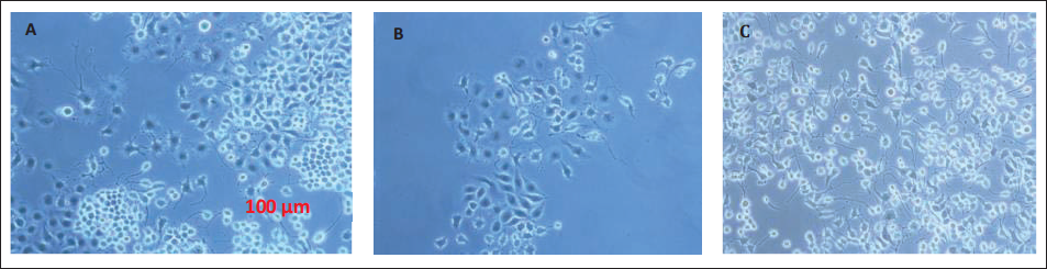
The neuron-astrocyte co-culture cell development and structural changes have been observed using a confocal microscope with 40× magnification, and photos were taken before and after OGD (Figure 1).
The Morphological Changes of Neurons and Astrocytes Under a Confocal Microscope on Day 4 (A), 14 (B) and 20 (C), Respectively.
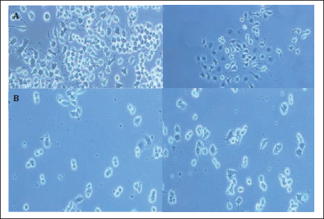
After 24 hours of OGD induction and 72 hours of reperfusion, was lead to cell necrosis and axonal degeneration (Figure 2) and cell death has also been noticed.
Morphological Changes of the Neurons and Astrocytes Before (A) and After (B) OGD Induction.
Cell Viability
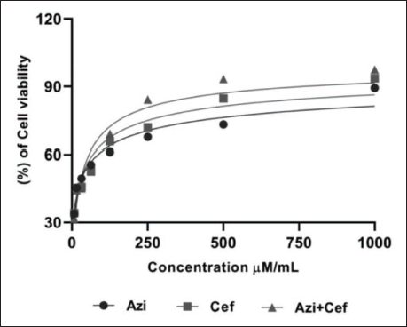
The cell viability of neuron-astrocytes after 24 hours of OGD induction was increased with the treatment of Azi, Cef, and both combinations (Figure 3). EC50 values of the Azi, Cef, and the novel combination were found to be 41.78 µM/mL, 29.43 µM/mL, and 5 µM/mL, respectively.
The Comparative Plot of Half-maximal Effective Concentrations (EC50) of Azilsartan, Ceftriaxone, and the Novel Combination Azilsartan + Ceftriaxone in Neuron-astrocyte Co-culture.
The novel combination of Azi
ROS Assay
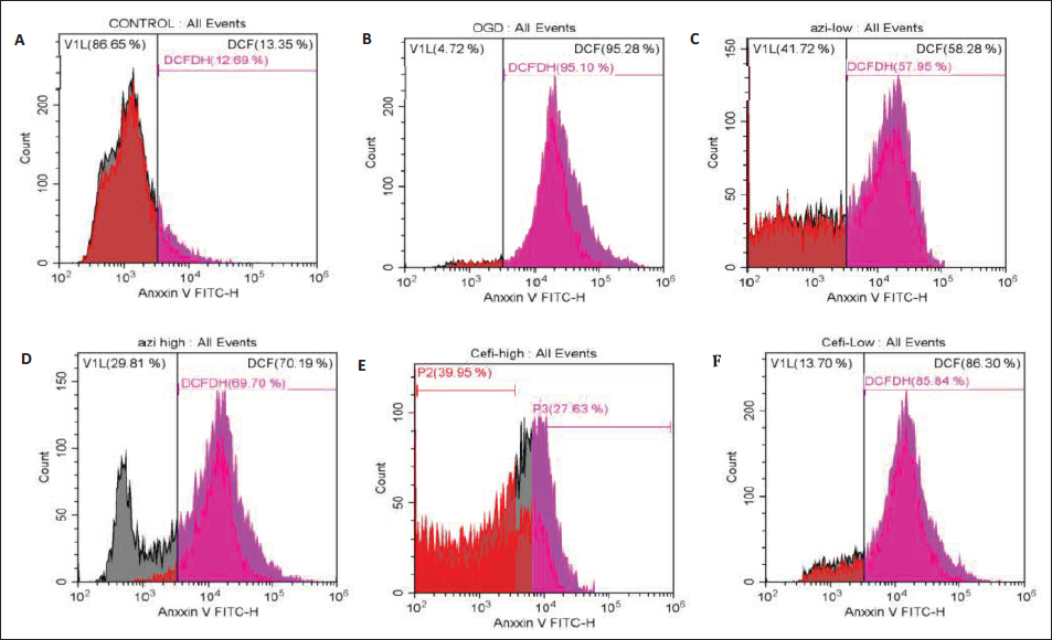
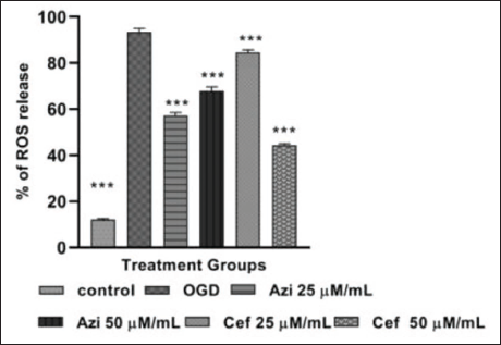
The ROS assay was performed in neuron-astrocyte co-culture with six different treatment groups, such as control, OGD, Azi 25 µM/mL, Cef 25 µM/mL, Azi 50 µM/mL and Cef 50 µM/mL. The ROS level in control group was found to be 12.69%, OGD group has expressed high amount of ROS content when compared to rest of the groups and it was found to be 95.10% and the treatment group (Figure 4) which received the of Azi 25 µM/mL, 50 µM/mL, Cef 25 µM/mL and 50 µM/mL after OGD induction has significantly inhibited the ROS levels against the OGD group, the level of ROS for all treatment groups were found to be Azi 25 µM/mL 57.95%, Azi 50 µM/mL 69.70%, Cef 25 µM/mL 85.84% and Cef 50 µM/mL 44.60%.The low dose of Azi and high dose of Cef treated groups have shown lesser ROS levels than the OGD (Figure 5) and Azi high dose, Cef low dose treatment groups. The Azi 25 µM/mL and Cef 50 µM/mL treated group significantly reduced the ROS levels and exhibited a neuroprotective effect.
Plots of ROS Expression in Neuron-astrocyte Co-culture With Azilsartan (Azi) and Ceftriaxone (Cef) Treatments in Different Concentrations Using Flow Cytometry (DCFH-DA Assay), Plots of ROS Expression in Cells (A) Control Group, (B) OGD, (C) Azi Low Dose (25 µM/mL) and (D) Azi High Dose (50 µM/mL), (E) Cef Low Dose (25 µM/mL), (F) Cef High Dose (50 µM/mL).
The Percentage of ROS Release in Different Treatment Groups, Control (p < .0001), OGD (p < .05), Azi 25 µM/mL (p < .0001), Azi 50 µM/mL (p < .0001), Cef 25 µM/mL (p < .0001) and Cef 50 µM/mL (p < .0001). Data Expressed as Mean ± SEM (n = 3) ***p < .0001, **p < .001, *p < .05 When Compared With OGD Group.
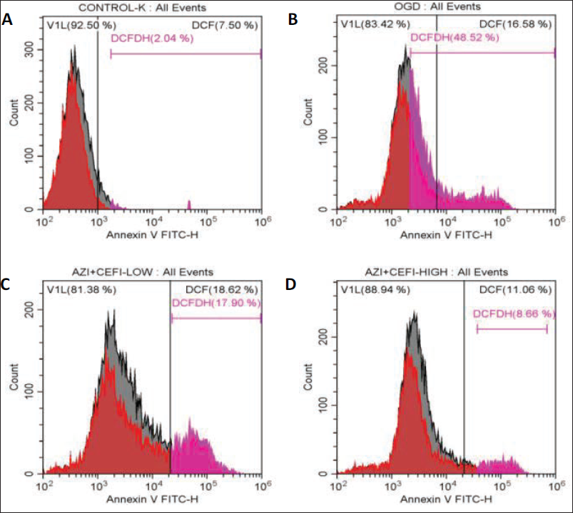
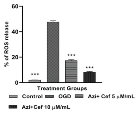
The ROS assay was performed in neuron-astrocyte co-culture with four different treatment groups, including control, OGD, Azi + Cef 5 µM/mL and Azi + Cef 10 µM/mL. The level of ROS in the control group was found to be 2.04%, the OGD group expressed a high amount of ROS content as compared with the rest of the groups, and it was found to be 48.52% and the treatment group (Figure 6), which received the novel combination of Azi + Cef 5 µM/mL after OGD induction, significantly inhibited the ROS levels against the OGD group; the level of ROS was found to be 17.90%. The high dose of Azi + Cef 10 µM/mL has shown lower ROS levels than the OGD and Azi + Cef 5 µM/mL treated groups. The Azi + Cef 10 µM/mL treated group remarkably reduced the ROS levels; the percentage of ROS levels in the Azi + Cef 10 µM/mL treated group (Figure 7) was found to be 8.66%. The novel combination-treated groups have exhibited good neuroprotection in dose dependent manner.
Plots of ROS Expression in Neuron-astrocyte Co-culture With Azilsartan (Azi) and Ceftriaxone (Cef) Treatment in Different Concentrations Using Flow Cytometry (DCFH-DA Assay), Plots of ROS Expression in Cells (A) Control Group, (B) OGD, (C) Azi + Cef Low Dose (5 µM/mL) and (D) Azi + Cef High Dose (10 µM/mL).
The Percentage of ROS Release in Different Treatment Groups, Control (p < .0001), OGD (p < .05), Azi + Cef 5 µM/mL (p < .0001) and Azi + Cef 10 µM/mL (p < .0001). Data Expressed as Mean ± SEM (n = 3) ***p < .0001, **p < .001, *p < .05 When Compared With OGD Group.
Apoptosis Assay
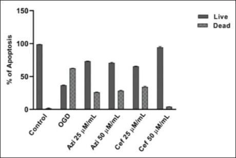
The apoptosis assay was performed with neuron-astrocyte co-culture in six different groups, such as control, OGD, Azi 25 µM/mL, Azi 50 µM/mL, Cef 25 µM/mL and Cef 50 µM/mL. The percentage of apoptosis in the control group was found to be 1% with 98.64% of viable cells (Figure 8). The OGD group has the highest number of apoptotic cells in comparison to the rest of the groups, and it was found to be 62.83% of apoptotic cells with 37.15% of viable cells. The treatment group, which received Azi 25 µM/mL after OGD induction, significantly inhibited cell apoptosis, surpassing the OGD group. The apoptotic and viable cells were found to be 26.09% and 73.54%. The high dose of Azi 50 µM/mL has shown lesser apoptosis than the OGD group; the level of apoptosis and viable cells were found to be 29.95% and 71.04%. The Cef 25 µM/mL treated group reduced the level of apoptosis 34.85% with 65.15% viable cells, and Cef 50 µM/mL potentially turned down the apoptosis in dose dependent manner, exhibited neuroprotective activity (Figure 9) and the percentage of apoptotic and viable cells of the Cef 50 µM/mL treated group were found to be 3.32% and 95.82%, respectively.
Quadrant Plots of Apoptosis Assay in Neuron-astrocyte Co-culture With Azilsartan (Azi) and Ceftriaxone (Cef) Treatment in Different Concentrations Using Flow Cytometry. Quadrant Plot of Apoptotic Cells in (A) Control Group, (B) OGD Group, (C) Azi 25 µM/mL Group, (D) Azi 50 µM/mL Group, (E) Cef 25 µM/mL Group and (F) Cef 50 µM/mL Group.
The Percentage of Live and Dead Cells in Different Treatment Groups, Control, OGD, Azi 25 µM/mL, Azi 50 µM/mL, Cef 25 µM/mL and Cef 50 µM/mL.
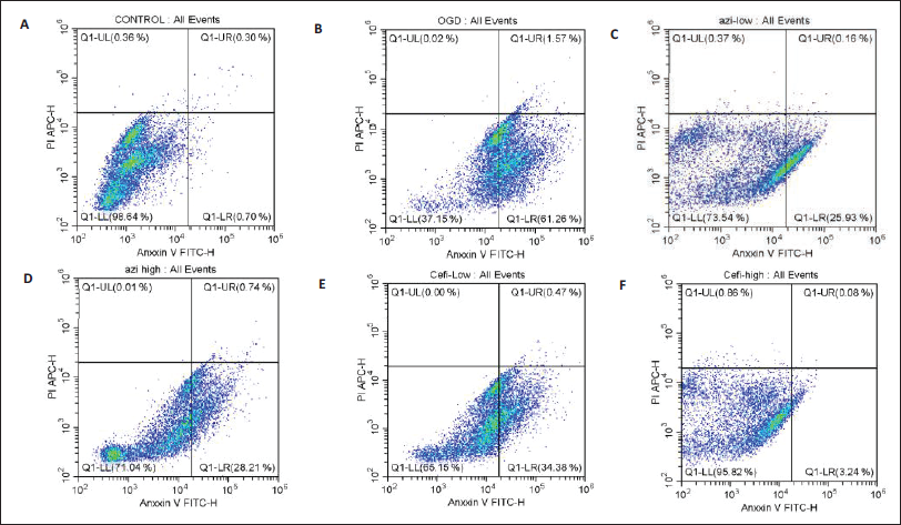
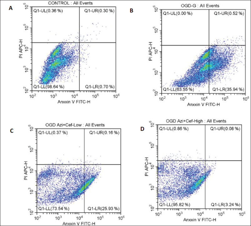
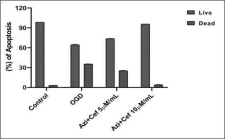
The apoptosis assay was performed in neuron-astrocyte co-culture with four different treatment groups, such as control, OGD, Azi + Cef 5 µM/mL and Azi + Cef 10 µM/mL. The percentage of apoptosis in the control group was found to be 1%, with 98.64% of viable cells. The OGD group has the highest number of apoptotic cells as compared with the rest of the groups, and it was found to be 36.46% of apoptotic cells with 63.55% of viable cells (Figure 10). The treatment group, which received the novel combination of Azi + Cef 5 µM/mL after OGD induction, significantly inhibited cell apoptosis, surpassing the OGD group. The apoptotic and viable cells were found to be 26.9% and 73.54%. The high dose of Azi + Cef 10 µM/mL has shown less apoptosis than the OGD and Azi + Cef5 µM/mL treatment group. The Azi + Cef 10 µM/mL treated group potentially reduced the apoptosis in dose dependent manner, exhibited good neuroprotection and the percentage (Figure 11) of apoptotic and viable cells for the Azi + Cef 10 µM treated group was found to be 3.32% and 95.82%, respectively.
Quadrant Plots of Apoptosis Assay in Neuron-astrocyte Co-culture With Azilsartan (Azi) and Ceftriaxone (Cef) Treatment in Different Concentrations Using Flow Cytometry. Quadrant Plot of Apoptotic Cells in (A) Control Group, (B) OGD Group, (C) Azi + Cef Low Dose (5 µM/mL) and (D) Azi + Cef High Dose (10 µM/mL).
The Percentage of Live and Dead Cells in Different Treatment Groups, Control, OGD, Azi + Cef 5 µM/mL and Azi + Cef 10 µM/mL.
AO/EB Fluorescent Staining
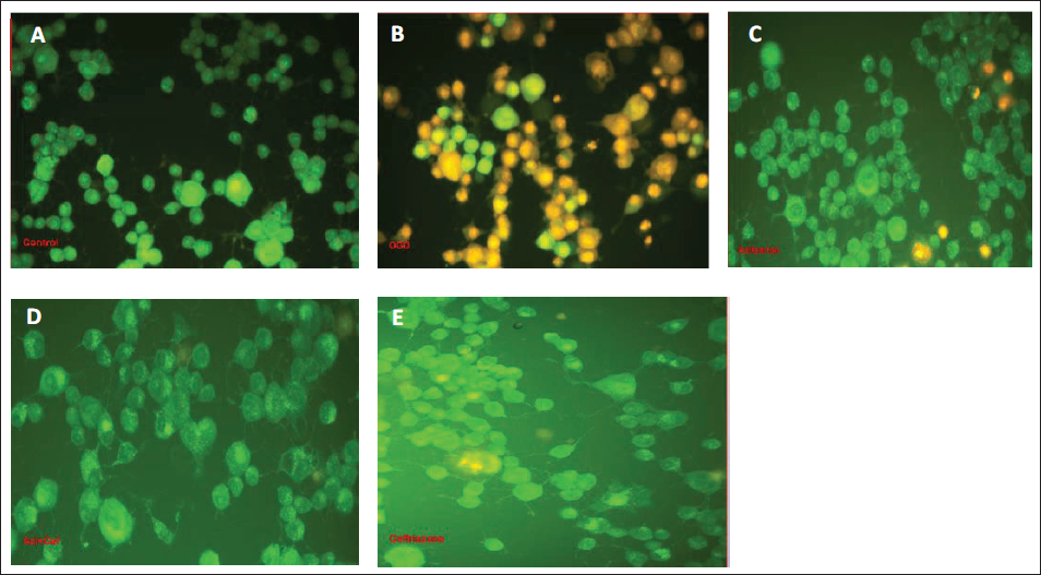
The necrotised and dead cells were found in the OGD group in comparison to the rest of the treatment groups, and the novel combination outstandingly decreased oxidative stress-mediated astrocyte and neuronal cell deterioration and exhibited good neuroprotection when compared to Azi and Cef alone-treated groups (Figure 12).
The AO/EB Staining of Neuron-astrocyte Co-culture After Respective Treatments (A) Control, (B) OGD, (C) Azilsartan, (D) Ceftriaxone, (E) the Novel Combination of Azilsartan + Ceftriaxone.
Oxidative Stress Parameters
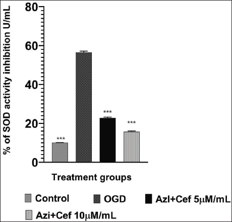
1. Superoxide dismutase
The antioxidant enzyme SOD activity was drastically inhibited in the OGD group when compared to the rest of the groups, such as control, Azi + Cef 5 µM/mL and Azi + Cef 10 µM/mL. The enzyme activity was inhibited in the OGD group due to oxidative stress and gradually increased (Figure 13) in the novel combination-treated groups, Azi + Cef 5 µM/mL, Azi + Cef 10 µM/mL, in dose dependent manner and exhibited neuroprotective potential.
The Percentage of SOD Activity Inhibition in Different Treatment Groups in Neuron-astrocyte Co-culture, Control (p < .0001), OGD (p < .05), Azi + Cef 5 µM/mL (p < .001), Azi + Cef 10 µM/mL (p < .0001). Data Expressed as Mean ± SEM (n = 3) ***p < .0001, **p < .001, *p < .05 When Compared to OGD Group.
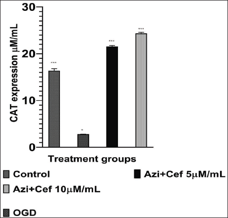
2. Catalase assay
The antioxidant enzyme CAT activity was significantly increased in novel combination-treated groups Azi + Cef 5 µM/mL, Azi + Cef 10 µM/mL, and it was diminished in the OGD group (Figure 14) due to oxidative stress in comparison to control and treatment groups. The novel combination exhibited a potential neuroprotective effect in dose dependent manner.
The Release of Catalase in Different Treatment Groups in Neuron-astrocyte Co-culture, Control (p < .0001), OGD (p < .05), Azi + Cef 5 µM (p < .0001), Azi + Cef 10 µM (p < .0001). Data Expressed as Mean ± SEM (n = 3) ***p < .0001, **p < .001, *p < .05 When Compared to OGD Group.
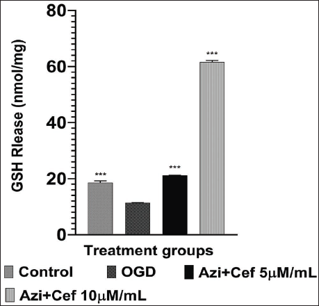
3. Reduced glutathione
An antioxidant enzyme, GSH release was significantly increased in novel combination-treated groups in comparison to the OGD group. Azi + Cef 5 µM/mL, Azi + Cef 10 µM/mL groups notably upregulated the levels of GSH when compared to control and OGD groups (Figure 15). The novel combination exhibited the neuroprotective effect in dose dependent manner.
The Percentage of GSH Release in Different Treatment Groups in Neuron-astrocyte Co-culture, Control (p < .0001), OGD (p < .05), Azi + Cef 5 µM/mL (p < .0001), Azi + Cef 10 µM/mL (p < .0001). Data Expressed as Mean ± SEM (n = 3) ***p < .0001, **p < .001, *p < .05 When Compared to OGD Group.
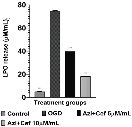
4. Lipid peroxidation
The catalytic enzyme lipid peroxidase concentration was inhibited in treatment groups in comparison to the OGD group, due to the oxidative stress OGD group has more amount of LPO (Figure 16) when compared to control and treatment groups. The novel combination-treated groups Azi + Cef 5 µM/mL, Azi + Cef 10 µM/mL have significantly decreased LPO in dose dependent manner and exhibited the neuroprotective effect.
The Release of Lipid Peroxidase in Different Treatment Groups in Neuron-astrocyte Co-culture, Control (p < .0001), OGD (p < .05), Azi + Cef 5 µM/mL (p < .0001), Azi + Cef 10 µM/mL (p < .0001). Data Expressed as Mean ± SEM (n = 3) ***p < .0001, **p < .001, *p < .05 When Compared to OGD Group.
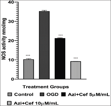
5. Nitric oxide synthase
The pro-inflammatory mediator NOS release was found to be high in the OGD group (Figure 17), and notably decreased in the novel combination-treated groups Azi + Cef 5 µM/mL, Azi + Cef 10 µM/mL. The novel combination exhibited the neuroprotective effect in dose dependent manner.
The Release of NOS in Different Treatment Groups in Neuron-astrocyte Co-culture, Control (p < .0001), OGD (p < .05), Azi + Cef 5 µM/mL (p < .001), Azi + Cef 10 µM /mL (p < .0001. Data Expressed as Mean ± SEM (n = 3) ***p < .0001, **p < .001, *p < .05 When Compared to OGD Group.
Estimation of Cytokines Intensity
1. Interleukin-6
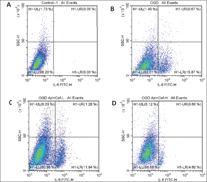
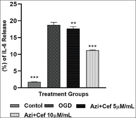
The IL-6 releasing assay was performed in neuron-astrocyte co-culture with four different treatment groups, such as control, OGD, Azi + Cef 5 µM/mL and Azi + Cef 10 µM/mL. The percentage of cytokine IL-6 in the control group was found to be 1.81%, the OGD group showed elevated IL-6 concentration in comparison to the control and the intensity of cytokines was found to be 19.15% (Figure 18). The treatment group which received the novel combination of Azi + Cef 5 µM/mL after OGD induction has shown 17.99%. The high dose of Azi + Cef 10 µM/mL has expressed less IL-6 than the OGD and Azi + Cef 5 µM/mL treatment groups. The Azi + Cef 10 µM/mL treated group potentially inhibited the IL-6 intensity 11.32% in dose dependent manner (Figure 19) and exhibited good neuroprotection through an anti-neuroinflammatory mechanism.
Quadrant Plots of IL-6 Intensity in Neuron-astrocyte Co-culture With Azilsartan (Azi) and Ceftriaxone (Cef) Treatment in Different Concentrations Using Flow Cytometry. Quadrant Plots of IL-6 Release in (A) Control Group, (B) OGD Group, (C) Azi + Cef Low Dose (5 µM/mL) and (D) Azi + Cef High Dose (10 µM/mL).
The Percentage of IL-6 Release in Different Treatment Groups, Control (p < .0001), OGD (p < .05), Azi + Cef 5 µM (p < .01) and Azi + Cef 10 µM (p < .0001). Data Expressed as Mean ± SEM (n = 3) ***p < .0001, **p < .001, *p < .05 When Compared to OGD Group.
2. Interleukin-1β
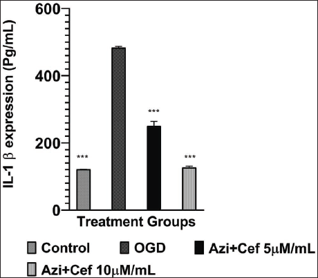
The intensity of IL-1β was significantly inhibited in both novel combination-treated groups, Azi + Cef 5 µM/mL, and Azi + Cef 10 µM, by manifesting the antioxidant and anti-neuroinflammatory mechanisms. The OGD group have shown a high level (Figure 20) of cytokine IL-1β in comparison to the rest of the groups. The novel combination Azi + Cef exhibited neuroprotection in dose dependent manner.
The Intensity of Cytokine IL-1β in Different Treatment Groups in Neuron-astrocyte Co-culture, Control (p < .0001), OGD (p < .05), Azi + Cef 5 µM/mL (p < .0001), Azi + Cef 10 µM/mL (p < .0001). Data Expressed as Mean ± SEM (n = 3) ***p < .0001, **p < .001, *p < .05 When Compared to OGD Group.
3. Tumor necrosis factor-alpha
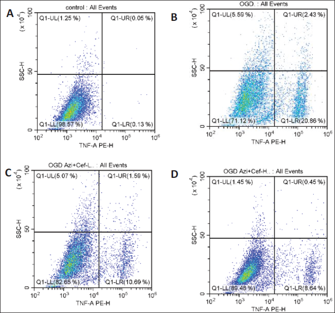
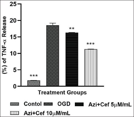
The TNF-α releasing assay was performed in neuron-astrocyte co-culture with four different treatment groups, such as control, OGD, Azi + Cef 5 µM/mL and Azi + Cef 10 µM/mL. The percentage of cytokine TNF-α in the control group was found to be 1.43%, the OGD group have shown highest TNF-α concentration (Figure 21) in comparison to the rest of the groups, and it was found to be 28.88%. The treatment groups which received the novel combination of Azi + Cef 5 µM/mL after OGD induction have shown 17.35%. The high dose of Azi + Cef 10 µM/mL has expressed less TNF-α than the OGD and Azi + Cef 5 µM/mL treatment groups. The Azi + Cef 10 µM/mL treated group significantly inhibited the TNF-α level (11.32%) in dose dependent manner (Figure 22) and exhibited good neuroprotection through anti-neuroinflammatory mechanisms.
The Quadrant Plots of TNF-α Release Assay in Neuron-astrocyte Co-culture With Azilsartan (Azi) and Ceftriaxone (Cef) Treatment in Different Concentrations Using Flow Cytometry. Quadrant Plot of TNF-α Release in (A) Control Group, (B) OGD Group, (C) Azi + Cef Low Dose (5 µM/mL) and (D) Azi + Cef High Dose (10 µM/mL).
The Percentage of TNF-α Release in Different Treatment Groups, Control (p < .0001), OGD (p < .05), Azi + Cef 5 µM/mL (p < .0001) and Azi + Cef 10 µM/mL (p < .0001). Data Expressed as Mean ± SEM (n = 3) ***p < .0001, **p < .001, *p < .05 When Compared to OGD Group.
RT-PCR
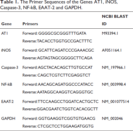
The Primer Sequences of the Genes AT1, iNOS, Caspase-3, NF-kB, EAAT-2 and GAPDH.
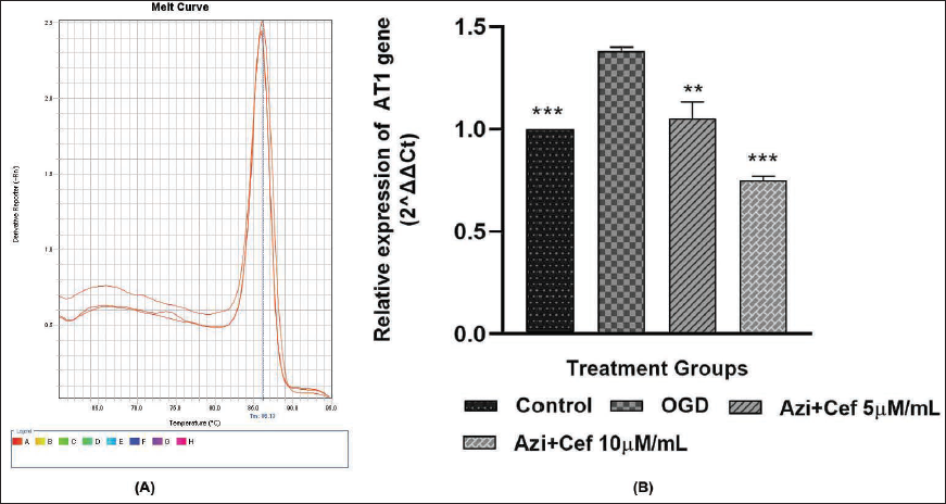
The relative gene expression of AT1 receptor was detected using the 2-∆∆CT method after OGD induction in neuron-astrocyte co-culture (Table 1). The level of gene expression was gradually decreased (Figure 23B) with the treatment of the novel combination Azi + Cef 5 µM/mL and Azi + Cef 10 µM/mL in comparison to the OGD group. The novel combination down-regulated the Ang-II/AT1 receptor gene expression.
(A) Melt Curves of AT1 Receptor Protein Corresponding Gene Expression Using 2-∆∆CT Method in OGD-induced Neuron-astrocyte Co-culture After the Treatment With the Novel Combination of Azilsartan and Ceftriaxone in Different Concentrations. (B) The Relative Gene Expression of AT1 Receptor in Neuron-astrocyte Co-culture With Treatment of the Novel Combination Azilsartan and Ceftriaxone, Different in Concentration Azi + Cef 5 µM/mL (p < .0001), Azi + Cef 10 µM/mL (p < .0001), OGD (p < .05) and Control (p < .001). Data Expressed as Mean ± SEM (n = 3) ***p < .0001, **p < .001, *p < .05 When Compared to OGD Group.
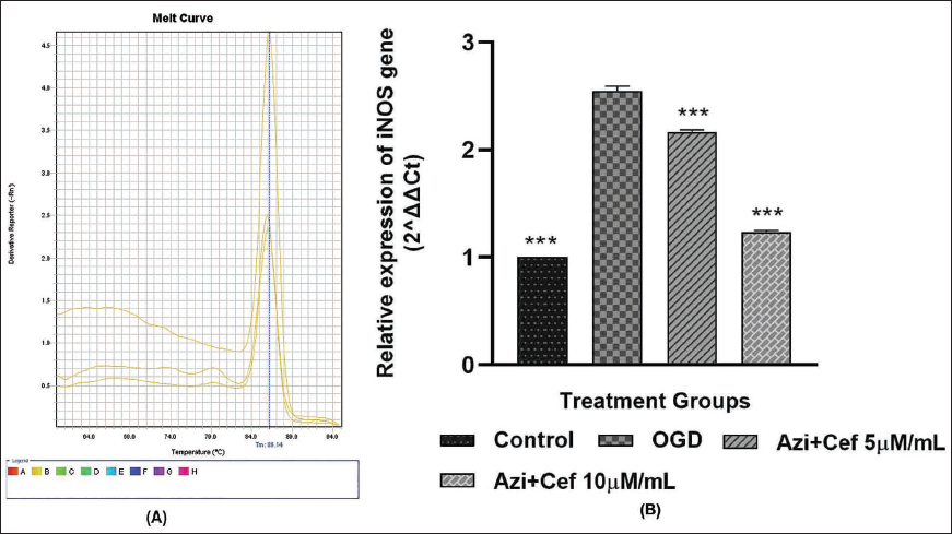
The catalytic enzyme inducible NOS (iNOS) gene expression has been found to be lower (Figure 24B) in novel combination-treated groups, Azi + Cef 5 µM/mL and Azi + Cef 10 µM/mL, as well as the control group, than the OGD group and OGD group have shown a high level of gene expression when compared to the rest of the groups.
(A) Melt Curves of the Catalytic Enzyme iNOS Corresponding Gene Expression Using the 2-∆∆CT Method in OGD-induced Neuron-astrocyte Co-culture After the Treatment With the Novel Combination of Azilsartan and Ceftriaxone in Different Concentrations. (B) The Relative Gene Expression of Catalytic Enzyme iNOS in Neuron-astrocyte Co-culture With the Treatment of Novel Combination Azilsartan and Ceftriaxone in Different Concentrations Azi + Cef 5 µM/mL (p < .0001), Azi + Cef 10 µM/mL (p < .0001), OGD (p < .05) and Control (p < .0001). Data Expressed as Mean ± SEM (n = 3) ***p < .0001, **p < .001, *p < .05 When Compared to OGD Group.
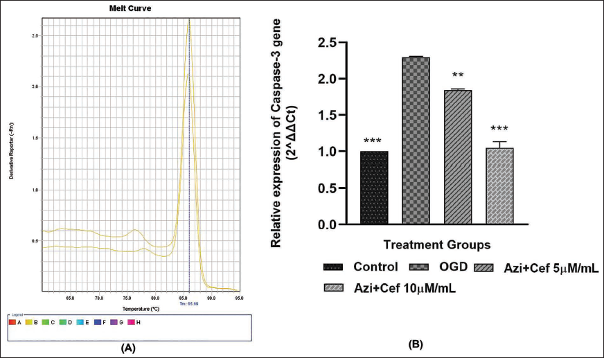
The apoptotic marker and the enzyme Caspase-3 have been down-regulated with the treatment of novel combination Azi + Cef 5 µM/mL and Azi + Cef 10 µM/mL. The level of gene expression was very high in the OGD group in comparison to the rest of the groups (Figure 25). Novel combination treatment turned down the apoptosis gene expression and exhibited neuroprotection.
(A) Melt Curves of the Enzyme Caspase-3 Corresponding Gene Expression Using the 2-∆∆CT Method in OGD-induced Neuron-astrocyte Co-culture After the Treatment With the Novel Combination of Azilsartan and Ceftriaxone in Different Concentrations. (B) The Relative Gene Expression of Enzyme Caspase-3 in Neuron-astrocyte Co-culture With the Treatment of the Novel Combination Azilsartan and Ceftriaxone in Different Concentrations Azi + Cef 5 µM/mL (p < .0001), Azi + Cef 10 µM/mL (p < .0001), OGD (p < .05) and Control (p < .0001). Data Expressed as Mean ± SEM (n = 3) ***p < .0001, **p < .001, *p < .05 When Compared to OGD Group.
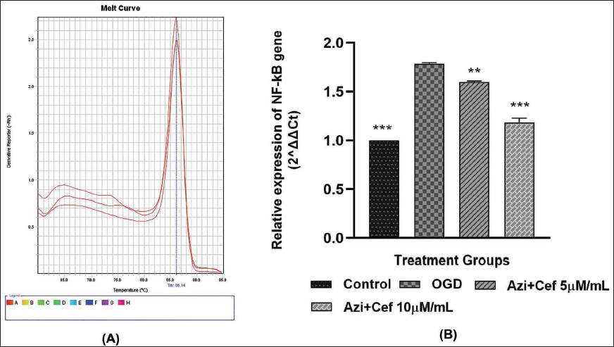
The nuclear transcription factor-kappa beta (NF-kB) gene expression has been down-regulated in the novel combination-treated groups Azi + Cef 5 µM/mL, Azi + Cef 10 µM/mL, as well as the control group, when compared to the OGD group. The novel combination has significantly decreased (Figure 26) the level of NF-kB gene expression and thereby regulated the transcription of pro-inflammatory mediators and cytokines.
(A) Melt Curves of the Nuclear Transcription Factor-kappa Beta (NF-kB) Corresponding Gene Expression Using the 2-∆∆CT Method in OGD-induced Neuron-astrocyte Co-culture After the Treatment With the Novel Combination of Azilsartan and Ceftriaxone in Different Concentrations. (B) The Relative Gene Expression of NF-kB in Neuron-astrocyte Co-culture With the Treatment of Novel Combination Azilsartan and Ceftriaxone in Different Concentrations Azi + Cef 5 µM/mL (p < .0001), Azi + Cef 10 µM/mL (p < .0001), OGD (p < .05) and Control (p < .0001). Data Expressed as Mean ± SEM (n = 3) ***p < .0001, **p < .001, *p < .05 When Compared to OGD Group.
EAAT-2
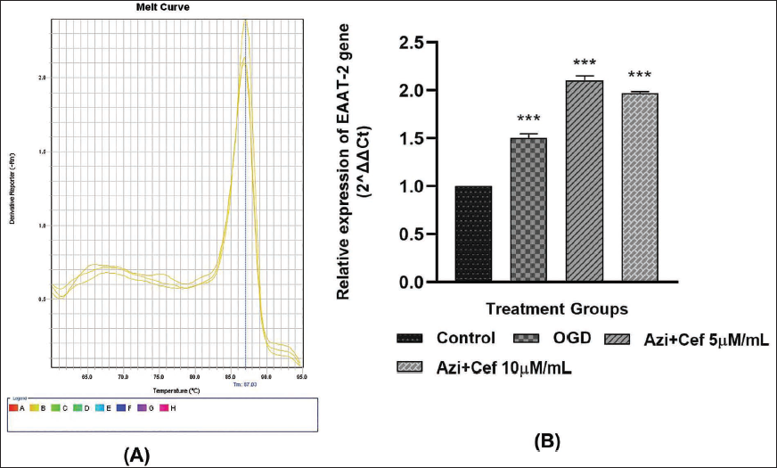
The EAAT-2 relative gene expression has been found to be slightly higher in the novel combination-treated groups Azi + Cef 5 µM/mL, Azi + Cef 10 µM/mL than in the control and OGD groups (Figure 27). The novel combination helps to protect the neurons by upregulating the expression of the EAAT-2 gene to transcribe and express the more number of glutamate transporter pumps during excitotoxicity conditions such as ischemic stroke.
(A) Melt Curves of the Excitatory Amino Acid Transporter-2 (EAAT-2) Corresponding Gene Expression Using the 2-∆∆CT Method in OGD-induced Neuron-astrocyte Co-culture After the Treatment With a Novel Combination of Azilsartan and Ceftriaxone in Different Concentrations. (B) The Relative Gene Expression of EAAT-2 in Neuron-astrocyte Co-culture With the Treatment of Novel Combination Azilsartan and Ceftriaxone in Different Concentrations Azi + Cef 5 µM/mL (p < .0001), Azi + Cef 10 µM/mL (p < .0001), OGD (p < .0001) and Control (p < .001). Data Expressed as Mean ± SEM (n = 3) ***p < .0001, **p < .001, *p < .05 When Compared to OGD Group.
In Vivo Neuroprotective Evaluation of Azi and Cef Novel Combination in the MCAo Rat Model
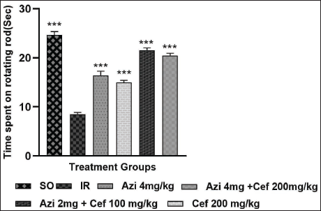
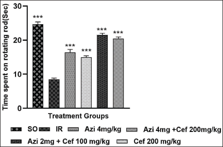
Rotarod
The muscle coordination was remarkably decreased in the IR group animals when compared to the rest of the groups. The treatment groups notably increased the time spent on the rotating rod (Figure 28) in Azi 4 mg/kg, Cef-200 mg/kg, Azi 2 mg + Cef 100 mg/kg, and Azi 4 mg + Cef 200 mg/kg. The novel combination Azi 2 mg + Cef 100 mg/kg treated group have shown better improvement and good muscle coordination among the all treatment groups which is indicating that the novel combination of neuroprotection and its synergistic effect.
The Assessment of Muscle Coordination in Ischemic Animals Using Rotarod, Data Are Expressed as Mean ± SEM (n = 6). p < .0001 When Compared to the IR Group. SO (p < .0001), IR (p < .05), Azilsartan 4 mg/kg (p < .0001), Ceftriaxone 200 mg/kg (p < .0001), Azi 2 mg + Cef 100 mg/kg (p < .0001) and Azi 4 mg + Cef 200 mg/kg (p < .0001).
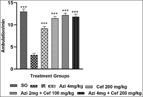
Actophotometre
The mobility of animals was drastically decreased in the IR group in comparison to the SO and treatment group animals. The locomotor activity was significantly increased in (Figure 29) Azi 4 mg/kg, Cef 200 mg/kg, Azi 2 mg/kg + Cef 100 mg/kg and Azi 4 mg/kg + Cef 200 mg/kg groups. The novel combination-treated group (Azi 2 mg/kg + Cef 100 mg/kg) have shown better improvement in locomotor activity and shown good neuroprotection.
The Assessment of Motor Coordination in Ischemic Animals Using an Actophotometre, Data Are Expressed as Mean ± SEM (n = 6). p < .0001 When Compared to the IR Group. SO (p < .0001), IR (p < .05), Azilsartan 4 mg/kg (p < .0001), Ceftriaxone 200 mg/kg (p < .0001), Azi 2 mg + Cef 100 mg/kg (p < .0001) and Azi 4 mg + Cef 200 mg/kg (p < .0001).
Superoxide Dismutase
(A and B). The Assessment of SOD Activity in the Ischemic Animal Hippocampus and Cortex, Data Are Expressed as Mean ± SEM (n = 6). p < .0001 When Compared to the IR Group. SO (p < .0001), IR (p < .05), Azilsartan 4 mg/kg (p < .008), Ceftriaxone 200 mg/kg (p < .0002), Azi 2 mg + Cef 100 mg/kg (p < .0001) and Azi 4 mg + Cef 200 mg/kg (p < .0001).
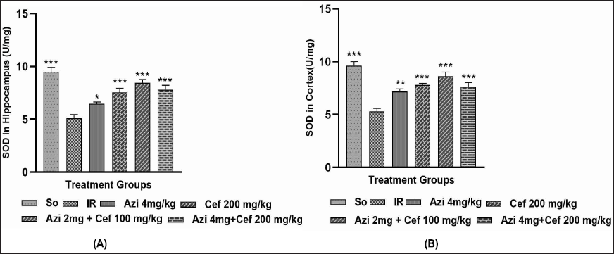
The antioxidant enzyme SOD levels were elevated in the treatment groups as compared to the ischemic reperfusion group animals. Ischemic reperfusion animals have shown low SOD levels in both hippocampus and cortex regions (Figures 30A and B) compared to the rest of the groups (Azi 4 mg/kg, Cef 200 mg/kg, Azi 2 mg + Cef 100 mg/kg, Azi 4 mg + Cef 200 mg/kg). The novel combination-treated groups have significantly upregulated the SOD levels. The Azi 2 mg + Cef 100 mg/kg group has exhibited a more potent neuroprotective effect than all treatment groups.
Catalase
(A and B). The Assessment of Catalase Activity in Ischemic Animal Hippocampus and Cortex, Data Are Expressed as Mean ± SEM (n = 6). p < .0001 When Compared to the IR Group. SO (p < .0001), IR (p < .05), Azilsartan 4 mg/kg (p < .03), Ceftriaxone 200 mg/kg (p < .0001), Azi 2 mg + Cef 100 mg/kg (p < .0001) and Azi 4 mg + Cef 200 mg/kg (p = .0004).
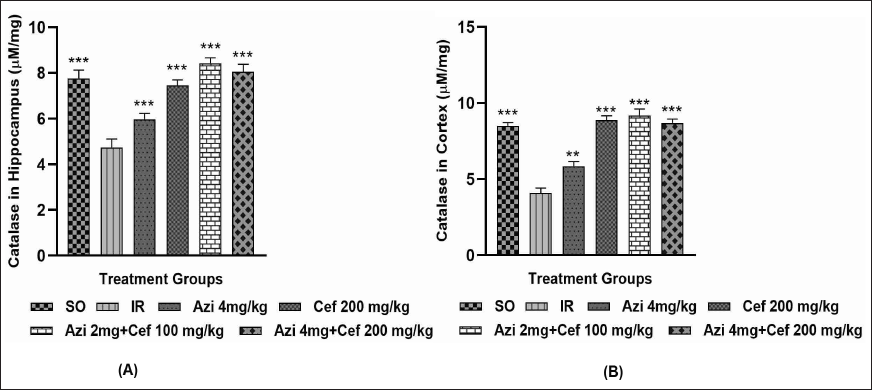
The antioxidant and hydrogen peroxide scavenging enzyme CAT levels were elevated in the treatment groups as compared to the IR group (Figures 31A and B). The ischemic reperfusion animals have shown lower CAT levels in both the hippocampus and cortex regions than the rest of the treatment groups. the treatment groups such as Azi 4 mg/kg, Cef 200 mg/kg, Azi 2 mg + Cef 100 mg/kg, Azi 4 mg + Cef 200 mg/kg treated groups have shown significant upregulation in the CAT activity, and especially Azi 2 mg + Cef 100 mg/kg treated group CAT levels was notably increased in cortex region in comparison to hippocampus, also exhibited potential neuroprotective effect than all treatment groups.
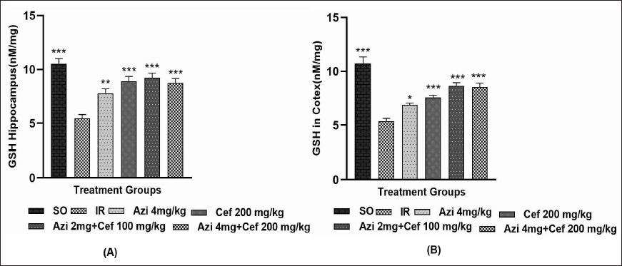
Reduced Glutathione
An intracellular antioxidant enzyme, GSH concentration was notably increased in the treatment groups when compared to the ischemic reperfusion group. The GSH levels were decreased (Figures 32A and B) in both hippocampus and cortex regions of the IR animals in comparison to the rest of the treatment groups (SO, Azi 4 mg/kg, Cef 200 mg/kg, Azi 2 mg + Cef 100 mg/kg, Azi 4 mg + Cef 200 mg/kg). The novel combination-treated groups have significantly upregulated the GSH levels. Azi 2 mg + Cef 100 mg/kg groups have exhibited a more potent neuroprotective effect than all treatment groups.
(A and B). The Assessment of GSH Activity in Ischemic Animal Hippocampus and Cortex, Data Are Expressed as Mean ± SEM (n = 6). p < .0001 When Compared to IR Group. SO (p < .0001), IR (p < .05), Azilsartan 4 mg/kg (p < .003), Ceftriaxone 200 mg/kg (p < .0004), Azi 2 mg + Cef 100 mg/kg (p < .0001) and Azi 4 mg + Cef 200 mg/kg (p < .0001).
Lipid Peroxidation
(A and B). The Assessment of LPO Activity in the Ischemic Animal Hippocampus and Cortex, Data Are Expressed as Mean ± SEM (n = 6). p < .0001 When Compared to the IR Group. SO (p < .0001), IR (p < .05), Azilsartan 4 mg/kg (p < .004), Ceftriaxone 200 mg/kg (p < .0001), Azi 2 mg + Cef 100 mg/kg (p < .0001) and Azi 4 mg + Cef 200 mg/kg (p < .0001).
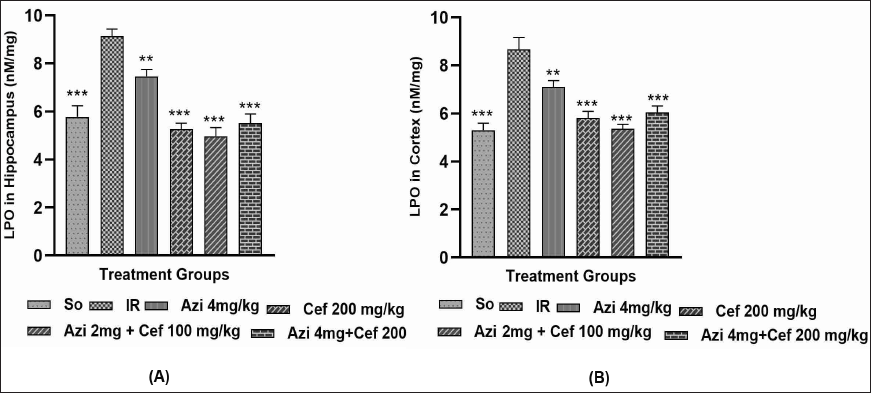
The catalytic enzyme lipid peroxidase concentration was inhibited in treatment groups as compared to the ischemic reperfusion group animals. Ischemic reperfusion animals have sown more amount of LPO levels (Figures 33A and B) in both hippocampus and cortex regions than the rest of the treatment groups (Azi 4 mg/kg, Cef 200 mg/kg, Azi 2 mg + Cef 100 mg/kg, Azi 4 mg + Cef 200 mg/kg) due to elevated oxidative stress. The novel combination-treated groups have significantly down-regulated the LPO levels, and Azi 2 mg + Cef 100 mg/kg groups have exhibited a more potent neuroprotective effect than all treatment groups.
Glutamate Assay
(A and B). The Estimation of Glutamate Concentration in Ischemic Animals With Respect to the Different Treatments, Data Are Expressed as Mean ± SEM (n = 6). p < .0001 When Compared to the IR Group. SO (p < .0001), IR (p < .05), Azilsartan 4 mg/kg (p < .008), Ceftriaxone 200 mg/kg (p < .0002), Azi 2 mg + Cef 100 mg/kg (p < .0001) and Azi 4 mg + Cef 200 mg/kg (p < .0001).
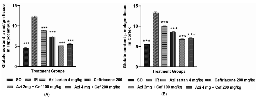
The excitatory amino acid glutamate concentration was very high in ischemic reperfusion animals when compared to the rest of the treatment groups (Figures 34A and B), especially the novel combination-treated groups, which notably turned down the glutamate storm and prevented the excitotoxicity-mediated neurodegeneration and proved the neuroprotective potential of the novel combination Azi and Cef.
Intensity of Cytokines
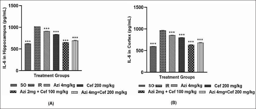
1. Interleukin-6
The intensity of the cytokine IL-6 was significantly inhibited in the treatment groups in comparison to the ischemic reperfusion group. The treatment groups have shown low levels (Figures 35A and B) of IL-6: Azilsartan 4 mg/kg, Ceftriaxone 200 mg/kg, Azi 2 mg + Cef 100 mg/kg and Azi 4 mg/kg + Cef 200 mg/kg. The novel combination-treated groups exhibited a potential neuroprotective effect and also showed a synergistic effect.
(A and B). The Intensity of IL-16 in the Ischemic Animal Hippocampus and Cortex Data Are Expressed as Mean ± SEM (n = 6). p < .0001 When Compared to the IR Group. SO (p < .0001), IR (p < .05), Azilsartan 4 mg/kg (p < .001), Ceftriaxone 200 mg/kg (p < .0001), Azi 2 mg + Cef 100 mg/kg (p < .0001) and Azi 4 mg + Cef 200 mg/kg (p < .0001).
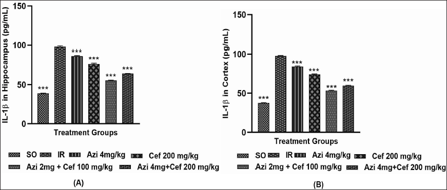
2. Interleukin-1β
The intensity of IL-1β was significantly inhibited in the treatment groups (Figure 36A and B) in comparison to the ischemic reperfusion group. The treatment groups have shown low levels of IL-1β (Azi 4 mg/kg, Cef 200 mg/kg, Azi 2 mg + Cef 100 mg/kg and Azi 4 mg + Cef 200 mg/kg). The novel combination-treated groups exhibited a more potent neuroprotective effect than the rest of the treatment groups.
(A and B). The Intensity of IL-1β in Ischemic Animals, Data Are Expressed as Mean ± SEM (n = 6). p < .0001 When Compared to IR Group. SO (p < .0001), IR (p < .05), Azilsartan 4 mg/kg (p < .001), Ceftriaxone 200 mg/kg (p < .0001), Azi 2 mg + Cef 100 mg/kg (p < .0001) and Azi 4 mg + Cef 200 mg/kg (p < .0001).
Histopathology
1. 2,3,5-triphenyltetrazolium Chloride Staining
The ischemic and infarcted area of MCAo-induced animals was assessed using TTC stain; the IR group, animal hemispheres, especially the cerebral cortex and hippocampus regions, were affected mostly (Figure 37). The treatment groups, such as Azi 4 mg/kg, Cef 200 mg/kg, reduced the infarction of neuronal cells, and the novel combination-treated groups have shown better recovery (Figure 38), significantly reduced the neuronal loss and exhibited good neuroprotective potential.
The Histopathological Observations of Ischemic Rats in Different Treatments With TTC Staining (A) SO, (B) IR, (C) Azilsartan 4 mg/kg, (D) Ceftriaxone 200 mg/kg, (E) Azi 2 mg + Cef 100 mg/kg, (F) Azi 4 mg + Cef 200 mg/kg.
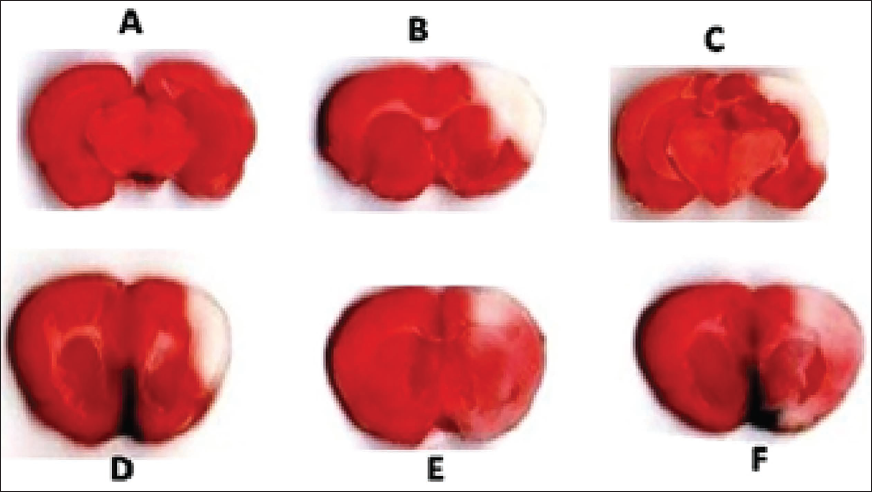
The Histopathological Observations Using TTC Stain in Ischemic Rats With Different Treatments, Data Are Expressed as Mean ± SEM (n = 6). p < .0001 When Compared to IR Group. SO (p < .0001), IR (p < .05), Azilsartan 4 mg/kg (p < .0001), Ceftriaxone 200 mg/kg (p < .0001), Azi 2 mg + Cef 100 mg/kg (p < .0001) and Azi 4 mg + Cef 200 mg/kg (p < .0001).
Cresyl Violet Staining
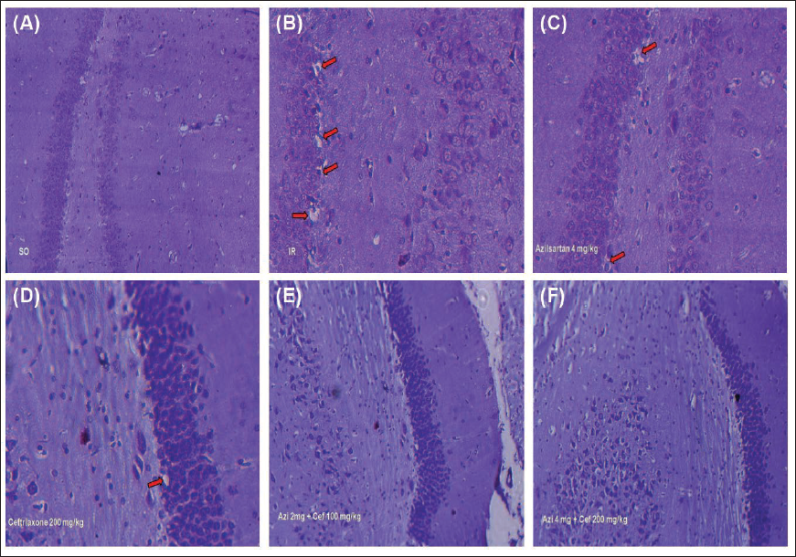
The microscopic examination of the hippocampus CA1 region was observed by staining with cresyl violet. The ischemic reperfusion group have shown various morphological changes (Figure 39) like multifocal moderate neurodegeneration with pyknotic nuclei, when compared to the rest of the treatment groups. The novel combination-treated groups significantly reduced the neuronal loss and exhibited good neuroprotective potential.
The Histopathological Observations of Ischemic Rats With Different Treatments in CA1 Region of the Hippocampus With Cresyl Violet Staining (A) Sham Operated (SO), (B) Ischemic Reperfusion (IR), (C) Azilsartan 4 mg/kg, (D) Ceftriaxone 200 mg/kg, (E) Azi 2 mg + Cef 100 mg/kg, (F) Azi 4 mg + Cef 200 mg/kg.
Haematoxylin and Eosin Staining
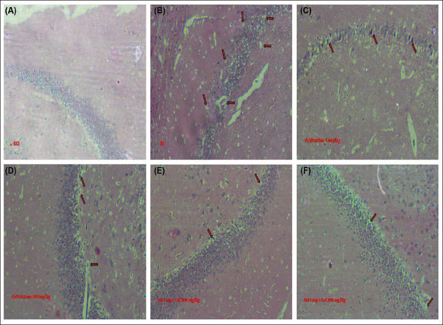
The microscopic examination of the hippocampus CA1 region was observed by staining with haematoxylin and eosin. The ischemic reperfusion group have shown various morphological changes like multifocal neurodegenerative lesions with pyknotic nuclei (Figure 40), when compared to rest of the treatment groups. The novel combination-treated groups notably reduced the neuronal loss and exhibited good neuroprotective potential.
The Histopathological Observations of Ischemic Rats With Different Treatments in CA1 Region of the Hippocampus With Haematoxylin and Eosin Staining (A) Sham Operated (SO), (B) Ischemic Reperfusion (IR), (C) Azilsartan 4 mg/kg, (D) Ceftriaxone 200 mg/kg, (E) Azi 2 mg + Cef 100 mg/kg, (F) Azi 4 mg + Cef 200 mg/kg.
Discussion
Cerebral ischemia is a leading cause of morbidity and mortality in the world; more than 80% of stroke incidences are ischemic type of strokes only. Sedentary lifestyle and preexisting diseases such as diabetes, hypertension, and hyperlipidaemia increase the risk of cerebral ischemia. At present, recombinant tPA’s are being used for the treatment of acute ischemic stroke. The efficacy of tPAs is very limited, and most of the time it depends on the administration of the drug within the onset of stroke symptoms development; generally, these drugs have to be administered within 3–4.5 hours. Although the administration of tPA could recanalise the cerebral arterial circulation, it cannot interfere with the neuroinflammatory mechanisms which will be initiated by the oxidative stress, excitotoxicity, cytokine storm and insufficiency of energy metabolites. Apart from that, treatment with anticoagulants and antiplatelet agents is not widely used due to their high incidence of secondary stroke and haemorrhage. As of now, osmotic diuretics such as Mannitol are being used for the treatment of acute ischemic stroke and cerebral oedema. However, the benefits of the osmotic diuretics in cerebral ischemia are still elucidative. In order to fulfil the lacuna of existing treatments, we designed a novel combination for cerebral ischemia, which will minimise the post-ischemic complications and act as an adjuvant therapy.
Ang-II plays a critical role in neuroinflammation, especially through AT1 receptor; activation of AT1 receptor during the ischemic condition initiates the inflammatory cascade, especially through the receptors that are situated on the neurons and microglia. The microglial cells will be activated by the Ang-II/AT1 axis, which further leads to activation of NF-kB, thereby causing release of inflammatory cytokines like IL-6, IL-1β and TNF-α. Earlier studies suggested that the Ang-II overexpression during ischemic conditions augmented the neuronal loss in an in vivo ischemic mouse model, and another research stated that overexpression of ACE-II ameliorates the neuronal deterioration by converting the Ang-II into its subforms, as well as declining the AT1 receptor activation.7, 8 Regulation of AT1 receptor activation using ARBs exhibited neuroprotective effects mediated through downregulation of inflammatory cytokine TNF-α and MCP-1 levels in the MCAo rat model.29, 30 The AT1 receptor blockers are already being used as anti-hypertensive agents in clinical practice. Interestingly, in earlier reports, telmisartan, valsartan, irbesartan, olmesartan, candesartan, and Azilsartan were found to have neuroprotection through their anti-inflammatory and antioxidant mechanisms, suggesting that the blockade of central AT1 receptors decreased the severity of cerebral ischemic injury in a murine model of transient focal ischemia. 14 Cef has been screened as a potential neuroprotective clinical agent that acts as a transcriptional activator for EAAT-2 expression on astroglial cells via the NF-kB signalling pathway, 17 and it significantly protected neurons against global ischemia via upregulating EAAT-2/GLT-1 expression in in vivo animal models, as upregulation of EAAT-2 expression increases glutamate clearance. Thus, it regulates the concentration of glutamate below the excitotoxic level, 18 these research findings were considered as substantial evidence for the current research.
In the present study, the designed novel combination was evaluated for the neuroprotective effect in an in vitro cell line and in vivo MCAo animal model. In vitro neuron-astrocyte co-culture was developed and kept for OGD for 24 hours to mimic the ischemic reperfusion injury. Treatment with Azi, Cef and both combination was significantly shown neuroprotective effect in comparison to the OGD group in terms of downregulating the ROS levels, apoptosis, and turning down the cytokine storm in dose dependent manner. The combination has also exhibited a synergistic effect, especially EC50 concentrations of the combination found to be five folds lesser than the individual dosing. The ROS levels were found to be low when compared to individual dosing of Azi 25 µM/mL, 50 µM/mL, Cef 25 µM/mL, 50 µM/mL, and the novel combination improved the oxidative stress-mediated excitotoxicity and microglial activation by exhibiting the antioxidant effect. The Azi + Cef 10 µM/mL treated group have remarkably prevented the neurodegeneration more than other treatment groups, as well as the Azi + Cef 5 µM/mL treatment group. The treatment groups significantly decreased the apoptosis of neuron-astrocytes and also enhanced the levels of antioxidant enzymes such as SOD, CAT and GSH; oxidative enzymes like NOS, LPO were diminished in novel combination-treated groups. The cytokine-mediated abruption of glutamate clearance and neuroinflammation was found to be lower in treatment groups than in the OGD group; the Azi + Cef combination significantly protected the neurons by exhibiting the anti-inflammatory mechanisms, and these results were comparable with earlier research findings. Gene expression studies were carried out to elucidate the intra molecular level changes which were happened with particular treatment groups. Azi and Cef combination alleviated the inflammatory gene expressions in the brain, iNOS, AT1, NF-kB, Caspase-3 and also EAAT-2/GLT-1 gene expression was found to be increased with the novel combination treatment. Upregulation of EAAT-2 expression through Cef is the greatest advantage to prevent the excitotoxicity-mediated neuroinflammation in ischemia.
The MCAo-induced ischemic rats were treated with a designed treatment regimen after the ischemic insult, where all animals received three days of consecutive treatment daily. The behavioural assessments were done using rotarod and actophotometre; mobility of the animal and motor coordination were notably increased in novel combination-treated animals than in the rest of the treatment groups. Oxidative stress parameters such as LPO, SOD, CAT and GSH were remarkably improved, and also excitatory amino acid glutamate concentration was found to be normal. The levels of cytokines IL-6 and IL-1β were significantly decreased in the treated groups compared to the control. The ischemic penumbra region was notably recovered in Azi 4 mg/kg, Cef 200 mg/kg, Azi 2 mg + Cef 100 mg/kg, and Azi 4 mg + Cef 200 mg/kg treatment groups. The Azi 2 mg + Cef 100 mg/kg combination-treated group animals exhibited an excellent recovery compared to the rest of the treatment groups. TTC, Cresyl violet, and Haematoxylin & Eosin staining histopathological observations strongly evidenced the neuroprotective potency of the Azi and Cef combination.
Azi attenuated the oxidative stress and malfunctioning of mitochondria, thereby alleviating the risk of excitotoxicity and cytokine storms by inhibiting the Ang-II/AT1 receptor axis; and also exhibited neuroprotection through antioxidant and anti-neuroinflammatory mechanisms. Generally, AT1 blockers can diminish oxidative stress and downregulate the production of cytokines that will be released from activated microglia, but they cannot influence EAAT-2 pumps for the rapid clearance of overly secreted glutamate during ischemic conditions. Cef has significant potential for glutamate clearance by enhancing EAAT-2 expression on the astrocyte plasma membrane through the NF-kB signalling pathway. Unfortunately, Cef does not have any role in the regulation of glutamate secretion from the presynaptic cleft region. Both Azi and Cef exhibited good neuroprotective activity in in vitro and in vivo ischemic models by acting on different targets through different mechanisms.
The drawbacks of Azi were compensated by Cef, and the drawback of Cef was compensated by Azi. The novel combination exhibited two different mechanisms on different targets. Azi arrested the neuroinflammatory pathway in activated microglial cells, which mediates through AT1 receptor activation and inhibits cytokine storm-induced EAAT-2 dysfunction. Cef enhanced the EAAT-2 expression through gene transcription in astrocytes via the NF-kB signalling pathway and helped to lessen excitotoxicity through the rapid clearance of glutamate through EAAT-2 pumps. Hence, targeting central AT1 receptors with its antagonist Azi and EAAT-2 transcriptional and expressional enhancement with Cef potentiated the neuroprotection in combination with individual dosing.
Conclusion
The repurposing of anti-hypertensive, AT1 receptor blocker Azi and β-lactam antibiotic Cef novel combination, demonstrated neuroprotective potential, mediated through antioxidant and anti-inflammatory mechanisms in in vitro OGD-induced astrocyte-neuron co-culture as well as MCAo rat model. Excitotoxicity-mediated neuroinflammation was significantly down-regulated in both in vitro and in vivo models by the novel combination of Azi and Cef. However, Preclinical data will not be enough to bring the resolution to repurposing the existing clinical candidate, like Cef and Azi, for use in medical conditions like acute ischemic stroke and other neurodegenerative disorders. The pilot study has to be initiated to rule out the safety and efficacy of the designed combination in a systematic approach.
Footnotes
Authors’ Contribution
Gaddam Narasimha Rao and Antony Justin have developed the concept, have done a literature survey, and written the manuscript; Srikanth Jupudi, Antony Justin, and Devarakonda Krishna Prasad reviewed and revised for necessary changes in the manuscript; Jeyaram Bharathi J has supported in carrying out the cell culture and animal studies; All authors contributed equally to the manuscript preparation and revision.
Availability of Data and Materials
The datasets generated and analysed during the current study are not publicly available but are available from the corresponding author on reasonable request.
Consent to Participate
The human subjects were not included in the current research.
Declaration of Conflict of Interests
The authors declared no potential conflicts of interest with respect to the research, authorship and/or publication of this article.
Funding
The authors disclosed receipt of the following financial support for the research, authorship and/or publication of this article: The authors acknowledge the JSS Academy of Higher Education & Research, Mysuru, India (JSSAHER/REG/RES/URG/54/2011-12/10419) for financial assistance to this work. The authors also thank the DST-FIST, New Delhi, India, for the support towards infrastructure development of the Department of Pharmacology, JSS College of Pharmacy, Ooty, Tamil Nadu, India.
Statement of Ethics
This study was performed in accordance with the principles of the Guide for the Care and Use of Laboratory Animals. Approval was granted by the Institutional Animal Ethical Committee, Anurag University, Hyderabad, on 15 June 2023 (IAEC No. I/IAEC/AU/016/2023 WR♂.