
Abstract
Background:
Parkinson’s disease is a neurodegenerative disorder and is marked by inflammation and death of neurons in the striatum region of the midbrain. It has been reported that expression of NF-κB increases during Parkinson’s disease, which promotes oxidative stress, stimulates release of proinflammatory cytokines, and induces expression of nitric oxide. Therefore, in this study, we have used mangiferin a specific NF-κB inhibitor. Mangiferin is a polyphenolic compound traditionally used for its antioxidant and anti-inflammatory properties.
Methods:
The study utilized male Wistar rats weighing 200–250 g (56 rats; n = 8/group). On day “0,” stereotaxic surgery of rats was done to induce 6-hydroxydopamine lesioning in rats. Coordinates for substantia nigra were anteroposterior-2 mm, mediolateral-5 mm and dorsoventral-8.2 mm. After 14 days, those rats which show at least 210 contralateral rotations after administration of apomorphine (0.5 mg/kg S.C.) were selected for the study and were given treatment for 28 days. On day 28 of treatment, rats were subjected to behavioral studies to evaluate the effect of mangiferin and their brains were taken out after euthanasia to perform biochemical, molecular and immunological studies.
Results:
Treatment with mangiferin significantly improves the key parameters of locomotor activity and oxidative stress and reduces the parameters of inflammatory stress. Also, the activity of caspases was reduced. Significant decrease in activity of both cyclooxygenase 1 and 2 was also observed. Maximum improvement in all parameters was observed in rats treated with grouping of mangiferin 45 µg/kg and levodopa 10 mg/kg. Treatment with levodopa alone has no significant effect on biochemical and molecular parameters though it significantly improves behavioral parameters.
Conclusion:
Current treatment of Parkinson’s disease does not target progression of Parkinson’s disease. Results of this study suggest that mangiferin has protective effect in hemi-Parkinsonian rats. Therefore, the combination therapy of mangiferin and levodopa can be helpful in management of Parkinson’s disease.
Graphical Abstract
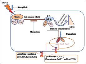
Mangiferin prevents inflammation by inhibiting tumor necrosis factor-alpha (TNF-α) mediated nuclear translocation and activation of NF-κB, which is required for the activation of cyclooxygenase and toll-like receptors. Mangiferin also prevents phosphorylation of NF-κB by promoting its proteasomal degradation, which further decreases the secretion of interleukins (IL-1 and IL-12) and chemokines, such as monocyte chemoattractant protein-1 (MCP-1), and regulated upon activation, normal T cell expressed and presumably secreted. Mangiferin further decreases the activation of caspases by inhibiting TNF-α, NF-κB, and mitogen-activated protein kinase pathway.
Introduction
Parkinson’s disease (PD) is a progressive locomotor disorder characterized by death of neurons in the nigrostriatal area of basal ganglia. Bradykinesia, muscular rigidity, rolling tremors, postural abnormalities, and gait issues are some of the clinical features of PD. 1–4
Although PD is an idiopathic disorder with its etiopathology not fully known, after decades of research, researchers believe that dysregulation of transcription factors controlling inflammation is the key reason behind the advancement of PD and PD like symptoms. Various studies have suggested that the expression of transcription factor NF-κB increases during inflammation. Increased expression of transcription factor NF-κB then results in downstream activation of toll-like receptors (TLRs) and Interleukin 1 receptors (IL1R), which then triggers myeloid differentiation factor 88 (Myd88) gene to recruit Interleukin-1 receptor activated kinase 1 (IRAK1) to this receptor signaling complex for phosphorylation. After phosphorylation, IRAK1 form a complex with tumor necrosis factor receptor-associated factor 6 (TRAF6). The IRAK-1 TRAF-6 complex thus formed then activates NF-κB signaling pathway through transforming growth factor-β-activated kinase 1 (TAK1)/TAB1/TAB2, NF-κB essential modulator (NEMO)/IKKβ/IKKα, and IκB/p50/p65 complexes,5–9 which then subsequently leads to gene induction of proinflammatory cytokines by NF-κB. Thus, increased expression of NF-κB mediated proinflammatory cytokines, such as Interleukin-1 (IL-1) and tumor necrosis factor-alpha (TNF-α) and transcription factors like TLR-4 then result in microglial activation and further aggravate the inflammatory response by increasing the expression of inducible nitric oxide synthase in glial cell along with increased cytokine production. Furthermore, activation of the microglia also increases reactive oxygen species (ROS) production because of NADPH oxidase induction and causes oxidative stress and free radical-induced cell injury. 10–17 All these factors then further increase the expression of NF-κB, which then regulates apoptosis through the nuclear buildup of RelA. Nuclear translocation of RelA increases the membrane permeability of mitochondria by chaperon-mediated activation of Bax onto the outer membrane of mitochondria and inhibiting the antiapoptotic protein B-cell lymphoma (Bcl-XL). 18 Increased ROS production causes activation of p38/mitogen-activated protein kinase (MAPK) pathway. Activation of p38/MAPK then triggers NF-κB activation, which then activates p53 gene. 19 This ROS-mediated activation of p38/MAPK then induces the expression of caspase-3 and caspase-9. 20–32
In this study, we have used a 6-OHDA model of PD. 6-OHDA is a neurotoxin which is unable to cross blood brain barrier and therefore is administered stereotaxically direct into the nigral region of brain. 6-OHDA exerts its neurotoxic effects through upregulation of oxidative stress pathways such as unfolded protein response and increases mitochondrial permeability that triggers release of cytochrome c and activates caspases. 6-OHDA also up regulates the expression of p38MAPK and p53-upregulated mediator of apoptosis.20–23,33
In recent years, research interest in natural compounds has been re-verified owning to their slighter toxic effect as compared to chemical compounds. In our study, we have used polyphenolic compound mangiferin obtained from plants belonging to Anacardiaceae and Gentianaceae family. In our previous study, we have shown that mangiferin has protective effect in rheumatoid arthritis and it significantly restores complete Freund’s adjuvant (CFA)-induced changes in arthritic parameters of rats. 34
Mangiferin subdues the inflammatory reaction primarily by interfering with NF-κB activation, which further aggravates ferocious cycle of inflammation by activating cytokines, such as TNF-α, IL-1, and IL-4, and signaling pathways, such as signal transducer and activator of transcription proteins and TLRs. Mangiferin first and foremost impedes NF-κB activation as it bind with NEMO, IKK-α, and IIK-β complex, which forestall its auxillary phosphorylation and consequent degradation and translocation of this complex into nucleus.35–38 Mangiferin also has diminutive effects on (a) TRAF6, (b) TNF-receptor-associated death domain and the receptor interacting protein, and (c) nitric oxide producing property of macrophages and phagocytes.39, 40
Thus, owing to its anti-inflammatory and antioxidant effect, mangiferin could be useful in pharmacotherapy of PD to rescue the neurons from cell death pathways.
Methods
Animals
Male Wistar rats (200–250 g) were used in the study. Rats (56 rats; n = 8/group) were kept in the Institutional Animal Facility of the University under 12-h light/dark cycles with free access to food and water. Before starting the study, necessary approval from the Institutional Animal Ethics Committee (IAEC) of King George’s Medical University (KGMU), Lucknow, India, was obtained with approval letter No. 84/IAEC/Pharma/2017.
Induction of PD in Rats
6-OHDA lesions were performed in rats using stereotactic frame. Rats were anesthetized with ketamine–xylazine (ketamine 60 mg/kg and xylazine 7.5 mg/kg) cocktail before placing them into stereotactic chamber. Top of rats’ heads was shaved and cleaned through 70% ethanol. Stereotactic coordinates for substantia nigra region was then determined using Paxinos and Watson, The Rat Brain in Coordinates. 41 For substantia nigra pars compacta (dorsal part) lesion coordinates in reference to bregma were anteroposterior (A/P-2 mm, mediolateral (M/L)-5 mm, and dorsoventral-8.2 mm. A midline incision was done and lambda and bregma were identified at the intersection of coronal and saggital sutures. A burr hole was then drilled into rat brain using these coordinates. After hole was drilled, rats were cannulated with a 20 gauge cannula. Length of the cannula was kept at 8 mm. After implanting the cannula, it was then fixed using denture material.
5 µg/2 µL of freshly prepared 6-OHDA solution was then injected at the rate of 1 µL/min. After 14 days of 6-OHDA injection, rats which show at least 210 contralateral rotations in 30 min when challenged with apomorphine (0.5 mg/kg S.C.) were selected for further study.
Proposed treatment from mangiferin (15 µg, 30 µg, and 45 µg) was done for 28 days, from day 14 to day 42 through the cannula. After treatment with mangiferin, rats were subjected to behavioral studies to evaluate the effect of mangiferin on locomotor changes in 6-OHDA-lesioned rats. After completion of the study, rats were sacrificed with high dose of anesthesia (pentobarbital 100 mg/kg) and their brain were taken out to perform biochemical and molecular studies.
Drugs and Chemicals
6-OHDA, mangiferin, levodopa, 4-(2-hydroxyethyl)-1-piperazineethanesulfonic acid, magnesium chloride (MgCl2), ethylenediaminetetraacetic acid, hexadecyl-trimethyl-ammonium bromide, o-Dianisidine dihydrochloride, phenyl-methyl-sulfonyl fluoride (PMSF), tripyridyltriazine, and apomorphine were purchased from Sigma (Millipore Sigma, Burlington, MA, USA). Enzyme-linked immunosorbent assay (ELISA) kits for measuring Th1 and Th2 cytokines, NF-κB, TNF-α, IL-1β, IL-4, and IL-6 were purchased from Cloud Clone Corp., Katy, TX, USA. Caspase-3 and caspase-9 assay was done using commercially available assay kit for caspases (BioVisionInc., Milpitas, CA, USA). Assay for cyclooxygenase concentration in rat brain tissue was done using Elisa kit (CUSABIO, Houston, TX, USA).
Behavioral and Locomotor Analysis
Animal Activity Meter: Opto-Varimex-5 Auto-Track
Opto-Varimex-5 (Columbus Instruments, Columbus, OH, USA) is modern software that helps in quantification of locomotive parameters, such as total distance travelled (cm), average speed (cm/s), total ambulatory time (s), resting time (s), and stereotypic time (s).
ANY-Maze Video Tracking System
Rats’ behavioral and locomotive activity were evaluated utilizing Any-Maze video tracking system (Stoelting, Chicago, IL, USA). Rats were placed for a short time in an open field corner and the required parameters, including the distance traveled, average speed, freezing duration, and episodes of freezing, were chosen through ANY-maze software and were recorded with the aid of overmounted camera movement of rats.
Cylinder Test
The magnitude of forelimb activity of rats was measured when the rat places its entire paw on the cylinder wall for body support while rearing. A total of 20 such forelimb contacts were, measured for each rat. The numbers of impaired and nonimpaired forelimb contacts were calculated as a percentage of total contacts. 42
Grip Strength Meter
Forelimb grip strength was measured using Grip Strength Meter (Columbus Instruments, USA).
Cook’s Pole Climbing Test
In pole climbing test, rats are accustomed to climb onto the pole to steer clear of the shock. A tone of 50 Hz and current of 1 mA was passed onto wooden floor to condition the rats, which is then succeeded by current of 0 A as unconditioned stimuli. Time taken by the rats to climb the wooden pole (shock-free zone) in the middle of instrument was then recorded. 43
Stepping Test
In this test, the number of adjustment steps taken by rats was recorded while rats travel sideways on a 60-cm-wide flat surface with one of his forelimb being restrained. 44
Estimation of Oxidative Stress Markers
Malondialdehyde (MDA) Levels in Brain Tissue Homogenate
After completion of the study, animals were euthanized (pentobarbitone sodium 100 mg/kg i.p.) and their brains were isolated and homogenized. The homogenate thus obtained was assayed for the MDA concentration by the method of Ohkawa et al. 45 and was expressed as nmol/mg protein. Protein estimation was done by using the method of Lowry et al. 46
Myeloperoxidase (MPO) Assay in Brain Tissue Homogenate
MPO activity was evaluated in brain tissue to assess microglial activity as described by Barone et al. 1992. 47 Rats were sacrificed by high dose of pentobarbitone sodium 100 mg/kg i.p and brains were taken out and homogenized. Supernatant thus obtained was then assayed for MPO at 460 nm and was represented in mU/g weight of wet tissue.
Superoxide Dismutase (SOD) Activity in Brain Tissue Homogenate
The analysis of SOD activity in brain tissue homogenates was done by the method of Marklund and Marklund 48 and expressed in U/gm of protein. Protein estimation was done by Lowry’s method. 46
Catalase Assay in Brain Tissue Homogenate
Catalase activity in rat brain tissue homogenate was determined as per method described by Sinha et al. and Aebi et al. and was expressed as U/mg of protein.49, 50
Total Antioxidant Capacity (TAC) Assay
Evaluation of TAC in striatum was done by ferric reduction antioxidant power assay. 51
Th1/Th2 Cytokine Assay
Solid phase sandwich ELISA kits obtained from Cloud Clone Corp. were used in Th1 (IFN-γ) and Th2 (IL-4) cytokine assay. In this assay, monoclonal antibody specific for rat IL-4 and interferon (IFN-γ) were coated on to the wells of the microtiter strips. Antigen and antibodies were then incubated simultaneously at 37ºC for 1 h. Streptavidin horseradish peroxidase and chromogen 3,3’, 5,5- tetramethylbenzidine were used in the revelation step. Rest of the protocol was followed as described in the assay kit. The plates were read on Microscan-5405A (ECIL) and results were expressed in pg/mL.
Proinflammatory (TNF-α, IL-1β, IL-4, and IL-6) Cytokines Estimation
At the end of study, all animals were sacrificed using high dose of anesthesia and their brains were isolated and homogenized. Tissue levels of proinflammatory cytokines TNF-α, IL-1β, IL-4, and IL-6 were determined using commercially available ELISA kits (Cloud Clone Corp.) and was represented as pg/µg tissue.
NF-κB Estimation
At the end of the study, rats were sacrificed using high dose of anesthesia and their brains were isolated and homogenized in 10% phosphate buffer saline (PBS). NF-κB was then determined in brain tissue homogenates using commercially available ELISA kit (Cloud Clone Corp.) and results were expressed in pg/µg tissue.
Caspase-3 Activity
Caspase-3 activity was assayed using fluorometric assay system (BioVisionInc.) brain tissue homogenates as per the manufacturer’s protocol. Final reading was taken at 360/460 nm and was expressed as pg/µg tissue.
Caspase-9 Activity
Caspase-9 activity was calculated using the ready to use fluorometric assay system (BioVisionInc.). Aliquots of the brain tissue homogenate were resuspended in lysis buffer and subjected to further homogenization and were assayed for caspase-9 activity as per the manufacturer’s protocol. Final reading was taken at 400/505 nm and expressed in pg/µg tissue.
Cyclooxygenase (Cox) Activity
Cyclooxygenase (Cox1 and Cox 2) activity was measured in rat brain tissue homogenates using Cox-1 and Cox-2 ELISA kit (CUSABIO) and expressed as pg/µg tissue.
Statistical Analysis
The data obtained were analyzed by two-way ANOVA followed by Newman–Keuls posthoc test for multiple group analysis by using Graph Pad Prism 6.0. The P value < .05 was considered as significant in all parameters. The data were analyzed and represented as Mean ± SEM.
Results
Effect of Mangiferin on 6-OHDA-Induced Changes in Behavioral and Locomotor Changes
Effect of Mangiferin on Ambulatory, Stereotypic, and Resting Time in 6-OHDA-Lesioned Rats
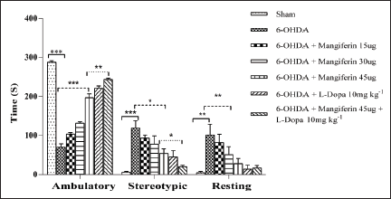
Ambulatory, stereotypic, and resting times were evaluated in rats after 42 days of 6-OHDA lesioning through activity meter (OptoVarimex-5, Columbus Instruments). 6-OHDA lesioning significantly reduces ambulatory time while increase in stereotypic and resting time was observed after 42 days of 6-OHDA-induced hemi-Parkinsonism. These, 6-OHDA-induced changes in hemi-Parkinsonian rats were significantly improved by treatment with mangiferin (15–45 µg). Mangiferin (15–45 µg) significantly increases ambulatory time and diminishes stereotypic and resting time (P < .005). Treatment with levodopa 10 mg/kg alone and in combination with mangiferin 45 µg considerably increases total ambulatory time while significant decrease in stereotypic and resting time was observed in hemi-Parkinsonian rats (F (2, 12) = 8.424; P = .0052) as shown in Figure 1.
Effect of Mangiferin on Ambulatory, Stereotypic, and Resting Time (s). Treatment With Mangiferin Reverses 6-OHDA-Induced Changes in Ambulatory, Stereotypic, and Resting Time in a Dose-Dependent Manner (All Data Obtained is Original and Presented as Mean ± SEM; *P < .005, n = 8/group).
Effect of Mangiferin on Distance Traveled, Average Speed, Time Mobile, and Number of Mobile Episodes in 6-OHDA-Lesioned Rats
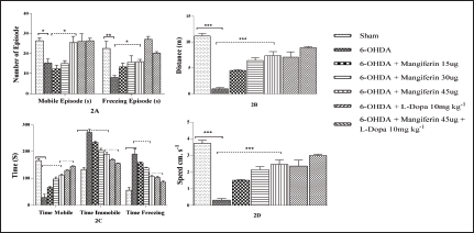
Effect of mangiferin (15–45 µg) on locomotor parameters in 6-OHDA-lesioned rats was assessed after 42 days of 6-OHDA lesioning. Significant decrease in locomotor parameters of number of mobile and freezing episodes, distance traveled, time mobile, time immobile, and time freezing, and average speed were observed in rats after 6-OHDA lesioning, while substantial increase in time freezing and time immobile was observed (P < .005). Treatment with mangiferin (15–45 µg) markedly enhances the locomotor activity. Significant increase in distance traveled, average speed, time mobile, and number of active and freezing episodes were observed in hemi-parkinsonism rats treated with mangiferin (15–45 µg). Treatment with levodopa 10 mg/kg also significantly improves these locomotor parameters (P < .005). Marked improvement in these parameters was also observed in rats treated with combinatorial therapy of mangiferin 45 µg and levodopa 10 mg/kg (F (6, 14) = 29.56; P < .0001; Figure 2a–2d).
(a) Effect of Mangiferin on 6-OHDA-Induced Changes in Mobile and Freezing Episodes. (b) Effect of Mangiferin on 6-OHDA-Induced Changes on Total Distance Travel. (c) Effect of Mangiferin on 6-OHDA-Induced Changes in Time Mobile, Time Immobile, and Time Freezing Time. (d) Effect of Mangiferin on 6-OHDA-Induced Changes in Mean Speed (All Data Obtained is Original and Presented as Mean ± SEM; *P < .005; n = 8/group).
Effect of Mangiferin on 6-OHDA-Induced Changes in Track Plot of Rats
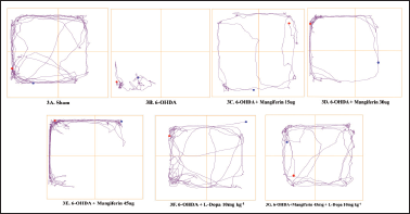
The heat map of hemi-Parkinsonian rats also showed that locomotor activity was markedly suppressed after 6-OHDA lesioning. Daily treatment with mangiferin (15–45 µg) for 28 days extensively improves locomotor activity, which was conspicuous through their heat map. Treatment with levodopa 10 mg/kg alone and in combination with mangiferin 45 µg also significantly improves locomotor activity as evident through their heat map (Figure 3a–3g).
(a) Track Plot of Sham Group. (b) Track Plot of 6-OHDA-Lesioned Rats. (c) Effect of Mangiferin (15 µg) on Track Plot of 6-OHDA-Lesioned Rats. (c) Effect of Mangiferin (30 µg) on Track Plot of 6-OHDA-Lesioned Rats. (d) Effect of Mangiferin (15 µg) on Track Plot of 6-OHDA-Lesioned Rats. (e) Effect of Mangiferin (45 µg) on Track Plot of 6-OHDA-Lesioned Rats. (f) Effect of Levodopa 10 mg/kg on Track Plot of 6-OHDA-Lesioned Rats. (g) Effect of Mangiferin 45 µg and Levodopa 10 mg/kg on Track Plot of 6-OHDA-Lesioned Rats.
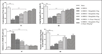
Effect of Mangiferin on Sensorimotor Forelimb Function in 6-OHDA-Lesioned Rats
The sensorimotor forelimb function was measured in 6-OHDA-induced hemi-Parkinsonian rats to reckon the asymmetry of forelimb function. Significant diminution in spontaneous use of contralateral front paw was observed in rats after lesioning with 6-OHDA. Upswing in the contralateral forelimb use was observed in lesioned rats after treatment with mangiferin (15–45 µg) on daily basis for 28 days. Treatment with levodopa 10 mg/kg also spurts contralateral forelimb use in Parkinsonian rats. Significant upturn in the use of contralateral forelimb was also observed in 6-OHDA-lesioned rats treated with combination of levodopa 10 mg/kg and mangiferin 45 µg (F (8, 18) = 20.70; P < .0001; Figure 4a).
Effect of Mangiferin on Grip Strength of 6-OHDA-Lesioned Rats
Grip strength of rats was recorded after 42 days post 6-OHDA lesion using Grip Strength Meter (Columbus Instruments). Grip strength of rats decreased significantly post 6-OHDA lesion. Daily treatment with mangiferin (15–45 µg) very much increases the grip strength in hemi-Parkinsonian rats. Levodopa 10 mg/kg significantly increases the grip strength in lesioned rats. Combination therapy of levodopa 10 mg/kg and mangiferin 45 µg largely increases the grip strength of hemi-Parkinsonian rats (F (6, 14) = 42.30; P < .0001) for all groups (Figure 4b).
Effect of Mangiferin on Cook’s Pole Climbing Test in 6-OHDA-Lesioned Rats
6-OHDA lesioning significantly increases the time to climb the pole as compared to rats in which sham surgery was performed. Daily treatment with mangiferin (15–45 µg) for 28 days significantly decreases the time to climb the pole in hemi-Parkinsonian rats. Significant decrease in time to climb the pole was observed in rats treated with levodopa 10 mg/kg. Combinatorial therapy with levodopa 10 mg/kg and mangiferin 45 µg also significantly decreases the time to climb the pole in hemi-Parkinsonian rats (F (6, 14) = 27.84; P < .0001; Figure 4c)
Effect of Mangiferin on Forelimb Akinesia in 6-OHDA-Lesioned Rats
6-OHDA lesioning significantly decreases the number of adjusting steps of contralateral forelimb, which is suggestive of forelimb akinesia in rats. Treatment with mangiferin (15–45 µg) significantly decreases forelimb akinesia by increasing the number of adjusting steps taken by contralateral forelimb in lesioned rats. Substantial decrease in forelimb akinesia was also observed in rats treated with levodopa 10 mg/kg alone and in combination with mangiferin 45 µg (F (8, 18) = 2.556; P < .0001; Figure 4d).
(a) Effect of Mangiferin on Sensorimotor Forelimb Function in 6-OHDA-Lesioned Rats. (b) Effect of Mangiferin on Grip Strength of 6-OHDA-Lesioned Rats. (c) Effect of Mangiferin on 6-OHDA-Lesioned Rats on Cook Pole Climbing Test. (d) Effect of Mangiferin in 6-OHDA-Lesioned Rats on Forelimb Akinesia (All Data Obtained is Original and Presented as Mean ± SEM; *: P <0.005; n = 8/group).
Effect of Mangiferin on 6-OHDA-Induced Changes in Oxidative Stress Parameters
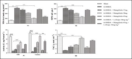
Effect of Mangiferin on MDA Concentration in 6-OHDA-Lesioned Rats
MDA, the end product of lipid peroxidation, was measured in brain tissue of 6-OHDA-lesioned rats to detect oxidative stress. Lesioning with 6-OHDA significantly increases the MDA concentration as compared to sham-operated rats. Treatment with mangiferin (15–45 µg) for 28 ameliorates this 6-OHDA-induced increase in MDA concentration. Treatment with levodopa 10 mg/kg has no significant effect on MDA concentration, whereas its combination with mangiferin 45 µg significantly reduces MDA concentration (F (6, 14) = 82.84; P < .0001; Figure 5a).
Effect of Mangiferin on MPO Activity in 6-OHDA-Lesioned Rats
MPO activity was assayed as an indicator of inflammatory response in PD. 6-OHDA lesioning substantially increases MPO activity as compared to sham surgery rats. Mangiferin (15–45 µg) treatment for 28 days significantly decreases MPO activity in hemi-Parkinsonian rats. Levodopa 10 mg/kg has no significant effect on MPO activity, whereas its combination with mangiferin 45 µg significantly decreases MPO activity (F (6, 14) = 81.57; P < .0001; Figure 5b).
Effect of Mangiferin on SOD and Catalase Activity in 6-OHDA-Lesioned Rats
SOD activity was measured to estimate oxidative stress in 6-OHDA-lesioned rats. SOD activity was observed to be significantly reduced in hemi-Parkinsonian rats. Mangiferin (15–45 µg) treatment for 28 days significantly increases SOD activity in hemi-Parkinsonian rats. Treatment with levodopa 10 mg/kg has no significant effect on SOD activity. Combination therapy of levodopa 10 mg/kg and mangiferin 45 µg substantially increases SOD activity in hemi-Parkinsonian rats (P < .005; Figure 5c).
Significant decrease in catalase activity was observed in rats after 6-OHDA lesioning. Treatment with mangiferin (15–45 µg) alone and in combination with levodopa significantly increases catalase activity in hemi-Parkinsonian rats (Figure 5c; F (1, 8) = 7.05; P < .029).
Effect of Mangiferin on TAC in 6-OHDA-Lesioned Rats
TAC assay was done to gauge any change in antioxidant status in nigrostriatal tissue of 6-OHDA-lesioned rats. 6-OHDA lesioning significantly reduces TAC thereby increasing total ROS capacity and oxidative stress. Daily treatment with mangiferin (15–45 µg) markedly increases TAC in lesioned rats. Levodopa 10 mg/kg has no significant effect on TAC in hemi-Parkinsonian rats. combination therapy with mangiferin 45 µg and levodopa 10 mg/kg significantly increases TAC in 6-OHDA-lesioned rats (F (6, 14) = 38.85; P = .0001; Figure 5d).
(a) Effect of Mangiferin on MDA Concentration in 6-OHDA-Lesioned Rats. (b) Effect of Mangiferin on MPO Activity in 6-OHDA-Lesioned Rats. (c) Effect of Mangiferin on SOD and catalase activity in 6-OHDA-Lesioned Rats. (d) Effect of Mangiferin on Total Antioxidant Capacity in 6-OHDA-Lesioned Rats (All Data Obtained is Original and Presented as Mean ± SEM; *P < .005; n = 8/group).
Effect of Mangiferin on 6-OHDA-Induced Changes in Inflammatory Parameters
Effect of Mangiferin on Cytokine Concentration in 6-OHDA-Lesioned Rats
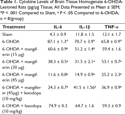
6-OHDA lesioning significantly increases concentration of proinflammatory cytokines (TNF-α, IL-6, and IL-1β) in Parkinsonian rats compared to sham surgery rats. Daily treatment with mangiferin (15–45 µg) attenuates this 6-OHDA-induced increase in concentration of these proinflammatory cytokines. Treatment with levodopa 10 mg/kg has no significant effect on concentration of proinflammatory cytokines. Levodopa 10 mg/kg in combination with mangiferin 45 µg significantly reduces concentration of these cytokines (F (2, 42) = 41.58; P < .0001; Table 1).
Cytokine Levels of Brain Tissue Homogenate 6-OHDA-Lesioned Rats (pg/µg Tissue; All Data Presented as Mean ± SEM; *P < .001 Compared to Sham, aP < .05 Compared to 6-OHDA; n = 8/group)
Effect of Mangiferin on 6-OHDA-Induced Changes in Immunological Markers
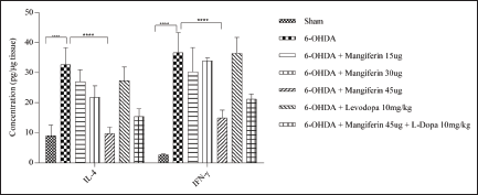
Effect of Mangiferin on 6-OHDA-Induced Changes in Th and Th Cytokine Assay
It was observed that 6-OHDA lesioning significantly increases Th1 and Th2 cytokines levels. Defense mechanism Th1 cytokines were more enhanced as compared to the Th2-cell-dependent defense mechanism. Treatment with mangiferin (15–45 µg) significantly reduces Th1 (IFN-γ) and Th2 (IL-4) cytokines levels (Figure 6). Levodopa 10 mg/kg has no significant effect on Th1 and Th2 cytokines levels (F (1, 28) = 12.94; P = .0012; Figure 6).
Effect of Mangiferin on Th1 and Th2 Cytokine Concentration in 6-OHDA-Lesioned Rats (All Data Obtained is Original and Presented as Mean ± SEM; *P < .005; n = 8/group).
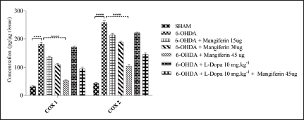
Effect of Mangiferin on Cox Activity in 6-OHDA-Lesioned Rats
Significant increase in Cox (Cox 1 and Cox 2) activity was observed in striatum of rats after lesioning with 6-OHDA. Treatment with mangiferin (15–45 µg) for consecutive 28 days significantly reduces both Cox 1 and Cox 2 activity in 6-OHDA-lesioned rats. Treatment with levodopa 10 mg/kg has no considerable effect on Cox activity. Combination of levodopa 10 mg/kg with mangiferin 45 µg reduces both Cox1 and Cox 2 activity (F (1, 28) = 445.9; P < .0001; Figure 7).
Effect of Mangiferin on 6-OHDA-Induced Changes on Concentration of Cox-1 and Cox-2 (All Data Obtained is Original and Presented as Mean ± SEM; *P < .005; n = 8/group).
Effect of Mangiferin on 6-OHDA Changes in Molecular Parameters
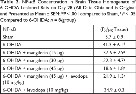
Effect of Mangiferin on NF-κB Concentration in 6-OHDA-Lesioned Rats
NF-κB is the master controller of inflammation. Increased oxidative stress and neuroinflammation increase NF-κB concentration in 6-OHDA-lesioned rats as compared to sham surgery rats. Canonical pathway of NF-κB signal transduction activated by 6-OHDA administration was inhibited by treatment with mangiferin (15–45 µg) for 28 days, as compared to normal saline treated 6-OHDA-lesioned rats. Treatment with levodopa 10 mg/kg has no effect on NF-κB concentration in 6-OHDA-lesioned rats. Treatment with combination of levodopa 10 mg/kg with mangiferin 45 µg significantly decreases NF-κB concentration in striatum of 6-OHDA-lesioned rats (F (6, 14) = 36.13; P = .0001; Table 2).
NF-κB Concentration in Brain Tissue Homogenate of 6-OHDA-Lesioned Rats on Day 28 (All Data Obtained is Original and Presented as Mean ± SEM; *P < .001 compared to Sham, aP < .05Compared to 6-OHDA; n = 8/group)
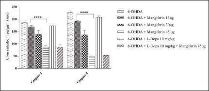
Effect of Mangiferin on Caspase-3 Activity in 6-OHDA-Lesioned Rats
Lesioning with 6-OHDA significantly increases activity of caspase-3 as compared to sham surgery rats. Daily treatment with mangiferin (15–45 µg) for 28 days significantly decreases caspase-3 activity to a large extent in striatum of lesioned rats. Treatment with levodopa 10 mg/kg has no significant effect on caspase-3 activity, whereas its combination with mangiferin 45 µg significantly decreases caspase-3 activity in 6-OHDA-lesioned rats (F (6, 14) = 36.13; P = .0001; Figure 8).
Effect of Mangiferin on Caspase-9 Activity in 6-OHDA-Lesioned Rats
Significant increase in caspase-9 activity was observed in striatum of rats after lesioning with 6-OHDA. Treatment with mangiferin (15–45 µg) for consecutive 28 days significantly reduces caspase-9 activity in 6-OHDA-lesioned rats. Treatment with levodopa 10 mg/kg has no considerable effect on caspase-9 activity. Combination of levodopa 10 mg/kg with mangiferin 45 µg substantially reduces caspase-9 activity (F (5, 24) = 87.14; P = .0001; Figure 8).
Effect of Mangiferin on 6-OHDA-Induced Changes in Concentration of Caspase-3 and Caspase-9 (All Data Obtained is Original and Presented as Mean ± SEM; *P < .005; n = 8/group).
Discussion
Levodopa is the mainstay of current pharmacotherapy of PD52, 53; however, owing to its auto-oxidant property, it could further aggravate the progression of disease. 54 Thus, because of its auto-oxidant nature, levodopa fails to stop the disease progression, rather it promotes the disease progression by increasing oxidative stress and stimulating TNF-α secretion which then leads to neuroinflammation and subsequent death of dopaminergic neurons.12, 55x–57 Therefore, in this study, we have tried to evaluate neuroprotective effect of NF-κB inhibitor mangiferin, which also has antioxidant property58, 59 and developed a combination therapy of mangiferin and levodopa to top off the dopamine in striatum and to arrest the disease progression.
Treatment with mangiferin (15–45 µg) significantly reverses 6-OHDA-induced changes in locomotor parameters of ambulatory time, stereotypic, and resting time. This could be attributed to the rescue of dopaminergic neurons from the death pathways due to its antiapoptotic, anti-inflammatory, and antioxidant property.60–62
6-OHDA injection in substantia nigra results in degeneration of about 60%–80% of dopaminergic neurons, which results in dopamine deficiency in caudate nucleus, nucleus accumbens, and ventral striatum along with other areas of the brain. 63 This deficiency of dopamine in key functional areas of the brain leads to the breakdown of the circadian rhythm of rats, which is responsible for the increase in some of the stereotypic behavior in rats after 6-OHDA lesioning. 64 Treatment with mangiferin decreases these behavior changes by preventing the apoptosis of dopaminergic neurons, which leads to an increase in the availability of dopamine. Supplementing mangiferin treatment with levodopa 10 mg/kg further improves this improvement in stereotypic behavior because of decreased apoptosis and increased dopamine availability, which results in restoration of the circadian rhythm.
Mangiferin also decreases resting time in 6-OHDA-lesioned rats due to increased availability of dopamine in the striatum region due to halt in degeneration of neurons from the striatum region.
6-OHDA lesioning substantially decreases locomotive parameters, such as total distance traveled, average speed, and number of mobile episodes and time mobile. 65 Treatment with mangiferin and levodopa substantially reversed this decrease in locomotive parameters. This improvement in locomotive parameters of time mobile, mobile episodes, average speed, and total distance traveled could be ascribed to increase availability of dopamine in striatum region, which then leads to partial restoration of indirect pathway of dopamine receptors in striatum. In 6-OHDA only group, freezing episodes were lesser compared to treatment groups. Mangiferin increases the number of freezing episodes. This increase in freezing episodes is associated with tardive dyskinesia, whereas the only logical conclusion of lesser freezing episodes in the 6-OHDA only group would be akinesia because of dopamine deficiency in the striatum.
6-OHDA lesioning results in a severe movement deficit in rats because of decreased availability of dopamine in the striatum and other regions of the brain.66–68 The heat map of rats showed that this 6-OHDA-induced movement deficit was significantly reversed by treatment with mangiferin (15–45 µg) alone and in combination with levodopa 10 mg/kg.
Decreased grip strength is one of the earliest measures of severity of disease progression. 69 ’ 70 The results of this study indicate that lesioning with 6-OHDA significantly reduces grip strength in rats and they have difficulty in grasping. 6-OHDA lesioning results in reduction of dopamine from nigrostriatal area, which then results in increased tonic inhibition of thalamus, which results in decreased excitation of cortex area. 71 Treatment with mangiferin significantly increases grip strength in 6-OHDA-lesioned rats, owing to its antiapoptotic and anti-inflammatory property. Treatment with levodopa also significantly reverses the 6-OHDA-induced changes in grasping capacity of rats. Maximum increase in grip strength was observed in rats treated with combination therapy of levodopa 10 mg/kg and mangiferin 45 µg as it ought to both replenish the depleted dopamine in the striatum and halts the disease progression by stopping neuroinflammation and subsequent degeneration of dopaminergic neurons.
On the expected lines, treatment with mangiferin significantly reduces the reaction time in Cook’s pole climbing test 72 because of increased motor activity in hemi-Parkinsonian rats. Maximum increase in this motor activity was observed in rats treated with combination of mangiferin 45 µg and levodopa 10 mg/kg.
Mangiferin also reverses 6-OHDA-induced changes in sensorimotor forelimb function and forelimb akinesia in hemi-Parkinsonian rats. 73 Maximum increase in these gait analysis parameters was observed in rats treated combination therapy of mangiferin 45 µg and levodopa 10 mg/kg.
Treatment with mangiferin 15–45 µg significantly reduces oxidative stress by decreasing MDA concentration because of its antioxidant property, which is due to its activation of NRF2-ARE pathway. Mangiferin promotes the nuclear translocation of NRF2 which thus results in increased nuclear expression of NRF2. Furthermore, treatment with mangiferin also upregulates the expression of NQO1 and promotes the binding NRF2 with NQO1-ARE complex.74, 75 Increased expression of NRF2 then stimulates the expression of antioxidant enzymes, such as SOD and catalase, and increases the total antioxidant capacity of 6-OHDA-lesioned rats.76, 77
Treatment with levodopa has no significant effect on these oxidative stress markers because of its auto-oxidant property. Metabolism of levodopa increases oxidative stress burden by generating free radicals and molecules like H2O2. 78
MPO activity, which measured index of inflammation and microglial activation, was significantly reduced by treatment with mangiferin 15–45 µg because of its property of inhibiting proinflammatory cytokine TNF-α and COX pathway. Mangiferin substantially reduces the transcription activity of both Cox-1 and Cox-2, 79 by inhibiting the nuclear translocation of NF-κB, 80 which is crucial for expression of proinflammatory factors, such as Cox-1, Cox-2, and TNF-α.
Levodopa has no significant effect on MPO activity as it promotes inflammation by stimulating TNF-α and IL-4 secretion.
6-OHDA lesioning considerably increased the concentration of proinflammatory cytokines TNF-α, IL-4, IL-6, and IL-1β. Mangiferin significantly reduces concentration of these proinflammatory cytokines. 81
Significant decrease in concentration of NF-κB was observed in striatum after treatment with mangiferin as it is a specific inhibitor of NF-κB. This decrease in NF-κB activation significantly reduces neuroinflammation as NF-κB is considered as “master regulator” of inflammation. After activation, NF-κB promotes activation of TLR-4, which in turn promotes NF-κB activation thus promoting nefarious cycle of inflammation.3, 4, 21 Mangiferin stops this cycle of inflammation by inhibiting NF-κB activation.82, 83
Balance between Th1/Th2 cytokines plays crucial role in inflammation. 84 GATA 3 and T-bet control the Th1/Th2 differentiation. GATA-3 is a regulator of Th2, whereas T-bet regulates the Th1expression. Increased expression of NF-κB promoted the GATA-3 mRNA and thereby increases the protein expression of GATA-3 while inhibiting the T-bet expression. This causes an imbalance between GATA-3 and T-bet which then results in Th1/Th2 imbalance and increased expression of Th2 cytokine. Treatment with Mangiferin restores the imbalance between Th1/Th2 cytokine.85, 86
Significant increase in activity of caspase-3 and caspase-9 expression was observed in 6-OHDA-lesioned rats, which indicate programmed death of dopaminergic neurons. Induction of p53 activates NF-κB expression that correlates with the ability of p53 to induce apoptosis. Inhibition or loss of NF-κB activity abrogated p53-induced apoptosis, indicating that NF-κB is essential in p53-mediated cell death. Activation of NF-κB by p53 was distinct from that mediated by TNF-α and involved MAPK/extracellular signal-regulated kinases (MEK1) and the activation of pp90rsk. 87 Treatment with mangiferin significantly reduces caspase-3 and caspase-9 activity because of its property of inhibiting proapoptotic transcription factor NF-κB and promotes transforming growth factor beta activity, which has anti-inflammatory and proliferative activity. 88 Treatment with levodopa has no significant effect on caspase-3 and caspase-9 activity, probably this is the reason why disease progression goes unchecked after treatment with levodopa.25, 89
Conclusion
The findings of this research indicate that mangiferin has a dose-dependent protective effect in 6-OHDA-lesioned rats. Mangiferin efficacy is amplified when combined with levodopa 10 mg/kg, which is the cornerstone of anti-Parkinsonism treatment. Thus, we may conclude that the combination of mangiferin with levodopa may have therapeutic benefit in PD therapy on our results.
Footnotes
Authors’ Contribution
PCT, MJC and RP conceived the experiments. PCT carried out experiments with assistance from SK. PCT wrote the manuscript with the help of MJC, and RP supervised the manuscript. All authors provided critical feedback and helped shape the research, analysis, and manuscript.
Statement of Ethics
Before initiating the study, necessary approval from the IAEC of KGMU, was obtained with approval letter No. 84/IAEC/Pharma/2017.
Declaration of Conflicting Interests
The authors declared no potential conflicts of interest with respect to the research, authorship, and/or publication of this article.
Funding
The authors disclosed receipt of the following financial support for the research, authorship, and/or publication of this article: Authors acknowledge Senior Research Fellowship provided to Prafulla Chandra Tiwari by Indian Council of Medical Research with file no. 45/34-2014/PHA-BMS.