
Abstract
Determination of age is essential in various civil and criminal circumstances. In the early part of life developmental processes like dentition, ossification, physical growth parameters are used and where as in later part of life aging processes like degenerative changes, calcification etc are used. Both these sets of assessment depend on multiple factors like ethnicity, genetics, sex, geography etc. Our study highlights the use of thyroid cartilage calcification in determination of age among middle and elderly individuals of Malnad region. Statistically significant progression of thyroid cartilage calcification (‘p’ of <0.001) with advancing age with an early calcification in females is observed in our study of 200 samples. This can be used as an additional parameter for radiographic assessment of age among adults and middle aged. However, it is advised not to use this criterion alone for age estimation in the middle aged and elderly.
Introduction
To get the benefits of various government social security schemes, such as widow pension, Manaswini, old age pension, and mandatory registration of marriages, citizens are approaching hospitals for their age certification. It is a known fact that there are scientifically acceptable methods available to determine age with an accuracy of ±1 year up to the age of 25 years and with an accuracy of ±5–10 years after the fifth decade. However, the majority of beneficiaries do not fall in this category, and, in our literature search, we did not find fully reliable methods that can be employed in the South Asian region to determine age between the second and fifth decades for our Malnad population. Calcification of hyaline cartilages of thyroid, cricoid, and arytenoids as a part of the normal aging process has been investigated with a note on individual and gender differences, but their application in forensic cases for age determination during the second to fifth decades using radiographic methods is sparingly employed. To use changes in the human body for age determination more accurately and efficiently, it is essential to study them in known individuals, document the frequencies, and variations with age and sex.
Objectives
To compare the radiographic percentage progression of thyroid cartilage calcification with documented age.
To study gender variations in the calcification of thyroid cartilage.
To observe for other degenerative radiographic markers.
Literature Review
Calcification of the main laryngeal cartilages was studied by Harijeet 1 on specimens obtained from 75 male and 75 female subjects varying in age from 14 to 80 years. A positive correlation between age and percentage calcification was noted, with a slower progression of calcification in females.
A review study to assess the degree of calcification of the thyroid cartilage at various ages on 200 patients from newborn to 50 years was carried out by Wenaas et al. 2 A significant relationship to age is reported with every age increase by one year, resulting in increased calcification by 1.5%–4% till 40 years.
A retrospective study on various degrees of calcification of laryngeal cartilages by Shenoy et al. 3 on 252 patients from 18 to 59 years reported varied ossification of the thyroid and cricoid cartilages from linear shadows to dense laminar calcifications in the third decade and beyond, and ossifications gradually increased with age in both sexes.
A study to evaluate age-related changes in the laryngeal cartilages for age estimation was carried out by Jadav et al. 4 on samples collected from 75 cadavers of age ranging from 17 to 65 years during postmortem examinations using a radiographic standard grading method, which showed statistically significant calcification positive correlation scores with chronological age. They also derived a regression model with a standard error of estimates ranging between 9.90 and 11.07 years for thyroid and cricoid cartilages.
Methodology
The present observational cross-sectional study was carried out after approval from the Institutional Ethics Committee on individuals approaching the Department of Forensic Medicine for age determination. The convenience sampling technique employed on individuals having documentary evidence of their chronological age in the form of a birth certificate, 10th marks card, school admission records, and those consenting to take part in the study were selected. Individuals aged between 20 and 70 years were included, and those with disorders known to induce early calcification, such as known malignancy or metabolic disorders, were excluded. A total of 200 subjects were studied.
After physical examination for senile changes—arcus senilis, wrinkling of facial skin, senile pigmentation, graying of body, and genital hairs—they were subjected to radiological examination of the neck lateral view.5–7 Computed Tomography (CT) scan was not employed as it may not be available at all the health centers, is not cost-effective, and a very accurate estimation of age for social security schemes is usually not required.
Metamorphosis of calcification of thyroid cartilage was categorized tentatively into five phases: Nil calcification, <25%, 26%–50%, 51%–75%, and >75% calcification. SPSS 22 software version was used, and a chi-square test was used to determine the significance, which is set at 0.05.
Results
Of the 200 samples studied, 9% (n = 18) were between 20 and 30 years, 16% (n = 32), 10% (n = 20), 29.5% (n = 59), 35.5% (n = 71) were between 31 and 40, 41 and 50, 51 and 60 and 61 and 70 years, respectively. Of these, 35% (n = 69) were males and 65% (n = 131) were females.
Arcus senilis was present in 68.5% (n = 137) of the subjects, while skin wrinkling and senile pigmentation were present in 59% (n = 118) and 11.5% (n = 23), respectively.
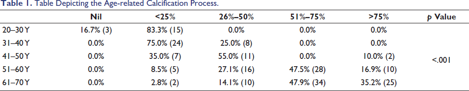
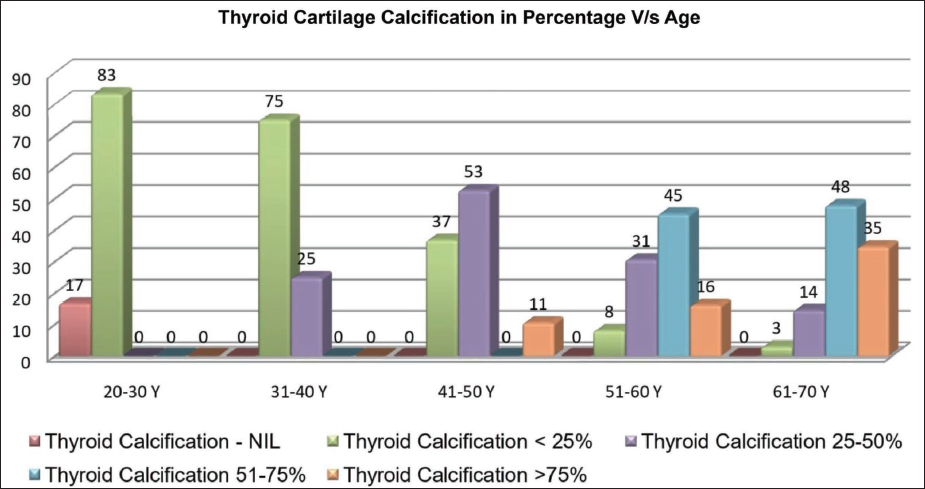
As the age increased, there was an increase in the calcification of thyroid cartilage (Figure 1) ranging from nil to <25% calcification among all those in the age group of 20–30 years to >50% calcification in 83% of individuals in the age group of 60–70 years with a “p” value of < .001 and hence, is statistically significant (Figure 2 and Table 1).
Table Depicting the Age-related Calcification Process.
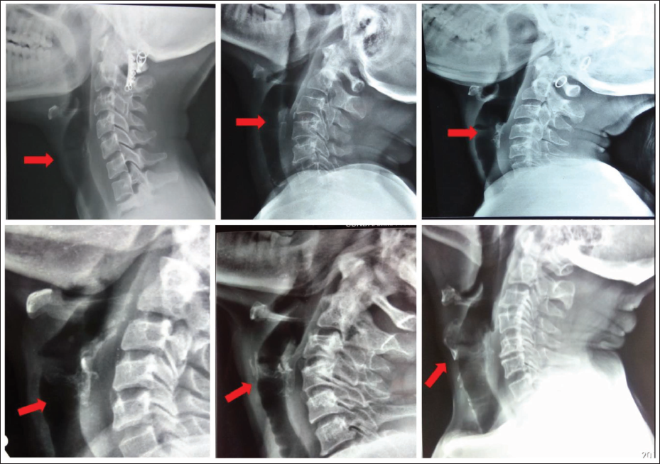
Progressive Calcification of Thyroid Cartilage (Arrow).
Calcification of Thyroid Cartilage Versus Age.
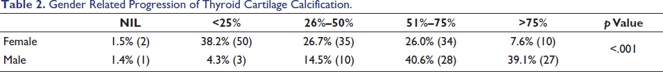
Gender variations are observed in the form of an early calcification in female and the same are depicted in Table 2. The same is statistically significant with a “p” value of < .001. Cramer V for progression of thyroid calcification in males was 0.335, and that for females was 0.2431, suggesting a weak association among females and a moderately strong association among males. Interobserver agreement for calcification staging ranged from 92% among the <25% group to 100% among the nil calcification group.
Gender Related Progression of Thyroid Cartilage Calcification.
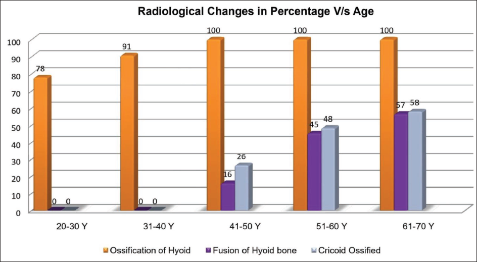
Other observations made in the same radiographs include ossification of the hyoid bone, fusion of the hyoid bone, and ossification of the cricoid cartilage. Ossification of the hyoid bone was seen as early as in the 20–30 years group (78%), but the fusion of the hyoid was evident only after 40 years, with calcification of cricoid cartilage, and the same is depicted in Figure 3. We also observed a reduction in the intervertebral space, osteophyte formation, and tracheal ring ossification in the same films.
Calcification of Hyoid and Cricoid Versus Age.
Discussion
The human body contains different types of cartilages and most of these initially calcify and later ossify. Similar to aging, the hyaline cartilages of the larynx calcify and ossify due to the normal process of mineralization. These changes can be radiographed, documented, and systematically analyzed by giving scores to estimate the approximate age of the individual. 8 Hately et al. 9 have given a detailed description of stages of progressive ossification of thyroid cartilage from anatomical samples, and accordingly, it is said to begin from the inferior portion of the posterior third of the lamina and in the inferior horn to involve the whole cartilage. Taking into account the effect of radiation on living samples, as in our study, the percentage of calcification was considered for analysis rather than the pattern, as in Hately et al. 9 Keen and Wainwright 10 or Turk and Hogg classification, 11 where they employed radiographic pattern progression analysis on samples removed from cadavers or following forensic autopsies. Since the details available in 2D lateral radiographs are limited, we feel it will not be appropriate to correlate these findings with the cadaveric analysis
Our study showed a positive statistically significant relationship between thyroid cartilage ossifications with increasing age, and is similar to the findings of Wenaas et al. 2 Shenoy et al. 3 and Sugiyama et al.12, 13 where they found calcifications to a greater extent in males compared to females,4, 13 suggesting calcification and ossification of cartilages as a natural aging process. Laryngeal cartilages dissected from cadavers of 17–65 years were radiographed, and calcification was studied using grading methods by Jadav et al. 4 showed a similar statistically significant positive correlation with chronological age (p < .05), with a standard error of estimates ranging between 9.90 and 11.07 years for thyroid and cricoid cartilages.
Keen and Wainwright 10 study developed five main and intermediate stages of calcification for both sexes, and as per them, the rate of calcification of laryngeal cartilages1, 10 was slower in females compared to males, and there was no positive correlation between age and the calcification pattern, which is in contrast to our study. This gender variation in the rate of calcification could be due to changes in the physiological hormonal milieu.
Though there is a statistically significant progression of thyroid calcification with age, rare cases of early and late calcification were observed in a few individuals in our study. One should be cautious while using calcification of thyroid alone or any degenerative process alone as a method of age estimation in medico-legal cases. However, these can add to age estimation when combined with the study of all other degenerative changes.
Our study employed lateral imaging of the thyroid cartilage, which generates 2D images in which it is difficult to differentiate calcification from other structures in the same anatomic plane, and it is not possible to know the volume of calcification when compared to 3D volumetric analysis, as in a CT scan.
Conclusion
Studies on various degenerative processes similar to developmental changes are essential for determining age after the second decade. There is no single best criterion available to find out age of an individual. If multiple methods are employed in the same individual, including the calcification of neck cartilages such as the thyroid cartilage, an estimation closer to the actual age of the individual can be deduced. Since aging is affected by various factors, including genetics, habits, geography, and innate studies are essentially required, and the findings of this study can be used as a reference standard for the Malnad region. However, further studies are needed with a larger sample and anterior-posterior or 3D studies so as to propose algorithms for age estimation.
Vertebral changes such as osteophyte formation, reduction in joint space, and tracheal ring calcification, as observed in our study, require further studies to draw conclusions.
Footnotes
Declaration of Conflicting Interests
The authors declared no potential conflicts of interest with respect to the research, authorship, and/or publication of this article.
Ethical Approval and Informed Consent
Ethical Approval: CDSCO Registered Institutional Ethical Committee (No.ECR/952/Inst/KA/2017), approved this study as per letter No. SIMS/IEC/439/2018-19 dated 11.11.2018 and participants were enrolled after obtaining ‘Informed consent’.
Funding
The authors received no financial support for the research, authorship, and/or publication of this article.