
Abstract
Sex determination and age estimation using bones have always been some of the core fundamentals in forensic medicine. This study was performed at a tertiary care military medical center in Uttar Pradesh, India, which included 100 subjects and measured the inter-lateral pterygoid plate distance from the base of the skull, the mandibular inter-condylar distance, the inter-gonial distance, and the ratio between the inter-condylar and inter-gonial distance. The data was then evaluated against the population’s gender to demonstrate any significant associations within the data. The results showed that all the parameters measured were higher in males as compared to females, except for the condylar-gonial ratio, which showed higher values in females. The results also showed that the inter-gonial distance (mm), condylar-gonial ratio, and inter-lateral pterygoid plate distance (mm) performed a more important role as an indicator of gender demarcation with respect to increasing age. Normalized inter-condylar distance (male: 117.8190 ± 6.756 mm; and female: 117.818 ± 6.644 mm) did not show a significant difference (level of significance α = 0.05) of mean with respect to male (n = 59) and female (n = 41) groups. Inter-gonial distance (mm) (male: 97.619 ± 6.8810 mm; and female: 89.173 ± 5.640 mm); inter-condylar-gonial distance ratio (male: 1.235 ± 0.077; and female: 1.273 ± 0.077) and inter-lateral pterygoid plate distance (mm) (male: 57.729 ± 5.852 mm; and female: 55.422 ± 3.934 mm) showed significant difference (α = 0.05) of mean with respect to the gender group. Our study postulates that the bony landmarks on the skull and mandible can exhibit sexual dimorphism and age-related changes, which can be used to identify genders with ethnic and regional variations. The application of these metrical parameters along with morphological features could be a useful tool for sex determination and age estimation.
Keywords
Introduction
Sex determination and age estimation from human skeleton remains have always maintained paramount importance not only in the field of forensic medicine but also in the subjects of anatomy, forensic odontology, and dental anthropology. 1 Chronological age and gender assessment form a critical part of medicolegal practice and involve a multitude of factors to reach a logical and positive conclusion. 2 Facial and skull bones are extremely resistant to fire and are usually the only remains left after an episode of mass casualty or an extended period of burial. Thus, forensic medicine and odontology can be a valuable tool in identifying these skeletal/dental remains. 3 Another bony landmark, like pterygoid plates at the base of the skull are stable bony structure that can be well preserved in scenarios of mass disasters due to their peculiar anatomical locations. These can also be used for body identification. 4 The mandible, being a dimorphic bone, has a dense layer of outer compact bone, making it a durable and well-preserved structure that can also be used as a valuable aid in the sex and age determination of casualties.
There have been numerous studies carried out worldwide involving the study of morphological features and metrical parameters in various populations of different races and ethnicities across the world; however, similar studies on the North Indian population have not yet been reported. Also, the evaluation and assessment of sexual variation and age differences of the pterygoid plates need attention, as they can produce valuable data for body identification.
The present study aims to evaluate the consistency of inter-pterygoid plates at the base of the skull and metric measurements of a few mandibular parameters for sex determination and age estimation in people residing in the Uttar Pradesh belt of North India, using non-contrast computed tomography (NCCT) images.
Materials and Methods
This study is a retrospective observational study that analyzed 100 randomly selected NCCT images involving both sexes in the age group of 2–90 years from the archives of the radiology department of a tertiary care military hospital. Images were previously recorded as part of emergency and routine diagnostic investigations on a 16-slice Siemens SOMATOM Emotion 16 machine (2010–2017).
The study did not involve any form of intervention that altered any treatment in the evaluated patients. It has been approved by the Institutional Ethics Committee.
The following criteria were followed during the course of data collection and evaluation in regard to radiographical measurements and diagnosis:
Knowledge of different anatomical structures like the condyle, coronoid, gonion, etc., as visualized on NCCT, depending on different planes and sections. Diagnostic accuracy of the condylar region and inter-condylar distance, inter-gonial distance, as well as that of the inter-lateral pterygoid plate.
NCCT images of the subjects were included and excluded based on the following criteria.
Inclusion criteria:
NCCT images taken with proper patient positioning and without any magnification errors. Patients are free from known morphological pathologies of bones.
Exclusion criteria:
NCCT images of patients with a past history of temporomandibular joint disorders and maxillofacial trauma/surgery.
Metric measurements in terms of inter-lateral pterygoid plates from the base of the skull, mandibular inter-condylar distance, and inter-gonial distance were measured on the axial sections of NCCT head scans (Figure 1).
Measurements of Axial Sections of NCCT Head Scans.
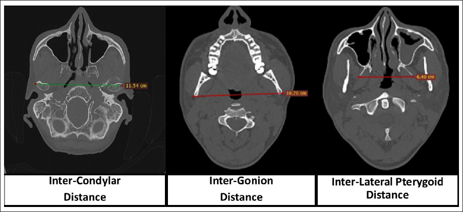
The ratio between the inter-condylar and inter-gonial distance was measured.
All the measurements taken were validated twice by a single expert radiologist on two different occasions with a single-day interval, and the mean of both values was considered.
The Institutional Ethical Committee of the hospital had waived the requirement for ethical approval.
The requirement for informed consent to participate had also been waived by the relevant Ethics Committee (i.e., it has been deemed that consent would be impossible or impracticable to obtain).
Results
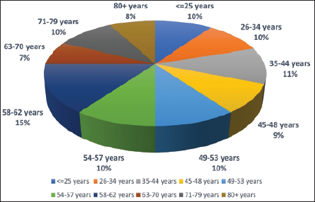
The distribution of the subjects showed that out of the 100 subjects, 59% were male and 41% were female. About 10% of the subjects were below 25 years of age, and 8% were above 80 years of age. The majority of the subjects were uniformly distributed across age groups (Figure 2).
Age Group Distribution.
Two independent sample t-tests (df = 98, α = 0.05) showed results which included normalized ages (male: 51.77 ± 17.94 years; and female: 52.94 ± 18.94 years).
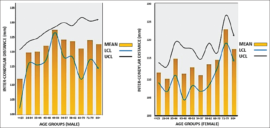
The normalized inter-condylar distance (male: 117.8190 ± 6.756 mm; and female: 117.818 ± 6.644 mm) did not show a significant difference (level of significance α = 0.05) of mean with respect to male (n = 59) and female (n = 41) groups (Figure 3).
Mean Inter-condylar Distance (mm) Distribution in Males and Females.
However, the inter-gonial distance (mm) (male: 97.619 ± 6.8810 mm; and female: 89.173 ± 5.640 mm), inter-condylar-gonial distance ratio (male: 1.235 ± 0.077; and female: 1.273 ± 0.077), and inter-lateral-pterygoid plate distance (mm) (male: 57.729 ± 5.852 mm; and female: 55.422 ± 3.934 mm) showed significant difference (α = 0.05) of mean with respect to the gender group (Figure 4).
Inter-gonial Distance (mm), Condylar-gonial Ratio and Inter-lateral Pterygoid Plate Distance (mm) Distribution in Males and Females.
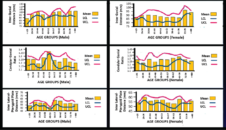
The results showed that the inter-gonial distance (mm), condylar-gonial ratio, and inter-lateral pterygoid plate distance (mm) performed a more important role as an indicator of gender demarcation with respect to increasing age (51.78 ± 18.958) (range = 81), which covers a wide range of age distribution (Table 1).
Showing Male–Female Statistics, df, and p Value of Different Parameters.
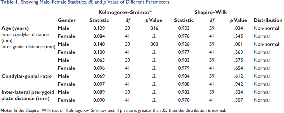
In males above the age of 48 years and in females above the age of 45 years, an irregular pattern and abrupt variation in the above-mentioned three indicating parameters (inter-gonial distance (mm), condylar-gonial ratio, and inter-lateral pterygoid plate distance (mm)) was observed, which could be a result of altered metabolism patterns and changing hormonal effects.
Discussion
Human skeleton remains can play a pivotal role in the determination of sex and age.5, 6 Morphological assessments and metric measurements of bony structures like the skull, mandible, and pelvic bones are crucial elements in the identification of sexual characteristics and age, as these structures can withstand gradients of extreme temperature, trauma, wear, and tear.7, 8 The base of the skull, orbital and nasal parameters, mandibular ramus breadth and width, bony landmarks like the gonion, condyle, coronoid, and angle of mandible have consistently been utilized in forensic sciences for sex and age determination because of sexual dimorphism and age-related changes.9, 10
There have been several studies on sex and age determination using cone beam computed tomography (CBCT), orthopantomogram (OPG), and X-rays of the human skull and mandible using morphologic and morphometric parameters, but there are very few studies done using NCCT. NCCT is a more widely available facility in a tertiary care military hospital, like in this study, in comparison to other sophisticated dental diagnostic investigations, like CBCTs and OPGs.
The mandible is known to be a sexually dimorphic bone because of the anatomical peculiarity of the bony landmarks. The stages of its development and growth are different in both sexes and thus become particularly useful in differentiating between sexes.11, 12
In our study, inter-condylar distance (mm) was slightly higher in the male group compared to the female group. This was not in consonance with similar studies carried out by Saraswathi et al., Gopal et al., and Kanjani et al.5, 13, 14 This indicated the fact that all the quoted studies were conducted on different regional populations that showed different anatomical diversity and peculiarity.
The results of our study showed that inter-gonial distance (mm) and inter-lateral pterygoid plate distance were higher in the male group as compared to the female group; however, the condylar-gonial ratio was lower in the male group (1.235 ± 0.077) as compared to the female group (1.273 ± 0.077). This finding in terms of sexual dimorphism in the condylar-gonial ratio was not in accordance with the study conducted by Ramesh et al., where they observed that the condyle to gonion (CG) height and coronoid to gonion (CoG) height values were significantly greater in males as compared to females. 15 The main reason for this variation may be the differences in imaging patterns, as well as the different ethnic origins in which the two studies were conducted. This study has utilized an NCCT 16-slice machine, and the quoted study was carried out on a dental OPG X-ray machine, which is known to have geometric distortion and positioning errors because of the narrow image layer. 16
This study was in relative consonance with the study carried out by Sairam et al., who compared the different mandibular measurements and devised that the condylar height was found to be significantly higher in males as compared to females, being 65.01 mm and 59.48 mm, respectively, on the right side and 65.71 mm and 59.65 mm, respectively, on the left side. 17
It has been established that socioenvironmental factors (nutrition, climate, food, etc.) influence the development and, eventually, the morphology of the bones. 18 Many studies have demonstrated that skeletal characteristics differ in each population and have stressed the need for population-specific osteometric and morphometric standards for gender determination and age estimation.
Positive Outcomes from the Study
The present study evaluated four different metrical parameters and statistically analyzed data correlation for the results, which provided valuable information pertaining to age variation and sexual dimorphism in the population of the Uttar Pradesh region in India, which has not been cited in any earlier study.
Limitations of the Study
Limitations of the study are a small sample size and a possible lack of exact metric measurements in cases where the age range was below the age of complete development of the skull and mandible. Also, as the study was conducted in a tertiary care military hospital on defense-serving personnel, their families, and dependents, a few of the subjects analyzed could belong to different demographic regions of Uttar Pradesh and thus were considered outliers. There is a definitive need to further this study on a larger sample size with a substantial number of subjects in every age group in the Uttar Pradesh population.
Conclusion
Medical and dental professionals can be valuable team members in the science of forensic medicine. The present study utilized four different metrical parameters. The application of these metrical parameters along with morphological features could be a useful tool for sex determination and age estimation. This study postulates that the bony landmarks on the skull and mandible can exhibit sexual dimorphism and age-related changes, which can be used to identify genders with ethnic and regional variations. All the parameters measured were higher in males as compared to females, except for the condylar-gonial ratio, which showed higher values in females. In this study, an attempt is made to use the more commonly available NCCT machines to analyze the morphological parameters.
Future Recommendations
Comprehensive knowledge about various parameters of the bony landmarks on the skull, pterygoid plates, orbital cavity, maxilla, and mandible will give us an insight into the crucial elements of age and sex determination in forensic dentistry. These will also enable us to create facial reconstruction using these anthropometric measurements and will ultimately provide a novel platform for further interpretations. It is recommended that more population‑specific studies with larger sample sizes be undertaken in the future to substantiate the usefulness of this study.
Footnotes
Authors’ Contribution
Sudip Indu: Data collection and patient evaluation. Umesh Kumar Mishra: Radiological evaluation and data collection. CP Shanthanu: Data collection and evaluation. Pathri Manjeera: Radiological evaluation and data collection.
Data Availability Statement
Data pertaining to the article is available at the Department of Medical Statistics and Records, No. 8 Air Force Dental Centre, Kanpur, Uttar Pradesh 208001, India.
Declaration of Conflicting Interests
The authors declared no potential conflicts of interest with respect to the research, authorship, and/or publication of this article.
Ethical Approval and Informed Consent
The Institutional Ethical Committee of the hospital (No. 8 Air Force Dental Centre, Kanpur, Uttar Pradesh 208001, India) had waived the requirement for ethical approval. The requirement for informed consent to participate had been waived by the relevant Ethics Committee (i.e., it has been deemed that consent would be impossible or impracticable to obtain).
Funding
The authors received no financial support for the research, authorship, and/or publication of this article.