
Abstract
Age estimation plays a critical role in forensic identification, particularly for legal and administrative purposes. Traditionally, age estimation relied on visual examination of cranial sutures, but modern radiological imaging techniques like computerized tomography (CT) scans have enhanced accuracy. This study aimed to assess cranial suture obliteration using a seven-stage scoring system on CT scans to estimate age in the Indian population. A total of 141 subjects were analyzed, and regression models were created for males, females, and the total population. The study found a significant correlation between chronological age and cranial suture oblite ration, with better prediction accuracy in females. Comparison with previous studies showed promising results, although caution is advised in using these models as standalone methods due to their relatively large standard error estimate (SEE). Further research is warranted to refine age estimation techniques from cranial sutures across diverse populations.
Keywords
Introduction
Age estimation is a crucial aspect of forensic identification. Until the age of 25, accurate estimation within a range of one year on either side is feasible through radiological assessment of secondary centers of epiphyses and dental examination.1, 2 This precision is essential for various legal, medical, social, and administrative matters concerning elderly individuals, such as employment regularization, pension settlements, and senior citizen benefits. 3 Beyond the age of 25, age estimation is typically expressed in terms of decades. Traditionally, evaluating cranial sutures involved invasive procedures, including the removal of soft tissues for visual analysis.
However, advancements in radiological imaging, particularly computerized tomography (CT) scans, have significantly improved the ability to estimate ages in both living and deceased individuals.4–6 Utilizing modern radiological techniques like CT allows for non-invasive evaluation of sutures, eliminating the need for tissue removal.
Numerous studies, such as those by Todd and Lyon in 1924 7 and Meindl and Lovejoy in 1985, 2 Singh et al., 8 Pardeep Singh et al., 9 Goyal et al., 10 Masih W et al., 11 Khandare et al., 3 and Mohammed Akbar N J et al., 12 have investigated forensic age estimation using cranial sutures, employing various techniques and scoring methods.
Despite this body of research, there remains a notable gap in CT-based cross-sectional imaging of cranial sutures for age determination, particularly in India. This study aims to address this gap by providing insight into the ages at which different cranial sutures fuse in the Indian population. Additionally, the study seeks to develop regression models for estimating age in this population by analyzing cranial suture obliteration in cross-sectional CT images, using a seven-stage scoring system developed by Harth et al. in 2009 13 and Chiba et al. in 2013. 14
Material and Methods
The study enrolled individuals aged 20 years and older who were referred to the Department of Radiology at Goa Medical College and Hospital, a tertiary healthcare facility in India. Patients were recommended by physicians or surgeons to undergo a head CT scan for diagnostic purposes. They were informed about the study’s scope, and inclusion of their CT images was contingent upon obtaining their informed consent and verifying their age and Goan ethnicity through Aadhaar card details. Cases were excluded if they exhibited motion blur or had a history of conditions or events that could affect cranial sutures, such as skull fractures, anomalies like stenocephaly, childhood abuse, or diseases impacting ossification.
Inclusion Criteria
Individuals aged 20 years and above.
Patients who underwent CT scans of the head for diagnostic purposes.
CT scan DICOM files with clear cross-sectional images of cranial sutures for analysis.
Exclusion Criteria
Patients with movement blur in CT scans.
Individuals with a history of conditions or events known to affect cranial sutures, such as skull fractures, cranial burns, cranial anomalies (e.g., stenocephaly), childhood abuse, or diseases affecting ossification.
Technical Information
CT scans were conducted using a Siemens Medical Solutions DSCT-SOMATOM Definition flash 256-slice CT scanner in Erlangen, Germany. The scan parameters were set to a tube voltage of 80 kV, tube current of 58 mAs, and slice thickness of 0.6 mm. Patient information displayed in the DICOM images was digitally concealed to maintain patient anonymity. Radiant DICOM software was utilized to generate orthogonal multiplanar reconstruction images and volume rendering technique images. The cross-sectional images were examined using a window width of 1,600 HU and a window level of 1,000 HU, specifically for bone visualization.
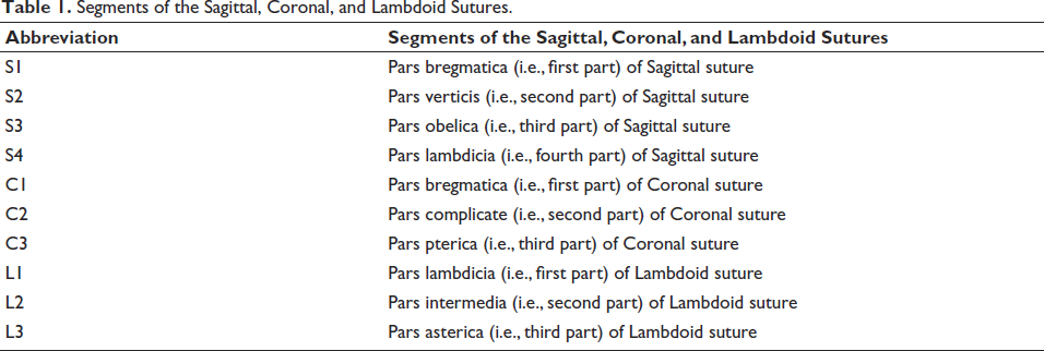
The sagittal, coronal, and lambdoid sutures were selected for evaluation. A three-dimensional multiplanar reconstructed image of the cranium containing all three sutures was divided into segments. The sagittal suture was divided into four segments, the coronal suture into three segments, and the lambdoid suture into three segments, as mentioned in Table 1.
Segments of the Sagittal, Coronal, and Lambdoid Sutures.
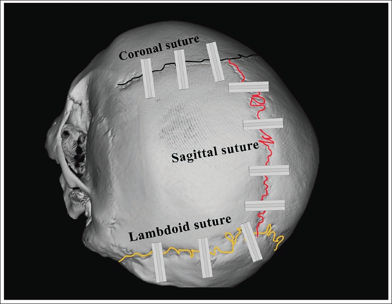
A 0.6-mm block was selected from the middle of each segment, as illustrated in Figure 1. Cross-sectional images perpendicular to the suture were examined. Axial, coronal, and sagittal sections were obtained at various levels in the bone window of the skull sutures.
3D-multiplanar Reconstruction Showing Coronal, Sagittal, and Lambdoid Suture and Their Segment for the Cross-sectional View to Observe the Fusion of Cranial Sutures.
Scoring of Cranial Suture Obliteration
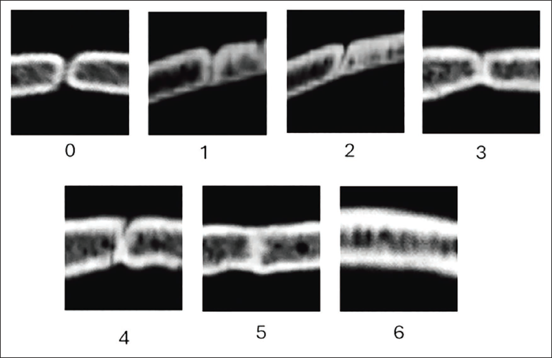
Cross-sectional slices of each segment of the sagittal, coronal, and lambdoid sutures were visually assessed and classified into one of seven stages, as shown in Table 2, which is based on the degree of closure, following the criteria outlined by Harth et al. 13 and Chiba et al. 14 the cross-section of the segment showing the degree of closure is shown in Figure 2.
Seven Stages According to the Degree of Closure by Harth et al. and Chiba et al.
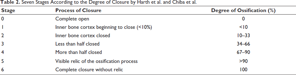
Image of the Seven Stages of Ossification on a Cross-section of Suture.
Results
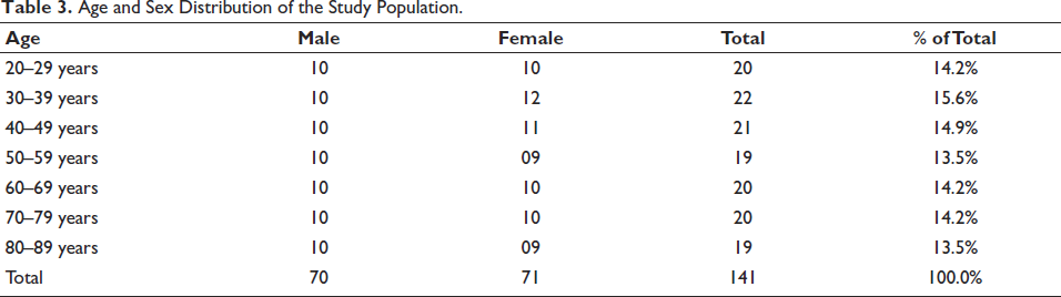
The study analyzed cross-sectional images of sutures from CT scan DICOM files to assess skull suture closure. The study included 141 subjects of known age and sex, comprising 70 males (mean age + standard deviation: 53.14 + 19.78 years) and 71 females (mean age + standard deviation: 53.29 + 19.65 years), ranging in age from 20 to 89 years, as illustrated in Table 3.
Age and Sex Distribution of the Study Population.
All statistical analyses were conducted using the Jamovi project software (2022), Version 2.3. As obliteration scores (ranging from 0 to 6) were ordinal values assigned to cranial suture obliteration, the dataset was recognized to follow a non-normal distribution, necessitating the use of non-parametric tests such as the Mann–Whitney U test and Spearman’s correlation. Spearman’s rho correlation was employed to determine the correlation between the suture obliteration score and chronological age.
The mean closure stage was calculated by summing the closure scores for each skull suture segment and dividing them by the number of segments. Simple linear regression models were constructed to estimate age using obliteration scores for each cranial suture part analyzed in this study, separately for males, females, and the entire study population.
Cohen’s κ was utilized to assess intra- and interobserver errors, based on 40 individual CT DICOM images and 40 randomly selected CT DICOM images from the participants’ scans. The significance level (α) was set to 0.005 for all observations. Intra-observer errors exhibited high agreement, with kappa scores of 0.98, indicating strong consistency between the principal investigator and secondary observations. Additionally, interobserver error kappa was 0.83, indicating substantial agreement between the lead investigator and each individual observer.
A statistically significant correlation (p < .001) was observed between chronological age and the mean cranial suture obliteration scores.
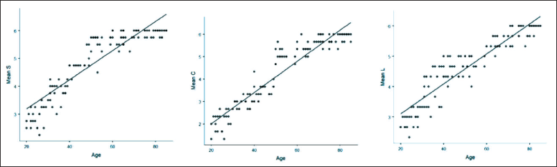
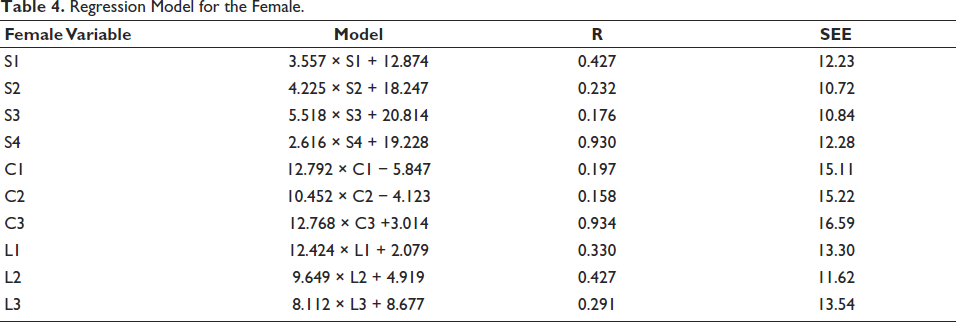
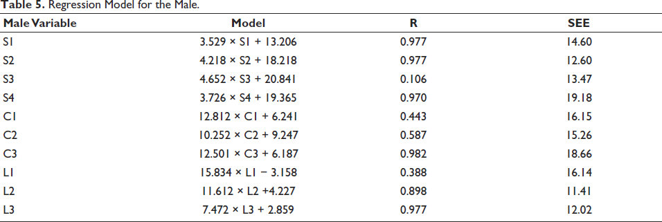
The multiple linear regression models were developed to estimate age using the obliteration scores of sagittal, coronal, and lambdoid sutures for males, females, and the total study population, as illustrated in Figure 3 and Tables 4, 5, and 6, respectively.
Scatter Diagram Showing the Slope and the Linear Regression Models of Age Estimation with Mean of the Respected Sutures.
Regression Model for the Female.
Regression Model for the Male.
Regression Model for the Total Study Population.
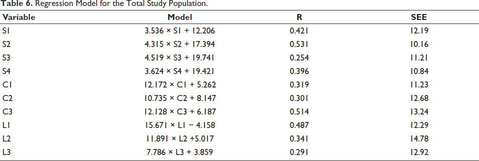
For the sagittal suture, the standard error of the estimate was 14.96 years for males, 11.40 years for females, and 11.10 years for the total sample. Regarding the coronal suture, the standard error of the estimate was 16.69 years for males, 15.64 years for females, and 12.38 years for the total sample. Finally, for the lambdoid suture, the standard error of the estimate was 13.19 years for males, 12.82 years for females, and 13.33 years for the total sample.
Discussion
Accurate estimation of age holds significant importance in forensic medicine, particularly concerning the identification of individuals from skeletal remains,15, 16 which is one of the fundamental aspects of forensic investigation. Once all teeth have erupted and most epiphyses have fused with the diaphysis, skull sutures become reliable indicators of age. While various changes such as bone lipping, greying of hair, arcus senilis formation in the cornea, lens opacity, atherosclerotic changes in arteries, and skin wrinkling, particularly on the face, occur with aging, these alterations are generally too nonspecific to serve as reliable markers for age determination in medical-legal contexts. 17
In forensic medicine and forensic anthropology, skeletal and dental indicators are commonly utilized to determine maturity, with most of these indicators reaching maturity by the age of 25. However, beyond the age of 25, age prediction becomes highly unreliable, and the use of skull sutures as indicators for age estimation remains a topic of debate among experts in the field. 18
Earlier studies predominantly relied on the fusion of both endocranial and ectocranial skull sutures using “The Acsádi and Nemeskéri method of scoring” for age estimation.17–23 However, caution is advised when interpreting ectocranial and endocranial suture closures, and they should be used in conjunction with other age indicators, as suggested by Meindl and Lovejoy et al. 2
In recent times, advancements in imaging modalities have led to the development and testing of multiple techniques for accurate age estimation.6, 10, 13, 24, 25 For instance, Postmortem CT imaging, has been extensively utilized in forensic medicine and forensic anthropology for age estimation in cases involving unidentified, decomposed, and skeletal remains.26–28
This study focuses on age estimation based on the cross-sectional analysis of cranial suture closure using CT scans in the Indian population, employing a seven-stage scoring system as proposed by Harth et al. 13 and Chiba et al. 14 A statistically significant correlation was observed between age and suture obliteration in males, females, and the total population.
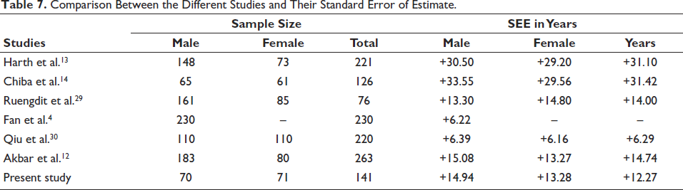
Harth et al. 13 conducted a study in 2009 to estimate age using flat-panel CT imaging of calvariae, observing 221 samples. The study utilized a seven-stage grading method for suture obliteration, with a standard error estimate (SEE) of 30.5 years in males and 29.2 years in females. However, the present study yielded lower SEE values, likely due to differences in sample size.
Similarly, in 2013, Chiba et al. 14 conducted a study focusing on the sagittal suture using cross-sectional CT imaging. Although their study reported higher SEE values compared to the present study, it highlighted the positive correlation between sagittal suture closure and age, particularly in adult women.
Ruengdit et al. 29 examined suture closure techniques in Thai crania in 2018, using three widely used methods - Acsádi and Nemeskéri (1970), Meindl and Lovejoy (1985), and Mann (1991). Their approach demonstrated an error of 13.3 years in males and 14.8 years in females. The Mann method showed higher accuracy in predicting age in older males but not in Thai females, contrary to the present study, which showed slightly higher accuracy in females than in males. Ruengdit et al. 29 also stated that inter-population variations exhibited biases and inaccuracies, with overestimation in younger adults and underestimation in older individuals, which is more likely in the present study as well.
Fan et al. 4 conducted a study in 2020 on the Chinese male population, employing CT scans for age estimation. Their study, utilizing a seven-stage grading system, reported slightly better accuracy compared to the present study, likely attributed to differences in population demographics and sample size.
Qiu et al. 30 conducted a study in 2020 on Han adults using thin-layer CT imaging for age estimation, achieving a mean absolute error of 6.39 years in males and 6.16 years in females. Although their standard error of estimate may be lower due to the usage of multiplanar reformation images, unlike the present study, which divided suture cross-sections into segments and employed multiplanar images.
In 2023, Akbar et al. 12 conducted a study on the Indian population, utilizing a three-stage scoring system for cranial suture closure assessment on 3D CT scans. Their study reported slightly higher SEE values compared to the present study.
A comparison of study characteristics between the available literature and the present study is provided in Table 7.
Comparison Between the Different Studies and Their Standard Error of Estimate.
Conclusions
The closure of cranial sutures progresses with age and can be evaluated using DICOM images obtained from modern computer tomography scans employing a seven-stage scoring method. The obliteration of cranial sutures demonstrates a statistically significant correlation with chronological age, with a stronger predictive value observed in females compared to males. Linear regression models based on stages of cranial suture obliteration offer valuable tools for forensic practitioners to estimate age.
However, given the relatively large SEE associated with the regression models developed using cranial suture obliteration scores in this study, caution must be exercised when utilizing these models. They should be employed in conjunction with other established age estimation techniques during the process of identification.
Age estimation based on morphological changes in bones has long been subject to debate due to its inherent variability, influenced by diverse factors such as climate, diet, genetics, nutrition, society, race, environment, and geography. Cranial sutures are no exception to this variability. Therefore, further research is warranted to refine age estimation methods based on cranial sutures across different populations.
Footnotes
Acknowledgements
We would like to express our gratitude to the doctors of the Department of Radiology and the Department of Forensic Medicine for their support during this study. Additionally, we extend our appreciation to Dr. Vishal KK for assisting in the statistical analysis of our data.
Declaration of Conflicting Interests
The authors declared no potential conflicts of interest with respect to the research, authorship, and/or publication of this article.
Ethical Approval
Ethical clearance is acquired from the Institutional Ethical Committee(letter number: ECR/83/INST/GOA 2013/RR-20). Participant confidentiality is safeguarded through encryption of their identities. Patients are provided with information and their Informed consent is obtained for the usage of CT scans DICOM files for study purposes.
Funding
The authors received no financial support for the research, authorship, and/or publication of this article.