
Abstract
Anatomical structures like the paranasal sinuses and the foramen magnum (FM) are typical and specific because they are found in the human body. In CBCT, these entities can be well demarcated and using specialized software, a three-dimensional (3D) volume model can be generated. This retrospective institutional study aimed to assess the reliability of volumetric measurements of paranasal sinuses in correlation with morphometrics of FM for age and gender determination using CBCT. Samples of 200 full skull CBCT images between 20 and 69 years were utilized and equally distributed among gender. ITK-SNAP 3.8.0 software was used to calculate the volumes of paranasal sinuses. The sagittal diameter and the transverse diameters were measured, and the circumference of the FM was then calculated. Using discriminant functional analysis and multivariate regression analysis, the correlation of paranasal sinus volumes and the FM were classified with respect to age and gender. Males exhibited higher mean values for the volumes of the frontal sinus, bilateral maxillary sinus, right sphenoid sinus, transverse diameter, and circumference of the FM in comparison to females. The overall accuracy rate to correctly classify gender was found to be 96.5%. In the multivariate regression model utilized for age estimation, the overall analysis of parameters in group-wise settings revealed that the significant predictors for age estimation included the volume of the left maxillary sinus, the circumference of the FM, and the volume of the right sphenoid sinus. The calculated regression analysis for the combined sample yielded a value of 43735.755, with a notably significant P value of .001. The study’s results were highly significant and deemed satisfactory.
Introduction
The necessity to identify individuals, whether before or after death, arises not only from social expectations but also from legal obligations. In forensic medicine, establishing the biological profile relies on four fundamental pieces of information: gender, age at the time of death, height, and ancestral background. 1 In general, most of the skeletal remains after catastrophes and natural calamities will be in fragment forms. Thus, the cranium, a denser skeletal part that holds the maxillofacial part comprising the teeth, protecting the structures such as the skull base and the paranasal sinuses can be used. 2 Through recent studies, it has been proved that in the case of recognition of an unidentifiable deceased individual, the use of dental and maxillofacial skeletal remains serves as an important key factor. 3 Paranasal sinuses possess specific features like unique nature, irregular shape, which makes it typical and specific. The enduring characteristics of these pneumatized structures render them essential for forensic applications. 4
CBCT has gained extensive usage due to its advantages in forensic cranium imaging, providing high resolution for skeletal examination at a relatively low cost. Its field of view is compatible with the area under investigation, and it offers portability and simplicity. 5 Numerous forensic research studies have utilized metric parameters such as the width, height, and length of paranasal sinuses.6, 7
Utilizing 3D imaging to discern sexual dimorphism is an emerging concept. Forensic determination of sex necessitates precise identification, for which population-specific baseline data is crucial. Numerous studies on the morphology of the maxillary sinus and nasal aperture have demonstrated that these parameters serve as effective indicators for gender and age estimation. 8
Despite numerous studies in the literature, the impact of age and gender on paranasal sinuses remains unclear. To address this, volume measurement has been employed to enhance accuracy and interest. In CBCT, the use of programs enabling segmentation and modeling through semi-automatic processing has facilitated volume measurement. Several studies have highlighted the utility of these sinuses in gender determination.9–11 However, other studies have argued that the paranasal sinuses do not possess the dimorphic gender character or age-related changes.12, 13 Thus, no clear consensus exists in this regard.
The foramen magnum (FM) is an intriguing structure of interest in anatomy, forensic medicine, and anthropology, given its unique location in the skull base, providing resistance to external physical insults. 14 Various studies have indicated that measurements related to the FM can be valuable for personal identification.15, 16 Correlating morphometric measurements of the FM with volumetric measurements of the paranasal sinuses holds the potential for achieving the highest accuracy in age and gender determination. Consequently, this study aims to assess the feasibility and accuracy of using volumetric measurements of paranasal sinuses and morphometric measurements of the FM for age and sex identification among the South Indian population attending an institution in Chennai. This specific analysis has not been reported in the existing literature.
Materials and Methods
This retrospective institutional-based study was conducted in the Oral Medicine and Radiology Department at Meenakshi Ammal Dental College. CBCT images of consecutive patients taken between 2014 and 2021 were retrieved from the dental archives. The scans were generated using the Planmeca Promax 3D Mid Proface CBCT machine. The study comprised 200 samples, evenly divided into 100 males and 100 females, within the age range of 20–70 years. The study population was further categorized into five age groups (i.e., Group I: 20–29 years, II: 30–39 years, III: 40–49 years, IV: 50–59 years, V: above 60 years), each consisting of 20 samples equally divided between both genders, and subsequent analysis was conducted.
The ITK-SNAP software version 3.8.0 was utilized for the analysis of CBCT images. All images were assessed, and measurements were taken by the primary researcher and two examiners, both trained and skilled Dento-maxillofacial radiologists specializing in radiographic analysis. Inter-examiner calibrations were performed to ascertain the reliability and reproducibility of the measurements. The entire image data was evaluated in coronal, sagittal, and axial sections for improved visualization.
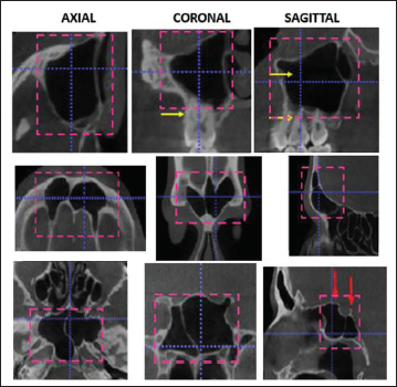
Due to discrepancies in measuring the volume and a lack of a feasible standardized method for ethmoidal sinuses, they were excluded from the study. In multiplanar images, the slice of interest was standardized for each sinus, and points were delimited based on the anatomical borders using their outlines (Figure 1). The active contour segmentation mode was employed to select the regions of the maxillary sinus, frontal sinus, and the sphenoid sinus for semi-automatic active contour segmentation.
Delimitation of the Sinuses in ITK-SNAP Viewer and Using Active Contour Segmentation Mode.
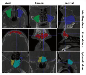
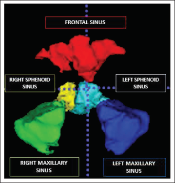
After the delineation, the sinuses were segmented and then the software updates the volumetric filling in the sinuses (Figure 2). The software updates the volume in cubic millimeters, which is then converted to cubic centimeters. Followed by, the 3D model was obtained based on their color coding (Figure 3).
Volumetric Filling (cm3) of Paranasal Sinuses in ITK SNAP Viewer.
Software Updated 3D Volumetric Image with Measurements.
The axial section of the CBCT image is reformatted for the measurement of FM diameters. The slice of interest is selected where the bony outline of the foramen is merely visible (Figure 4).
Axial Section of CBCT Showing Foramen Magnum Transverse and Sagittal Diameter.
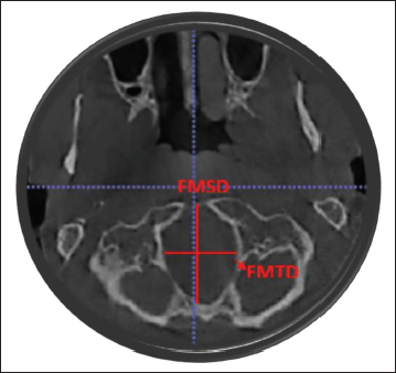
The greatest anteroposterior dimension was taken as the FM sagittal diameter. The maximum width in the mediolateral direction was taken as FM transverse diameter. The circumference of the FM was then calculated based on the standard formula for the circumference of a circle. CIRCUMFERENCE = 2×3.14× [(FMSD)2/(FMTD)2/2]1/2.
Results
Descriptive and inferential statistics were conducted using IBM SPSS version 20.0. Mean and standard deviation were employed to summarize the anthropometric parameter values within the groups. Discriminant analysis was utilized to formulate a discriminant equation for determining gender within the study population, while multivariate regression analysis was employed to establish a regression equation for age estimation among the study participants.
The mean age for both females and males were determined to be 45.4 ± 1.4 years. A comparison of each parameter between genders is presented in Table 1.
Comparison of Parameters Between Gender in the Study Group.
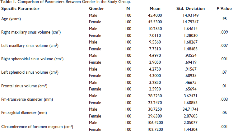
In men, the mean volume of the right maxillary sinus was 10.25 ± 1.6 cm3, significantly higher than in females, where it measured 7.01 ± 1.2 cm3, with a P value of .001. Similarly, the left maxillary sinus exhibited a statistically significant difference between genders, with mean volumes of 9.85 ± 1.68 cm3 in males and 7.73 ± 1.48 cm3 in females. The mean volume of the right sphenoid sinus in males was significantly greater at 4.2 ± 0.9 cm3 compared to females at 2.9 ± 0.6 cm3, with a P value of .001. However, no significant difference was observed in the volume of the left sphenoid sinus between genders. For the frontal sinus, males had a mean volume of 3.2 ± 0.46 cm3, while females had a mean volume of 2.59 ± 0.65 cm3, showing a statistically significant difference with a P value of .01.
Regarding the dimensions of the FM, males had a significantly larger mean transverse diameter at 28.32 ± 3.6 compared to females at 23.24 ± 1.6, with a P value of .003. The sagittal diameter exhibited an insignificant difference between males (31.6 ± 3.6 mm) and females (29.63 ± 2.8 mm) with a P value of .06. The mean circumference of the FM in males was 106.4 cm2, significantly larger than in females (102.7 cm2) with a P value of .001.
The independent sample t-test indicated significant differences in most anthropological parameters between gender groups, suggesting the potential for developing a discriminant analysis model for gender discrimination using these parameters. Canonical Correlation and Wilk’s lambda tests identified the variables suitable for the final model, including right and left maxillary sinus volume, right sphenoid sinus volume, frontal sinus volume, FM transverse diameter, and circumference of the FM.
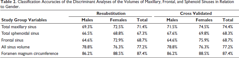
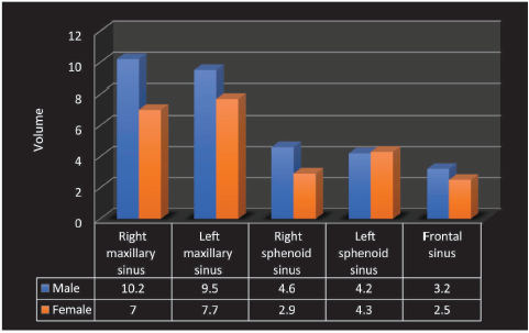
Table 2 shows discriminant analysis and classification accuracies for gender prediction using sinus volumes range from 64.6% to 78.8%, with higher accuracy for all sinus volumes (77.2%). The foramen magnum circumference achieves the highest classification accuracy (87.4%). Cross-validation confirms similar accuracy trends, ensuring model reliability. Figure 5 shows males have consistently larger paranasal sinus volumes than females across all sinus types. The right maxillary and left maxillary sinuses show the most significant differences (10.2 cm³ vs. 7 cm³ and 9.5 cm³ vs. 7.7 cm³, respectively). Frontal and sphenoid sinuses also follow this trend, highlighting gender-based anatomical variations.
Classification Accuracies of the Discriminant Analyses of the Volumes of Maxillary, Frontal, and Sphenoid Sinuses in Relation to Gender.
Distribution of Paranasal Sinuses Volume Among Gender.
Then the further tests of function were done and a significant P value was calculated, which shows that the model is significantly fitting. Using Canonical Discriminant Function Coefficients, all the parameters were analysed and a constant value of –43.886 was obtained and the gender estimation formula was derived.
Gender estimation formulae = –43.88 + (0.313 × right max sinus) + (0.208 × right sphenoid sinus) + (0.497 × left max sinus) – (0.408 × frontal sinus) + (0.148 × FM transverse DM) + (0.341 × circumference of FM)
If the estimated value using the above-mentioned formula is close to 1.986, then the gender is male, if it is close to –1.986 then the gender is female.
The accuracy rate on combining paranasal sinuses volume and FM for identification of males is 96% and for females is 97%.
The classification of results for both males and females show 96.5% of original grouped cases were correctly classified.
For the age estimation, the model summary analysing all the parameters in group-wise showed that predictors in age estimation are left maxillary sinus volume, the circumference of the FM, and right sphenoid sinus volume. Utilizing the ANOVA test, the Regression analysis for the combined sample yielded a calculated value of 43735.755, with a highly significant P value of .001. Consequently, the formulated regression equation is expressed as follows:
Age estimation formulae = –79.484 – (5.619 × left max sinus) + (1.766 × circumference of FM) – (2.556 × right sphenoid sinus)
The mean values of maxillary sinus volume decrease with increasing age in both genders. Notably, the volume of the maxillary sinus was found to be higher in the age group of 30–39 for both males and females. Similarly, the mean values of sphenoid sinus volume decrease with age for both genders, with the volume of the sphenoid sinus being higher in the age group of 20–29 for both males and females.
Examining the frontal sinus volume, it was observed that in males, the highest mean value was in the age group of 30–39 (2.8 cm3), while the lowest was in the age group of 50–59 (2.02 cm3). In females, the highest values were found in two age groups (20–29 and 50–59) at 2.5 cm3, and the lowest was in the age group of 30–39 (2.1 cm3).
Concerning the circumference of the FM, the mean value was higher in males in the age group above 60 years, measuring 107 cm2. In females, the highest value was also observed in the age group above 60 years, calculated as 103 cm2.
Analysing the transverse diameter of the FM, males had a higher mean value of 29.6 mm in the age group of 20–29 years, while females exhibited a higher mean value of 23.5 mm in the age group of 30–39 years. The mean value of the sagittal diameter of the FM was higher in the age group of 50–59 years for both males (31.2 mm) and females (30.4 mm).
Discussion
The challenging aspects of forensic odontology are the identification of the age and gender from the crania. Forensic specialists initially focus on morphology of the skeletal remains followed by morphometrics in the process of personal identification. 17 Therefore, research such as this, which is aimed at enhancing the accuracy of these areas, is of great value in the field of forensic odontology. Few Indian studies in different populations analysing the volumes of paranasal sinuses have been reported in the literature for the age and gender determination.18, 19 The accuracy of volume estimation in the tomographs is the main aim and requires sophisticated software. In the Indian population, studies supporting information on volumetric analysis of paranasal sinuses are scanty. The objective of this study was to utilize paranasal sinuses volumes in correlation with FM measurements for age and gender determination through discriminant analysis, assessing the effectiveness of CBCT as an imaging modality.
The fundamental result of this study is that on comparing the volumes of maxillary and frontal sinuses among gender, males had larger volumes compared to the females (P < .001). This is in accordance with the previous studies.
As per the findings of this study, the precision of sex determination based on the maxillary sinus was 71.5% for males and 74.5% for females, resulting in a combined accuracy rate of 74.4%. In 2014, Sharma investigated the maxillary sinus volume in the Indian population using CT scans and reported an accuracy rate of 69.81%. 20 In the current study, the frontal sinus exhibited an overall sex estimation accuracy rate of 68.7%. In 2010, Uthman et al. explored the morphometrics of the frontal sinus and skull using CT images in the Iraqi population. 21 The study revealed that using the frontal sinus, the ability to identify gender was 76.9%, and this accuracy rate further improved to 85.9% on adding skull measurements.20, 21 This is in congruity with our study.
Facial bones play a distinctive role with significant predictive value in the field of forensics. Research conducted by Hasham et al. in Karnataka, involving the analysis of 240 Osteo Meatal Complex CT scans, aimed to compare morphological measures of nasal bones between genders. The study findings indicated that certain morphometric parameters, such as the pyramidal angle, were more pronounced in males, while the distance between the nasion and tip of the nose was greater in females. 22
The efficacy of sphenoid sinus in gender determination is controversial since forensic identification studies in the literature exhibit both equal positive and negative predictability. In our study, the left sphenoid sinus volume showed that males and females had an insignificant difference. This aligns with the research conducted by Oliveira et al. in 2017. They reported that the mean values for males and females were not statistically significant, thus impeding their use in gender identification. 23
Ramos et al. in 2021, studied morphometrics and volumetrics of the sphenoid sinus for age and gender determination with 268 CBCT scans of the Brazilian population. He stated that comparing the morphometrics, the volumetric measurements have a higher predictability score for age and gender determination. 24 Alaettin Koc in 2020, studied maxillary and sphenoidal sinus volume in CBCT for gender determination and reported that there was a significant difference in both maxillary and sphenoidal sinus volume. 10 He further stated that the right sphenoid sinus volume had the best predictive power (65.9%) for gender. The findings of the current study align closely with these results.
The current research, which employed the Indian population, depicted that analysing the paranasal sinuses volume and FM measurements, estimating the male identification is 96% and 97% in female identification. The overall accuracy rate resulted in the final model was found to be 96.5%. The predictive accuracy obtained in the present study was found to be greater than other studies in different populations, such as the Japanese (84.1%), 25 South Africans (82%), 26 Black South Africans (91%), 27 the Egyptian population (83.9%), 28 and the European population (83.2%). 29 These studies correlated the FM and the mandible measurements for the gender identification. However, a study done by Wanzeler et al. in the Amazonian population in Brazil showed 100% accuracy in gender determination. These variations in literature may be due to the ethnicity, race difference, methodology of estimation, statistics used, type of radiograph and sample size. 11
Ominde et al. conducted a study involving the analysis of CT scans of complete skulls to examine maxillary sinus dimensions for sex determination. Their findings indicated a significantly larger size of the maxillary sinus in males compared to females. The height of the left maxillary sinus stood out as the most effective discriminating factor for determining sex, achieving an accuracy rate of 81.5%. 30
Chatra et al. conducted an analysis of frontal sinus volume to explore sexual dimorphism in full skull CBCT images within the Indian population. The study findings indicated a notable difference in volume between males and females, with males exhibiting a higher volume compared to females. 31
In our study, we observed that as age increases, there is a corresponding decrease in the volume of paranasal sinuses.
Moreover, using multivariate regression analysis, the positive predictors of age are found to be left maxillary sinus, right sphenoid sinus and circumference of FM. However, in a Turkish study done by Alaettin Koc et al. in 2020, analysing the maxillary and sphenoidal sinus volume in CBCT proved that both the sinus volumes have low predictivity value in age estimation. 11
Oded Cohen et al. in 2017, analysed full skull 201 CT images in the Israel population for age and gender differentiation using volumetric analysis of paranasal sinuses. 9 The results of the study showed that age differentiation in both the genders revealed that the maxillary and sphenoidal sinus volume decreases as the age increases. These results are similar to our current research results.
Ariji et al. in 1996, found that the absence of maxillary teeth did not impact the volume of the maxillary sinus. 32 This is in accordance with this current study that, even in older patients with a posterior edentulous maxilla, the volume of the maxillary sinus is lesser than compared to the younger age group. In 2018, K. O. Demiralp et al. conducted a study on paranasal sinus parameters in the dried skulls of the Turkish population. The results suggested that frontal sinus width and volume increased statistically with age, particularly above 60 years of age. In contrast, the current study’s findings indicate that frontal sinus volume did not show significant age differences. 33
The predictive accuracies of paranasal sinuses volumes in gender and age determination vary in the literature. These inconsistencies are possibly due to differences in patient populations, sample sizes, radiographic techniques, methodology, and different software.
Conclusion
The results of the study were highly significant and satisfactory. By correlating the paranasal sinus volume and FM and utilizing the formulae created in this research, a cranium of either male or female can be correctly classified with a 96.5% accuracy rate. The age estimation formulae calculated in this study found to be reliable and can be used in further studies. To summarize, the predictive role of FM in human identification is assessed again and analysed in combination with the human paranasal sinus volume measurements. Hence, this study provides, to date, the best classification figures for gender and age in the search for the ideal combination to predict the age and gender. The strength of the study is that estimation of volume of paranasal sinuses directly from the CBCT scans using semiautomatic segmentation without using the morphometrics of sinuses and formulas. In CBCT, comparing the stereological methods for volume estimation, that semiautomatic segmentation, by which three-dimensional modeling can be performed, is superior and can provide up to three times more practical volumetric estimations. 34 This current study analysed the CBCT samples of the patients reported to an institution in Chennai. The ethnicity of this population cannot be standardized as the Chennai population nor did the South Indian population, since the geographical birthplace of a patient differs and may belong to the other parts of India.
Hence, it is essential to recognize that the study results lack generalizability to the South Indian ethnicity and cannot be incorporated into the ethnic classification of the broader regional Indian population. This limitation needs acknowledgment.
Footnotes
Declaration of Conflicting Interests
The authors declared no potential conflicts of interest with respect to the research, authorship, and/or publication of this article.
Ethical Approval
The study and the protocol were approved by the Institutional Ethical Committee of Meenakshi ammal dental college and Hospital (MADC/ IEC -II/ 67/ 2021).
Funding
The authors received no financial support for the research, authorship, and/or publication of this article.
Informed Consent
This study was conducted according to the guidelines laid down in the Declaration of Helsinki and all procedures involving research study documents were approved by the Research Ethics Committee.