
Abstract
To harness the full potential of forensic odontology, specialized expertise and advanced knowledge in radiology are imperative. Maxillofacial forensic radiology employs both 2D and 3D imaging modalities to analyze oral and para-oral structures for identification and investigative purposes. Among the 3D imaging techniques, cone beam computed tomography (CBCT) has emerged as a promising forensic tool, provided its accuracy aligns with that of conventional post-mortem dental radiographs. Over recent decades, CBCT has gained significant traction in forensic practice due to its superior imaging capabilities, precision, and efficiency compared to traditional X-ray methods. This review consolidates contemporary evidence on the applications, advantages, and limitations of CBCT, with particular focus on its evolving role and future prospects in forensic odontology.
Keywords
Introduction
The word “forensic” originates from a Latin word “forum,” denoting the law home of legal disputes. 1 The Federation Dentaire Internationale has described forensic odontology as “that branch of dentistry, which in the interest of justice, deals with the proper handling and examination of dental evidence, and with the proper evaluation and presentation of the dental findings.” 2 Dr Oscar Amoeda is broadly considered the father of forensic odontology owing to his presentation of numerous identification techniques. 3 Forensic odontology has a wide range of specialized applications in various fields including personal identification, estimation of age, race, stature, and domicile, determination of sex, molecule-based analysis, subject construction-assisted knowledge assessment from criminal and civil actions, discerning of child abuse, or domestic violence, examination of the stomatognathic structure and its changes in mass disasters, and investigation of criminal activities with the aid of lip prints, bite marks, and dental charting.3, 4 This review aimed to present a comprehensive overview of the applications with emerging future trends of cone beam computed tomography (CBCT) in forensic dentistry.
For this review, literature searches were carried out in the PubMed and Scopus databases using the keywords CBCT, forensic dentistry, forensic odontology, human identification, and artificial intelligence. On Scopus, the targeted search phrase “CBCT forensic odontology” was specifically applied.
The inclusion criteria consisted of review articles published in English from 2010 onward. The exclusion criteria included articles that were not primarily focused on forensic topics, did not concentrate on the oral cavity or teeth, or dealt with specific pathologies or medical conditions. The initial keyword search retrieved 178 articles. Following the removal of duplicates and the application of inclusion and exclusion criteria by reviewing titles, abstracts, and full texts, 34 articles were finally included.
Need for Cone Beam Computed Tomography in Dental Forensic Identification
With the assistance of advanced radiographic techniques, forensic maxillofacial imaging is fast emerging as a field of economic importance. 2 Furthermore, the integration of CBCT into maxillofacial imaging represents a groundbreaking shift from two-dimensional (2D) to three-dimensional (3D) imaging. This transition is attributed to CBCT’s superior advantages compared to traditional radiography and medical computed tomography (CT). 5 The widespread availability and increasing use of CBCT in all sectors of dentistry have led to a rise in the frequency of forensic examinations of the craniofacial region. Due to advancements in technology and the greater accessibility of CBCT, this imaging method has progressively supplanted conventional radiography in forensic odontology (Table 1).6, 7
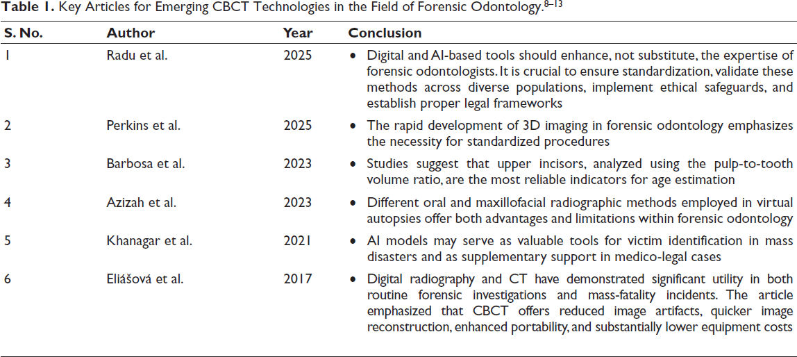
However, there is limited information available in the literature about the role of CBCT in forensic investigations. This informational gap was filled by conducting a computerized search in PubMed and several other search engines to precisely analyze the spatial relationship of teeth roots as well as the supporting alveolar structures on antemortem and post-mortem profiles provided by the technique of digital volumetric tomography. The present review provides a comprehensive narration of the varied literature on the significance of CBCT as a promising forensic tool. Additionally, it highlights the advancements in CBCT radiography, which enables us to capture the skeletal and anatomical details of the maxillofacial region.
Historical Perspectives of Dental Radiology
The use of X-rays for post-mortem investigations began in the year 1898. 14 Nevertheless, forensic identification using dental radiographs began in 1943. Owing to the improved digitalization in imaging techniques over the years, it is now possible to visualize jaws and associated alveolar structures in the form of a “truly reliable identity card” for forensic comparisons. 15
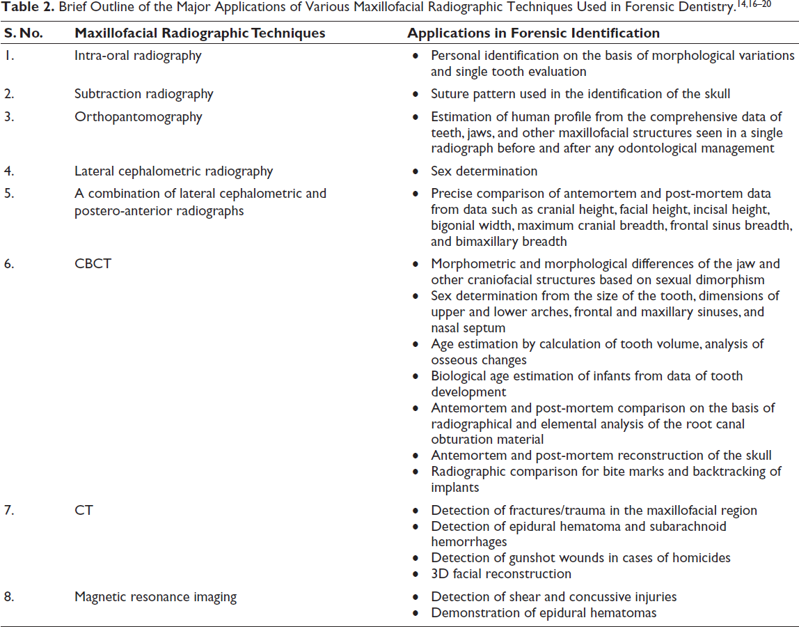
The radiographic techniques utilized for forensic identification include intra-oral and extra-oral radiographs in 2D and 3D. 16 Previously, bitewing and intra-oral periapical radiographic techniques were most frequently utilized in forensic identification methods to obtain post-mortem radiographs (Table 2). 15 However, literature evidence suggests the possibility of quality issues due to the chemical processing of the X-ray films, thereby necessitating the need for digitalization of the post-mortem data. 21 It is also important to note that the high-tech development of imaging modalities has brought a rapid expansion in forensic identifications. Currently, the forensic odontological identification and comparison system has been authorized by international criminal police organisation (INTERPOL) as one of the three primary identifiers in cases of multi-casualty incidents. 22 This is achieved by comparing radiographic images of the relevant region before and after death, followed by correlations made based on various characteristics of the stomatognathic system, such as tooth number and arrangement, osteopathology, periodontal bone loss, crown restorations, as well as osseous (anatomic) landmarks. 16 According to a study on the trends of forensic dentistry, Liu et al. (2016) have indicated significantly improved efficiency and accuracy rates with CBCT and orthopantomogram in personal identification of individuals when compared to the conventional X-ray methods. 4 There are different applications of maxillofacial techniques in forensic dentistry.
Applications of CBCT in Dental Forensic Identification
Previous evidence on age estimation has described the association between the deposition of secondary dentin in the walls of the pulp chamber. This process, which usually occurs over the course of a lifetime, was identified and measured by a sectioning procedure. To overcome this limitation, 2D and 3D radiographs are used nowadays to identify and measure these changes. However, the cylindrical shape of the pulpal structure renders its edges less visible in 2D radiographic techniques such as intra-oral X-rays or orthopantomograms, thereby resulting in inconsistencies and evaluation errors during its examination. Yet, the introduction of CBCT in dentistry provides a legitimate chance to collect precise measurements in 3D with superior image quality at a small radiation dose, thus affecting the role of CT and micro-CT to be less desirable for age estimations. Pinchi et al. conducted a pilot study in which they proposed a technique for determining age in a few minutes on the basis of perception of the pulp chamber and root canal to geometrical solids whose metric assessment was easier to obtain by CBCT image analysis. This radiographic approach also favored a good reproducibility rate due to the high inter-examiner agreement. 17 Considering age estimation with the aid of CBCT images by measuring the volume of pulp and tooth among the genders, Star et al. have suggested an association between age and volume of pulp and tooth in females. 23
To reliably estimate age, forensic teams opt for techniques based on the examination of osseous structures and developing teeth. The most common morphological approaches used are the evaluation of attrition, periodontal attachment, secondary dentin, cementum apposition, resorption of roots, and translucency of the apical zone. With the aid of CBCT images, Bayrak et al. suggested an association between the age in chronological order of an individual with cortication of mandibular condyles. He concluded that the stages of cortication increased subsequently with the chronological age in both males and females. 24
Apart from the dental findings, a major advantage of the craniofacial structures is that they are composed of hard tissues, which are relatively unbreakable and more likely to withstand physical trauma than other body parts. Therefore, these structures can be subjected to osteometric analysis for the identification of burned victims. Examples of such structures include the occipital condyle, mastoids, and foramen magnum. 25
Each tooth in the human dentition has a large variety of anatomical variants, including the presence of a supernumerary root in deciduous and permanent dentition, prevalence of more than double root canal in anterior teeth, occurrence of a mesial (middle) canal in mandibular molar teeth, and others. To facilitate comparative personal identification, a forensic odontologist must possess adequate knowledge of anatomical discrepancies and associated radiographic reference points that can be easily disseminated with the help of individual tooth examination by CBCT. Also, intracoronal/radicular restorations and broken instruments that have the capability of withstanding high temperatures can be used for comparison studies on the basis of analysis of elements of the obturative material. In his narrative review, Ahmed et al. recommended the need for endodontists to be equipped with the appropriate techniques required for a dental radiographic evaluation to ease the process of meticulous duplication of the radiographic images for forensic comparisons. 26
Advantages of CBCT in Forensic Maxillofacial Imaging
CBCT with a typically full field of view aids in the forensic examinations of the skull with an equivalent discerning efficacy comparable to CT, along with the advantage of low radiation dosage, wide availability, and cost-effectiveness than CT. 27 CBCT images are capable of detecting hidden pathologies and injuries associated with the maxillofacial region, thus allowing the focus of a regional autopsy on specific areas. 2 Murphy et al. investigated the precision of CBCT for dental forensic identifications using an analytical approach similar to that of Kirchhoff et al. 28 and concluded that radiographic images using CBCT had 96.6% and 98.4% of overall mean sensitivity and specificity, respectively. 29 The ability to acquire images in axial, sagittal, and coronal planes and their reconstruction in the absence of magnification are some of the reliable advantages of CBCT images. 24 One of the major limitations of CT is the presence of multiple streak artifacts, which prevailed despite the extended CT reconstruction scales. However, many studies determined the viability of comparing antemortem and post-mortem CBCT volumes of the maxillofacial region by introducing a CBCT reformatting approach, thereby resulting in images in a single scan with fewer streak artifacts than the CT images. 30
Limitations of CBCT in Forensic Maxillofacial Imaging
Some of the valid methods suggested for forensic dental identifications, such as CBCT and digital periapical radiography, also have certain inherent limitations. These methods lack standardized policies that safeguard the obtained radiographic images against manipulation. 26 One significant limitation of CBCT is the formation of artifacts in the presence of metallic objects, such as crowns or dental restorations, especially in regions above and below the X-ray source’s rotational plane during 3D image reconstruction. This leads to diminished image quality, which hampers the analysis of the maxillofacial region and restricts diagnostic accuracy, posing challenges since all of this information remains valuable for forensic identification when compared to antemortem data, even if it is not used as the primary basis for comparison. 8 Often, CBCT machines are not viable for radiographic imaging of deceased individuals as they require invasive detachment of half of the head or mandible from the body, which is not permitted ethically. Such circumstances necessitate the need for a supine CBCT unit or portable CT unit, which can be conveniently transported to the disaster scene for forensic identifications. 30 Also, literature evidence has suggested the employment of ultra-high resolution CT images for the qualitative analysis of endodontic and restorative materials using a virtual technique. Most of these authors have expressed a varied density of the endodontic and restorative materials in Hounsfield units, which is not reliable in CBCT imaging. 31
Research such as that by Chatra et al. (2020) supports the forensic reliability of frontal sinus volumetric analysis with CBCT, underscoring its role in sex determination. 32 Similarly, mandibular morphometric research by Yendriwati et al. (2022) indicates that measurements obtained from CBCT yield high levels of accuracy in distinguishing sex. 33 Age estimation research has also advanced, especially through pulp-volume assessment. The CBCT study by Sindhuja et al. (2024) highlighted this progress, demonstrating the effectiveness of volumetric regression models. 34 CBCT has also been used to assess skeletal maturation markers such as the spheno-occipital synchondrosis in a study by Vani et al. (2022), providing additional support for medico-legal age estimation. 35 Furthermore, morphometric studies of the orbit and paranasal sinuses by Shetty et al. (2021) underscore CBCT’s emerging role in biometric identification. 36
Recent Advancements in CBCT for Application in Forensic Identification
3D Printing
It is a process that is intended to create 3D objects with tangibly accurate depth information by establishing successive layers of material under computer control in the form of Standard File Format data. The use of 3D printing technology in forensic dentistry has opened several new fields, including analysis of lip prints and bite marks, 3D facial reconstruction, and estimation of dental age. 37
Advancements in Artificial Intelligence
Artificial intelligence (AI) has significantly improved the diagnostic efficiency of CBCT imaging. Depending on the specificity of the application, such as forensic identification and CBCT data characteristics, the necessary algorithm involves detection, classification, and segmentation using a deep learning approach. 38
Use of CBCT as a Non-invasive Imaging Modality
Used for craniofacial reconstruction cases to overcome the limitations of other 3D imaging techniques. 39 A study conducted by Meundi et al. to assess the effectiveness of CBCT over other imaging methods, such as CT, for measuring facial soft tissue thickness found a negligible difference between the two techniques, thereby suggesting the significant role of CBCT in forensic reconstructions. 40
Virtopsy
Combining medical imaging technologies is helpful for the detection of pathology in decomposing tissues. The resultant extracted information in the form of digital CBCT data may be utilized for forensic facial reconstruction. This approach forms the basis of “virtopsy,” which makes use of effective imaging methods such as CBCT to offer a 3D image of the crucial information regarding pathological diseases of the body. 31
Micro-computed Tomography
Primarily used in research, micro-CT also shows promise in forensics. It offers higher resolution than CBCT, allowing detailed microscopic examination of dental structures such as enamel, dentinal tubules, and cementum layers. These attributes are especially valuable for precise age determination and evaluating age-associated changes in adults. 8
Conclusion
Maxillofacial forensic radiology is an advancing branch of the forensic discipline with a favorable scope. The technology of CBCT is continually evolving with various CBCT unit manufacturers upgrading a distinct domain, such as increased gray-scale bit amounts, small voxel elements, and finer contrast resolution. These advancements provide a plethora of options for additional research on the efficacy of CBCT in forensics, making it a valuable tool. Advancing AI in forensic odontology requires the development of standardized, validated, and ethically responsible systems and clarifying legal guidelines surrounding CBCT data storage. The research gaps can be reduced by giving priority to building extensive, open-access dental datasets that represent diverse populations, ages, and dental conditions.
Footnotes
Declaration of Conflicting Interests
The authors declared no potential conflicts of interest with respect to the research, authorship, and/or publication of this article.
Ethical Approval
Not applicable.
Funding
The authors received no financial support for the research, authorship, and/or publication of this article.
Informed Consent
Not applicable.