
Abstract
The X-ray examination of corpses is the most useful tool in the field of medico-legal diagnosis and images allow clear visualization of fractures and radio-opaque foreign bodies within the deceased. In particular, the observations in the neck are quite challenging, as the anterior neck structures have anatomical variations which need differentiation from real pathologic or traumatic findings. Fractures of the hyoid laryngeal complex are commonly regarded as evidence of asphyxia but these can be too subtle to be detected during autopsy. Hyoid fractures in victims of strangulation and hanging are well documented. Plain radiographs of the hyoid laryngeal complex of the neck can be used to determine if fracture is present or if synchondrotic joint explains the apparent discontinuity in the bone or abnormal mobility. We present a study of postmortem radiological assessment of hyoid laryngeal complex of 12 medico-legal cases, which were conducted in the Department of Forensic Medicine & Toxicology, at a tertiary hospital, to find the exact cause and manner of death. Postmortem radiographs were helpful as an aid in the routine autopsy, and documented the signs of trauma to bony structures in the neck.
Introduction
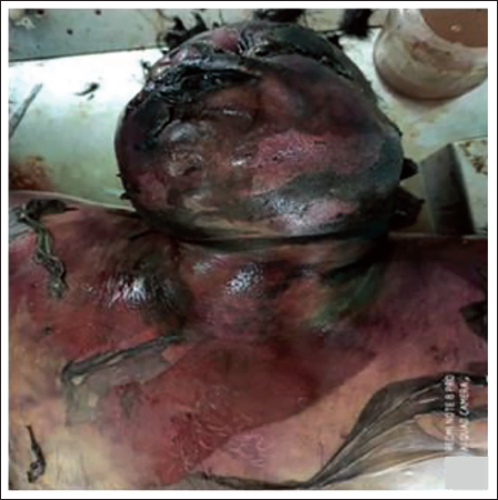
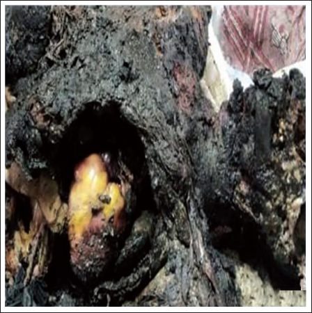
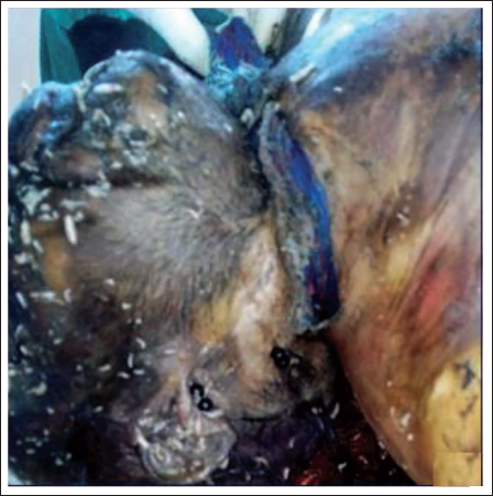
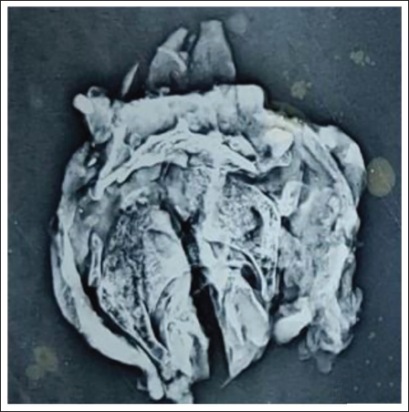
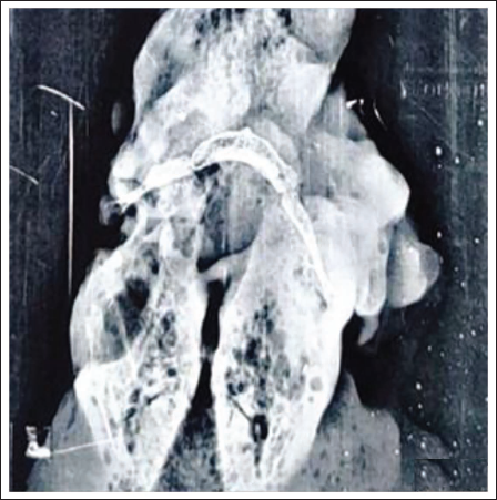
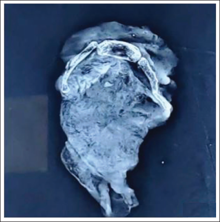
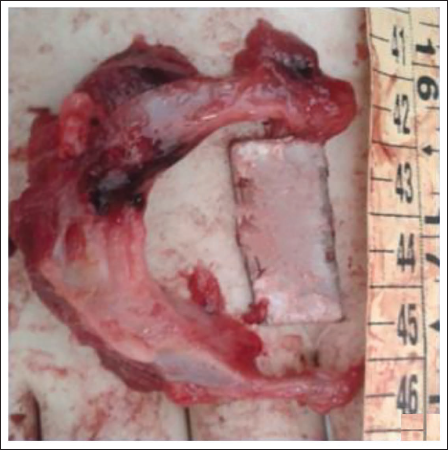
The objectives of a postmortem examination include establishing the identity of the body, to ascertain time since death, cause of death, and whether the death was natural or unnatural. 1 In postmortem examination of decomposed, charred (Figures 1–3) or highly infectious cadavers and in some countries due to religious practices, there should be a useful alternative way to examine injury in cervical part of cadaver, such as mechanical asphyxia, whiplash injury, vertebra injury, or vascular injury. 2 In addition, the charred tissues are difficult to dissect from the bones, and the traumatic fractures could be missed by the macroscopic analysis. 3 Similarly, in decomposed dead bodies where the neck skin is grossly discolored or lost, it is the internal damage to the neck tissue and hyoid bone, which tells the actual cause of death. 4 In suspected asphyxial deaths, the hyoid bone becomes the most integral part of internal examination to check whether it is a fracture, ante-mortem or postmortem in nature or just an artifact (Figure 4).
Swollen, Decomposed, Discolored, Peeling of Skin.
Charred Body with Neck Structures Charred.
Decomposed, Maggots, Ligature Material In Situ.
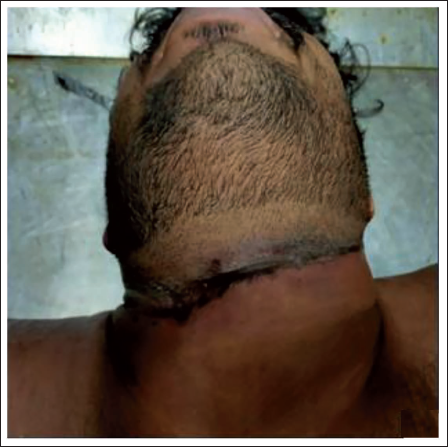
Oblique Ligature Mark on Neck.
Postmortem forensic radiology deals with the application of modern radiological methods in order to optimize postmortem diagnosis. The use of radiological imaging methods for postmortem purposes is nearly as old as radiology itself. 5 Such imaging methods can predict injuries in the internal body before autopsy with minimal or non-invasive techniques to the cadaver. 6 Keeping this view in mind, forensic medicine experts have used radiological findings in addition to support the autopsy findings in all gunshot wound cases, deaths of infants and young children, victims of explosions and if a body is decomposed, charred or unidentified. 7 Thus, radiology is a valuable tool in the identification of lesions in neck structures, enabling discrimination between strangulation and suicide. 8 The hyoid bone follows normal fracture characteristics of bone. A fracture line in the greater horn, dislocation and or angulation can be seen as a radiolucent line, interruption of the cortex or displaced fragments to diagnose a hyoid bone fracture. 9
Materials and Methods
During the routine postmortem examination in the Department of Forensic Medicine & Toxicology, cases with suspected asphyxia deaths which were referred, on the basis of either being decomposed/charred/suspicious history of asphyxia deaths or in the interest of justice. During dissection of the neck region, the hyoid laryngeal complex was subjected to radiological examination, to rule out any prior fracture or bony injury.
Results and Observation
The external, internal, and the radiological findings of HLC of 12 cases as and when were subjected to postmortem examination, are recorded, in tabulated format (Table 1).
Chart Showing the Findings and Radiological Images of Asphyxial Deaths.
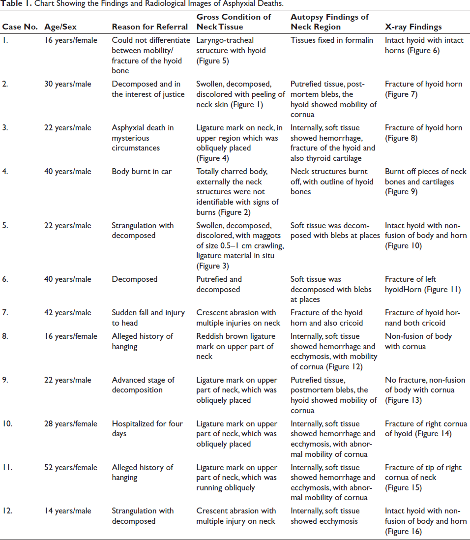
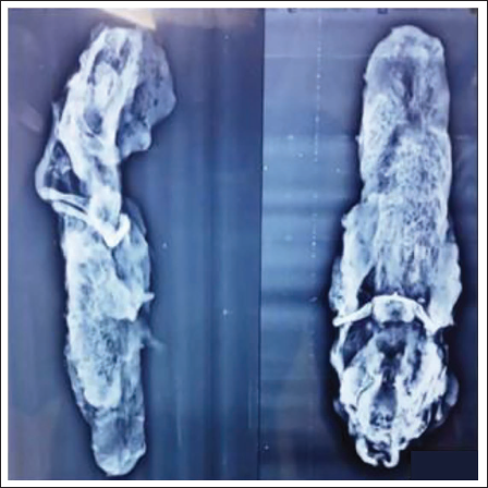
Thyro-Hyoid Complex in Formalin with Radiological View.
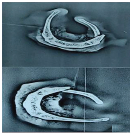
X-ray of Hyoid after Dissection of Soft Tissue.

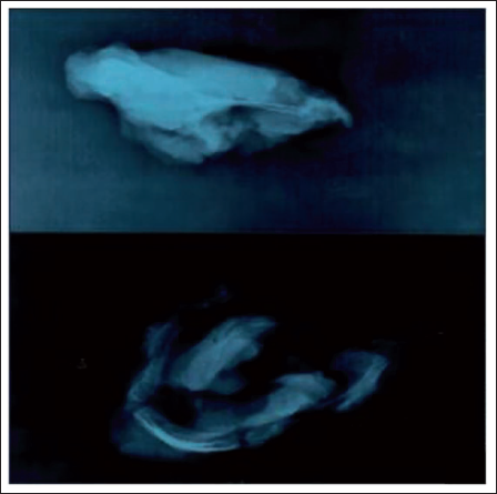
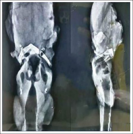
X-ray Showing Fracture of Hyoid Bone.
X-ray of Soft Tissue and Articulated Hyoid Showing Fracture.
X-ray of Charred Neck Tissue with Bony Structure and Soft.
X-ray of Neck Soft Tissue with Non Fused Hyoid Bone.
X-ray with Fracture on Left Side and Soft Tissue.
Fracture Hyoid with Haemorrhage and Ecchymosis.
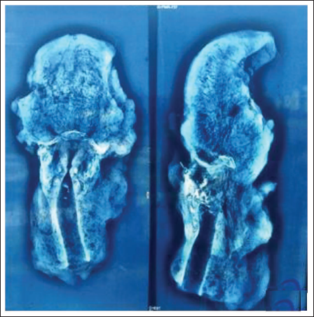
X-ray of Soft Tissue with Non Fused Hyoid.
X-ray with Soft Tissue and Fracture of Right Cornue of Hyoid.
X-ray of Soft Tissue with Fracture of Tip on Right.
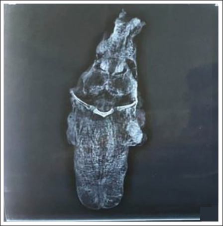
Hyoid Intact with Non Fusion of Body and Cornue.
Discussion
Hyoid bone is unique; being a sole bony structure, it is U-shaped and lies at the root of the tongue with a central body, two greater horns which sweep backwards and upwards and has natural joints between the body and the greater horns. In teenagers and young adults, the joints are cartilaginous and mobile, and they calcify irregularly as the age increases in middle and later life. These natural joints may be mistaken as fractures, if dissection is not done meticulously, as in Case No. 1 where mobility of the horns was doubtful, hence the radiography revealed no fractures as seen in Figures 5 and 6. The ante-mortem fractures are usually associated with extravasations of blood at the site 10 (Figure 16). There is also the possibility of fractures being postmortem, due to incorrect autopsy techniques, inexperienced forensic medicine experts, body transit trauma, improper handling in the mortuary. 11 While conducting autopsy, the hyoid bone fractures are difficult to detect and to avoid such misdiagnosis, a careful dissection, radiological examination, or seeking for evidence of hemorrhage is commonly recommended. 12
Isolated hyoid bone fractures are associated with accidental trauma (falls, car accidents), self-inflicted injuries (suicide by hanging) as well as assaulted injuries (manual or ligature strangulation, attack with a blunt instrument), but also artificial postmortem damage as a consequence of removal at autopsies. 13 Radiographs are typically taken in a lateral orientation and must reveal a radiolucent line, interruption of the cortex or displaced fragments to diagnose a hyoid bone fracture 14 (Figures 7, 8, 11, and 15), as was observed in Case No. 2 and 3, 6 and 11 where the radiographs of the neck structures were in coherence with the findings of the autopsy. Fractures are typically seen in the body or the greater horn of the hyoid. 15 As was observed in Case No. 2, 3, 6, 7, 10, 11, and corresponding Figures 9, 10, 11, 12, 14. Morlid also performed the radiography technique before dissection and found it to be very useful, as very fine bony structures of the small bones were revealed. 16 According to Polson G. J. (1955), the fractures of the hyoid ’bone in cases of hanging involve the greater horns, which are most likely to break at about the junction of their outer one third and inner two thirds. 17 It is used to document peri-mortem fractures (those caused at or around the time of death), confirm fractures of the hyoid bone in the neck due to hanging, and is also in identification of individuals by determining the age and sex of human remains. It establishes positive identification by means of comparison of ante-mortem and postmortem films. 18 A similar study proved that the X-ray assessment of the excised HLC proved to be the most reliable imaging technique for postmortem radiological evaluation of suspected violence to the neck 19 (Figures 9, 10, 13, and 14). Imaging can yield information about findings for later confirmation during autopsy and guide the forensic pathologist to focus on a structure of interest, but it can also visualize lesions that would be impossible to discover and diagnose via standard autopsy alone. 6 In light of the advantages of imaging, no autopsy of a trauma-related death can be complete without it. 20 It has been stressed, in a developing country like India, traditional postmortem cannot be completely replaced, but digital autopsy has also been increasingly prominent and recognized. 21 Legally for determining age or to prove and document injuries in victims of crime, radiologists understanding and forensic doctor’s knowledge of postmortem in a corpse must be synergized. 22 Hence application of imaging techniques can be used as the guidance and complimentary for the forensic autopsy in the identification of injury manner. 7
Conclusion
The evidence produced by radiologic methods can provide a scientific and documentary basis for the investigation agencies. It endorses precision, objectivity, critical thinking, careful observation and practice, repeatability, uncertainty management, and peer review. This will further encourage the use of multidisciplinary approach in the context of medico-legal evaluation of laryngo-hyoid injuries. At the periphery level it will guide to assess suspected laryngeal trauma.
Footnotes
Declaration of Conflicting Interests
The authors declared no potential conflicts of interest with respect to the research, authorship, and/or publication of this article.
Ethical Approval and Informed Consent
The Ethical Approval and Informed Consent has been obtained for the study.
Funding
The authors received no financial support for the research, authorship, and/or publication of this article.