
Abstract
Osteogenic matrix cell sheets (OMCSs) are ideal for bone regeneration. Transportation of OMCSs may be necessary, during which their osteogenic ability must be maintained. Here, we evaluated different media and temperatures for OMCS preservation. Bone marrow stromal/stem cells (BMSCs) were obtained from Fischer rats and analyzed for stem cell markers by flow cytometry. OMCSs were prepared from BMSCs by treatment with dexamethasone and ascorbic acid phosphate. After OMCS collection, they were stored in minimum essential medium (MEM) or Hank’s balanced salt solution (HBSS) at 37, 22, or 4°C for 24 hours. Cell viability and cytotoxic effects in the preservation conditions were determined by adenosine triphosphate (ATP) contents and lactate dehydrogenase (LDH) release, respectively. Osteogenesis was assessed by subcutaneously implanting preserved OMCSs around β-tricalcium phosphate ceramic disks into syngeneic rats. Implants were evaluated by alkaline phosphatase (ALP) activities, osteocalcin contents, and histology. Mesenchymal stem cells comprised 51% of primary cultured BMSCs. ATP contents were significantly different in OMCSs stored in MEM or HBSS at 22°C and 4°C. LDH release was significantly different in OMCSs stored in HBSS at 22°C and 4°C. The highest LDH release was observed in OMCSs stored in HBSS at 37°C. ALP activities and osteocalcin contents were the lowest in implanted OMCSs stored in HBSS at 37°C at four weeks after subcutaneous implantation. There was a significant difference in the osteocalcin levels of implanted OMCSs stored in MEM at 37°C and HBSS at 4°C. Abundant bone tissue around and inside disks was found in histological sections of OMCSs stored in all preservation conditions except for MEM and HBSS at 37°C. Maintaining the osteogenic ability of OMCSs during transport is important, and preservation of OMCSs in MEM or HBSS at 4°C or 22°C is a simple and inexpensive method.
Introduction
Cell-based therapy, carried out in a cell processing center (CPC), has been applied clinically 1 –3 . In particular, mesenchymal stem cells (MSCs) have attracted great interest for bone, tendon, muscle, and nerve regeneration because MSCs are multipotent and can differentiate into osteoblasts 4,5 , chondrocytes 2 , and neurons 6,7 . There is a huge demand for cell therapy using MSCs at general hospitals without a CPC. CPCs are generally established in university hospitals; the high expense of a CPC means that general hospitals cannot operate them. Recently, cell sheet technology has emerged as a cell transplantation system without a scaffold and has been used for regenerative treatments of the heart 8 , cornea 9 , and liver 10 . We have reported the utilities of osteogenic matrix cell sheets (OMCSs) derived from bone marrow stromal/stem cells (BMSCs) for bone reconstruction and treatment of pseudarthrosis 11 –15 . However, a CPC is necessary to prepare cultured cells and cell sheets, and cell sheets must be preserved and transported to general hospitals that have no CPC for clinical use. Transportation of OMCSs while conserving their activities enables the application of cell sheets in various diseases and injuries. There is currently no expedient and equable technique to conserve and transport cell sheets. The aim of this study was to develop a method for conserving and transporting cell sheets.
Materials and Methods
Ethics Statement
The care and handling of animals used in this study was approved by the institutional animal care and use committee of Nara Medical University and met the standards of the National Institutes of Health (protocol#: 11538).
Preparation of Bone Marrow Cells
Seven-week-old male Fischer 344 rats were purchased from CLEA Japan, Inc. (Tokyo, Japan) for use as donors and recipients. BMSCs were obtained by flushing out the rat femur shafts with 10 ml culture medium consisting of minimal essential medium (Nacalai Tesque, Kyoto, Japan) containing 15% fetal bovine serum and antibiotics (100 U/ml penicillin and 100 µg/ml streptomycin; Nacalai Tesque). The BMSCs were incubated at 37°C in a humidified atmosphere with 5% CO2. At confluency (approximately day 14), the primary cultured BMSCs were harvested using a 0.25% trypsin/EDTA solution (Nacalai Tesque) to obtain a cell suspension.
Analysis of Cell Surface Markers
To demonstrate MSCs among BMSCs after primary culture, cell surface CD markers were analyzed by flow cytometry (FACSCalibur, Becton Dickinson, Franklin Lakes, NJ, USA).
Single cell suspensions of BMSCs in phosphate-buffered saline (PBS) were exposed to antibodies directly coupled with a fluorochrome for 30 min on ice. The antibodies used were anti-CD90 (Becton Dickinson) or Mouse IgG1 κ isotype (control; Becton Dickinson) and anti-CD29 (Becton Dickinson) or Hamster IgM κ isotype (control; Becton Dickinson) directly coupled to phycoerythrin and anti-CD90 (Becton Dickinson) or Mouse IgG1 κ isotype (control; Becton Dickinson) coupled to fluorescein-5-isothiocyanate as representative stem cell markers. Similarly, anti-CD45 (eBioscience, San Diego, CA, USA) or Mouse IgG1κ isotype (control; eBioscience) directly coupled to allophycocyanin was used as s representative marker of hematopoietic and endothelial stem cells.
Cell Sheet Preparation and Preservation
We have previously reported a method to prepare OMCSs 11 . Briefly, BMSCs obtained from primary culture were seeded at a density of 1 × 10⁴ cells/cm2 onto 60-mm cell culture dishes for subculture in regular medium containing 10 nM dexamethasone (Sigma, St. Louis, MO, USA) and 0.28 mM ascorbic acid phosphate (AscP; L-ascorbic acid phosphate magnesium salt n-hydrate; Wako Pure Chemical Industries, Kyoto, Japan). OMCSs were formed by culture to confluency (approximately 2 weeks), and then rinsed twice with PBS (Gibco Life Technologies, Carlsbad, CA, USA). The sheet was lifted by a mechanical retrieval method using a cell scraper.
OMCSs were divided into seven groups according to the preservation method: fresh (control group), storage in culture medium at 37°C (37°C MEM group), storage in culture medium at room temperature (22°C MEM group), storage in culture medium at 4°C (4°C MEM group), storage in Hank’s balanced salt solution (HBSS; Gibco) at 37°C (37°C HBSS group), storage in HBSS at 22°C (22°C HBSS group), and storage in HBSS at 4°C (4°C HBSS group). For preservation, OMCSs were picked up using tweezers and placed into a 50-ml tube (Falcon) with 50 ml culture medium or HBSS (six OMCSs per tube). Tubes were then transferred to an incubator (37°C), a clean bench (room temperature: 22°C), or refrigeration chamber (4°C) and stored for 24 h.
Cell Viability and Cytotoxicity Assays
To investigate the viability of OMCSs before and after preservation, we employed a method based on adenosine triphosphate (ATP) content (TOYO B-NET, Tokyo, Japan). Furthermore, lactate dehydrogenase (LDH) activity in preservation media was measured to assess the cytotoxicity of OMCS preservation using a LDH Cytotoxicity Detection Kit (Takara Bio Inc., Shiga, Japan).
Total cellular ATP content was measured in an OMCS plated in a 60-mm dish. The OMCS was lysed with 1 ml lysis buffer and placed directly into the chamber of a luminometer (SpectraMax, Molecular Devices, San Jose, CA, USA). Light emission was recorded after addition of 1 ml luciferin-luciferase solution. ATP Standard Solution (TOYO B-NET) was used for the standard.
To evaluate LDH activity, preservation supernatants were collected and transferred to new tubes. The LDH activity was measured by determining absorbance at 490 nm with an automated microplate reader (SpectraMax M2, Molecular Devices). LDH Standard Solution (Roche, Tokyo, Japan) was used for the standard.
Implantation of Tricalcium Phosphate Constructs Wrapped with an OMCS
Sterilized porous β-tricalcium phosphate (TCP) ceramics (Superpore; disks: 5 mm in diameter; 2 mm thick; 65% porosity) were purchased from PENTAX (Tokyo, Japan). TCP constructs were prepared by wrapping OMCSs from each group around the TCP immediately prior to transplantation. TCP disk constructs were implanted subcutaneously into the backs of syngeneic rats.
Biochemical Analysis
The alkaline phosphatase (ALP) activities and osteocalcin contents of harvested constructs were measured as reported previously 16 . After homogenization of each TCP construct using a microhomogenizer in 1 ml of 0.2% Nonidet P-40 (NP-40), homogenized TCP was centrifuged at 12,000 rev/min for 10 min at 4°C. A 10 μl aliquot of the supernatant was combined with 56 mmol/l amino-2-methylpropanediol buffer containing 10 mmol/l p-nitrophenyl phosphate and 1 mmol/l MgCl2. The combined supernatant was incubated for 30 min at 37°C, and the reaction was stopped by addition of 0.2 N NaOH. The ALP activity was calculated by determining the absorbance of p-nitrophenol released at 410 nm using a spectrophotometer.
After extraction with 0.2% NP-40 in 4 ml of 20% formic acid for 2 weeks at 4°C, osteocalcin was extracted from the sediment. A 0.5 ml aliquot of the extract was applied to a prepacked Sephadex G-25 column (NAP-5 column; Amersham Pharmacia Biotech AB, Uppsala, Sweden) and eluted with 10% formic acid. The protein fractions were pooled and dried. After dissolution in 0.5 ml ELISA sample buffer, the osteocalcin content was assayed using a Rat Osteocalcin ELISA Kit (DS Pharma Biomedical, Osaka, Japan).
Histological Examination
Four weeks after subcutaneous implantation, the implanted TCP disks with OMCSs were harvested and bone formation was evaluated histologically. The disks were fixed in buffered formalin (Wako Pure Chemical Industries). Each disk was decalcified with a KCL solution (K-CX solution; Falma, Tokyo, Japan), embedded in paraffin, cut through the middle of the disk parallel to the base, and then stained with hematoxylin and eosin (HE) for histological evaluation.
Statistical Analysis
ATP content, LDH activity, ALP activity, and the osteocalcin level are presented as the mean and standard deviation. One-way analysis of variance with post-hoc multiple comparisons using Tukey’s test was conducted to determine statistical significance. Values of p < 0.05 were considered as statistically significant.
Results
BMSC Surface Markers
BMSCs were positive for CD29 and CD90, and negative for CD45 as identified by flow cytometry. The CD29+ CD90+ CD45- population comprised 51% of total cells in each experiment (Fig. 1).
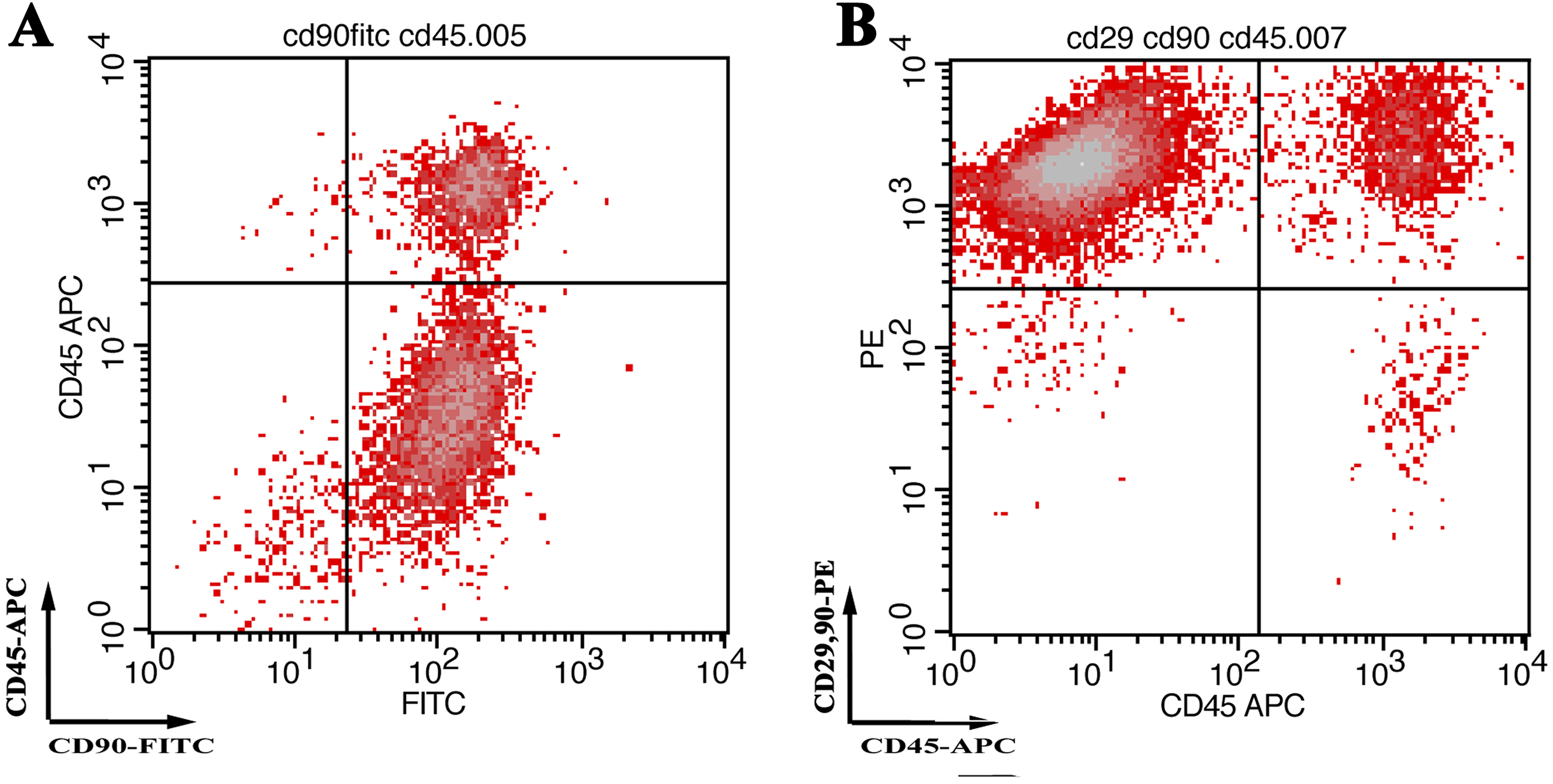
Profiling of bone marrow stromal/stem cells (BMSCs) for surface markers. Flow cytometry profiles of the BMSCs indicated the presence of >67% CD90+ and CD45– cells (A). Co-expression of CD29, CD90, and CD45 indicated the presence (>71%) of stem cells among BMSCs (B). PE: Phycoerythrin; APC: Allophycocyanin; FITC: Fluorescein isothiocyanate
ATP Contents and LDH Activities after 24 h of Preservation
There was no statistically significant difference in ATP content between the 22°C MEM and 4°C MEM groups or between the 22°C HBSS and 4°C HBSS groups (Fig. 2(A)). The OMCSs preserved at 22°C or 4°C retained 35–70% ATP content of the control group at 24 h. ATP content in the 37°C MEM and 37°C HBSS groups had largely decreased to 25% and 1% of those in the control group, respectively.

ATP content of OMCSs (A) and LDH activities (B) in the preservation solutions. There were significant differences in ATP content between the 22°C MEM and 4°C MEM groups, and 22°C HBSS and 4°C HBSS groups. For LDH activities, there were significant differences between the 22°C HBSS and 4°C HBSS groups. ATP: adenosine triphosphate; LDH: lactate dehydrogenase; MEM: minimum essential medium; HBSS: Hank’s balanced salt solution; ANOVA: analysis of variance
There were statistically significant differences in LDH release between the 22°C HBSS and 4°C HBSS groups (Fig. 2(B)). Significant LDH release was observed in the 37°C HBSS group.
ALP Activity and Osteocalcin Content of the Implanted TCP Wrapped with Preserved OMCSs
The ALP activities and osteocalcin content in the 37°C HBSS group were significantly lower than those in the other groups at 4 weeks after subcutaneous implantation (Fig. 3(A) and (B)). A significant difference was found in the osteocalcin level between the 37°C MEM and 4°C HBSS groups. There were no significant differences in the ALP activities or osteocalcin levels between the other two groups.

ALP activity (A) and osteocalcin levels (B) in TCP constructs. ALP activities and osteocalcin levels in the 37°C HBSS group were significantly lower compared with those in the other groups. There was a significant difference in osteocalcin levels between 37°C MEM and 4°C HBSS groups. ALP: alkaline phosphatase; MEM: minimum essential medium; HBSS: Hank’s balanced salt solution; ANOVA: analysis of variance
Histological Evaluation of the Implanted TCP Wrapped with Preserved OMCSs
Fig. 4 shows representative histological sections (HE staining) of the TCP constructs. Abundant bone tissue around and inside the disks was observed in the sections of all groups except the 37°C MEM and 37°C HBSS groups. Although mild new bone was found inside disks in the 37°C MEM group, only trace bone tissue was found.
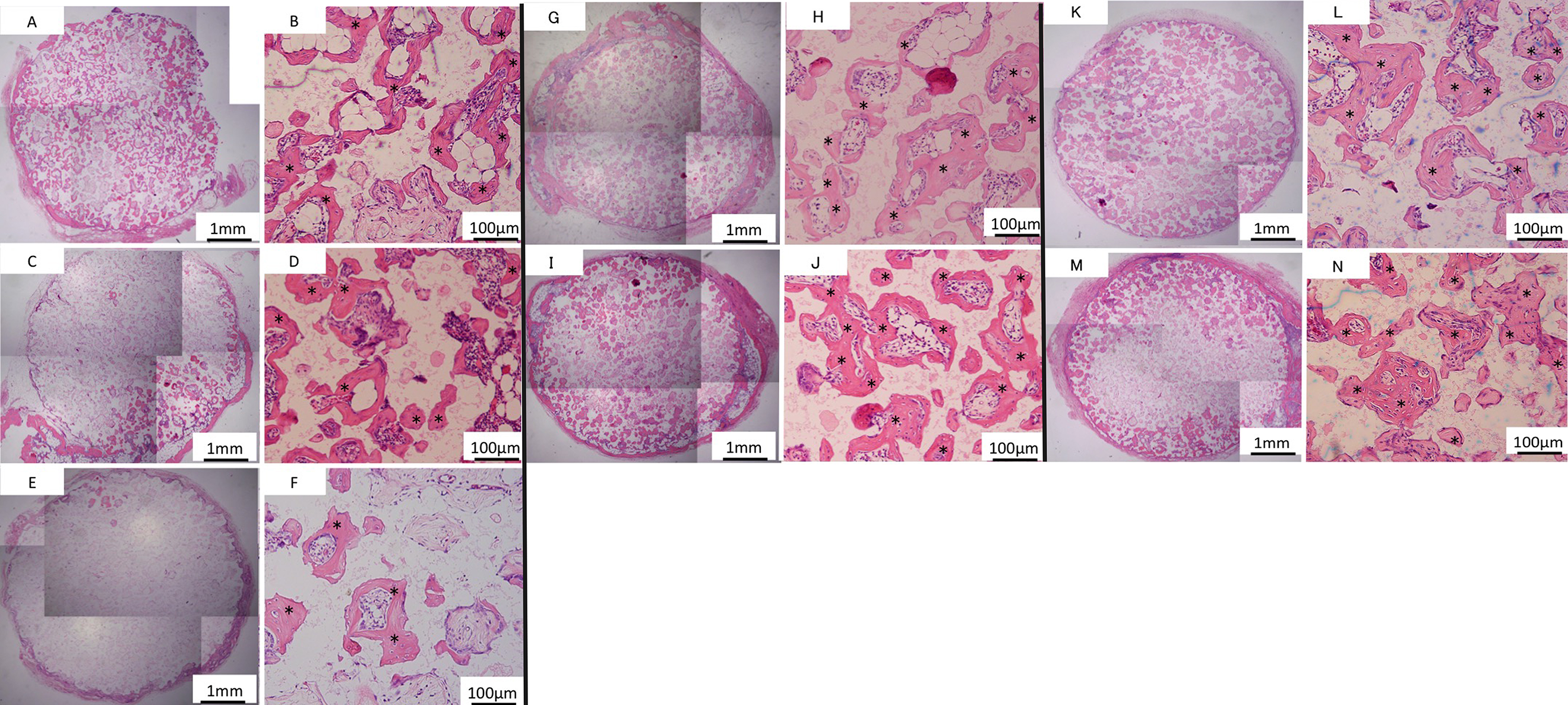
Hematoxylin and eosin-stained sections at 4 weeks after implantation into the subcutaneous layer of syngeneic rats. Poor bone formation was found in low magnification images of a β-tricalcium phosphate disk (TCP) in the 37°C MEM group (C and D). Low magnification images of a TCP disk in the 37°C HBSS group (E and F) showed negligible bone formation. Conversely, a high level of bone formation was visible in and around TCP in control (A and B), 22°C MEM (G and H), 22°C HBSS (I and J), 4°C MEM (K and L), and 4°C HBSS (M and N) groups. Asterisks indicate bone tissue. MEM: minimum essential medium; HBSS: Hank’s balanced salt solution
Discussion
Bone regeneration technology has undergone remarkable development and has been applied clinically 1 –3 . We have developed OMCSs as a novel cell transplantation technique and reported its effectiveness and utility for bone regeneration using several animal models 11,12,15,17,18 .
However, in our previous reports, we did not substantiate that BMSCs before subculture included MSCs. The results of flow cytometry in this study indicated that MSCs are an important cell source for OMCSs.
Because OMCSs abundantly produce bone growth factors and can differentiate to osteocytes, they are relevant materials for osteogenesis. However, cultured cells and cell sheets must be prepared in a CPC to ensure their safety. CPCs are established only in specific institutions such as universities and university hospitals. It is necessary to safely transport cell sheets cultured at a CPC to general hospitals to apply cell sheet-based tissue engineering and regenerative medicine. There have been several reports of transportation of cultured cells. Almost all the transportation techniques involved cryopreservation 19 –24 .
Because cryopreservation reduces the osteogenic abilities of OMCSs after thawing, it is not an ideal method for transportation within a short timescale 24 . Although a portable homothermal container has been developed to transport cells attached to culture dishes, it is a large, expensive device 25 . Therefore, the development of an easier and lower-cost method is anticipated.
In our present study, we investigated the osteogenic abilities of OMCSs preserved in culture medium or HBSS in plastic tubes for 24 h. After 24 h of preservation, OMCSs maintained their sheet form and could be wrapped around a TCP disk. In the viability assays assessing ATP activity and LDH release, OMCSs preserved at 22°C or 4°C showed low cell viabilities compared with fresh OMCSs. However, the OMCSs demonstrated similar osteogenic abilities to fresh OMCSs in biochemical analysis of ALP activities and osteocalcin content after subcutaneous implantation of TCP wrapped with the preserved OMCSs. A reason for this result is that, even in culture medium or HBSS, cells of OMCSs may slowly undergo apoptosis after detachment from culture dishes. However, a considerable number of cells may survive at 22°C. Therefore, the surviving cells could proliferate, differentiate, and secrete growth factors after implantation into the subcutaneous layer with TCP. Therefore, preserved OMCSs might maintain their osteogenic ability within 24 h. Because cellular metabolism of OMCSs does not decrease at 37°C, it had been speculated that nearly all preserved cells would not survive for 24 h. The current study suggests that OMCSs can be transported in plastic tubes containing culture medium or HBSS and applied to bone regenerative treatments. In our study, we evaluated osteogenesis of OMCSs on TCP that had been implanted into subcutaneous layers of syngeneic rats. The results indicate that OMCSs may promote osteogenesis at a necrotic bone site or bone defect as well as heal at a non-union.
Generally, temperature-responsive culture dishes are widely used to prepare cell sheets 26 . In our study, cell sheets were fabricated from BMSCs by addition of dexamethasone and AscP to the culture medium during subculture, and we harvested the cell sheets by retrieval with scrapers. Because dexamethasone and AscP are inductive differentiating factors, BMSCs can differentiate to osteoblasts in subculture. Moreover, dexamethasone and AscP promote secretion of extracellular matrix 27 . Therefore, the form of the cell sheet could be maintained for 24 h.
Our study has limitations that should be acknowledged. Six OMCSs were preserved in a 50 ml tube with 50 ml culture medium or HBSS. The amount of storage solution per sheet might be insufficient for 24 h of preservation. Second, we evaluated osteogenic potentials of OMCSs preserved for only 24 h. In a future study, it will be necessary to investigate how long an OMCS maintains its osteogenic ability. However, 24 h may be practically adequate for domestic transplantation of OMCSs, because the number of CPCs has been increasing. Last, although the current study employed animal experiments using rats, clinical application would be required to evaluate the osteogenic abilities of preserved OMCSs derived from human mesenchymal stem cells.
Conclusions
The results from this study clearly show that culture medium or HBSS maintain the osteogenic abilities of OMCSs within 24 h at 4°C or 22°C. Maintaining the osteogenic ability of OMCSs during transport is important, and preservation of OMCSs in MEM or HBSS at 4°C or 22°C is a simple, inexpensive, and useful method.
Footnotes
Acknowledgments
Author Contribution
TK conducted the animal experiments and wrote the manuscript; MA designed the study; TK, MA, and NO-S conducted biological analysis; SO, TI, and YT interpreted the results; TO conducted the data analysis.
Ethical Approval
This study was approved by the Nara Medical University Medical Ethics Review Committee (protocol#: 11538).
Statement of Human and Animal Rights
All procedures in the animal study were approved by the institutional animal care and use committee of Nara Medical University before experiments commenced.
Statement of Informed Consent
Statement of Informed Consent is not applicable for this article.
Declaration of Conflicting Interests
The authors declared no potential conflicts of interest with respect to the research, authorship, and/or publication of this article.
Funding
The authors disclosed receipt of the following financial support for the research and/or authorship of this article: this study was partially supported by the Translational Research Network Program of the Japan Agency for Medical Research and Development (AMED).