
Abstract
Scorpion envenomation is a serious public health issue. Androctonus mauretanicus (Am) and Buthus occitanus (Bo) are the most dangerous scorpions in Morocco. Despite their medical relevance, no study has yet related their kinetics of symptom apparition and the consequent tissue disorders at the same interval post-injection. This work achieved the first comparative pathophysiological and toxic-symptoms study between the Am and Bo venoms from a biochemical, toxicological and physiopathological standpoint. The activity of venoms and their subletal dose were determined by administration of increasing concentrations of the venoms. 30, 60 and 120 min following the experimental envenomation in mice, the profile of clinical symptoms was underlined and the main organs: brain, heart, lungs, liver and kidneys were removed for histological examination. The Am venom is a rich source of proteins and three-times more toxic than the Bo. The most observed clinical symptoms are neurological and cardiopulmonary. The Am venom caused histopathological alterations at 30, 60, and 120 min which were more important than the Bo. This study highlighted that both venoms exhibited a strong toxicity with variable intensities. Moreover, we showed the presence of correlation between the level of histopathological disorders observed and the intensity of signs appeared at the same time following venom inoculation.
Introduction
Scorpion envenoming is a relatively common occurence in the tropical and subtropical regions in North Africa, the Middle East, India, South, and Central America. 1 Annually, scorpion stings induce about 2600 deaths with an incidence of more than 1.5 million, most of which were reported in children. 2
In North Africa, Morocco has the greatest diversity of scorpion fauna, 3 with more than 50 species, explaining why Morocco has a high rate of endemism. 26 819 cases of scorpion stings were registered in 2019 by the Poison Control and Pharmacovigilance Center of Morocco. 4 The Androctonus mauretanicus (Am) and Buthus occitanus (Bo), belonging to the Buthidae family, are responsible for the majority of envenoming accidents.5,6,7
Scorpions are endowed with a venom apparatus that produces venom intended to attack, immobilize prey and defend against predators.
As for their composition, scorpion venoms are complex matrices, consisting of mucopolysaccharides, hyaluronidases, phospholipases, serotonin and histamine, but mainly of neurotoxins and small peptides 8 (<10 kDa) that target the ionic channels of excitable cells; especially sodium, potassium, calcium, and chloride.9,10,11 These neurotoxins are the most abundant components in the venoms of scorpions from the Buthidae family and may represent more than 73% of the venom composition. 12
During a scorpion sting, a very small amount of venom is inoculated causing multiple symptom that can be digestive, vegetative, neurological, cardiovascular, and/or respiratory signs13,14 and can eventually lead to death.
The major effects are mainly due to neurotoxins that disrupt the conduction of the nervous signal specifically by affecting the function of sodium and potassium ion channels, inducing the release of neurotransmitters, pro-inflammatory (IL-1β, IL-6, IL-8, and TNF-α) and anti-inflammatory cytokines (IL-10, IL-4), 15 that are responsible for all the alterations observed during scorpion envenomation. These neurotransmitters provoke pathophysiological actions, such as membrane destabilization, blocking of the central and peripheral nervous systems as well as alteration of muscle activity that may cause paralysis.
In order to improve the management of envenomed patients and anti-scorpionic therapy, an understanding of the mechanisms of action of venoms and their pathophysiological effects is necessary. In this context, we conducted a comparative pathophysiological study of scorpion envenomation in mice after intraperitoneal (IP) injection with sub-lethal doses of Am and Bo venoms. The main organs; brain, heart, lungs, liver, and kidneys were taken out at 30, 60, and 120 min after envenomation for histological analysis.
Methods
Venom milking
Am and Bo scorpions were collected from the region of Marrakech, Morocco (Figure 1). Captured specimens (200 scorpions for each species) were kept in the scorpionarium of the Institut Pasteur of Morocco (IPM). Venoms were milked by the electrical stimulation method.
7
The extracted venoms (40 mL for Am venom and 35 mL for Bo venom) were recovered using cold water and centrifuged for 20 min at 14,000 r/min at 4°C. The supernatant was lyophilized and then frozen at −20°C until use. Geographical localization of collected Androctonus mauretanicus and Buthus occitanus scorpions.
Estimation of protein
The protein concentration of Am and Bo venoms was estimated by the absorbance at 280 nm, according to the equation admitting that 1 unit of absorbance in a Quartz cuvette with 1 cm optical path is equal to 1 mg/mL protein concentration.16,17
Determination of Median Lethal Dose (LD50) and sub-lethal doses of Androctonus mauretanicus and Buthus occitanus venoms
As recommended by the World Health Organization,18,19 the Median Lethal Dose (LD50) was evaluated by the administration of increasing concentrations of the venoms adjusted in NaCl solution. The used concentrations of the Am and Bo venoms were 2, 5, and 8 μg/mice and 13, 16, 19, and 22 μg/mice, respectively. For each dose, six male Swiss mice (18–22 g) were used and observed to record the mortality rates for a period of 24 h after intraperitoneal injection (IP) of the venoms. The software package GraphPad Prism 5 was used for LD50 determination in accordance with the supplied algorithm. A non-linear curve was traced respecting the four-parameter logistical equation then constraints were set on minimum (0% mortality) and maximum values (100% mortality).
From the LD50, decreasing amounts of Am and Bo venoms were injected into six homogeneous groups of mice, to determine their corresponding sub-lethal doses (sLD). 20 The saline solution (NaCl 0.9%) was administered in mice control. All injections were made by IP route.
Experimental protocol
Male Swiss mice (18–22 g) were kept in cages and at room temperature with access to food and water at the animal unit of IPM.
Four groups of eight mice were used in this experiment; the control group received a physiological saline solution (NaCl 0.9%), while the other groups (2–4) were administered by each venom intraperitoneally.
Subsequently, the mice were euthanized and sacrificed at different times; 30, 60, and 120 min after envenomation. Then, the main organs: brain, heart, lungs, liver, and kidneys were taken out for histological analysis. During the experiment, the profile of clinical symptoms was underlined.
Histological analysis
The organs (brain, heart, lungs, liver, and kidneys) were carefully harvested for histological analysis and fixed immediately in 10% formaldehyde solution for 24 h. The tissues were subsequently dehydrated in ethanol, clarified with xylene and embedded in paraffin. They were then sectioned in a perpendicular plane across the epithelial layers at 4 μm thickness. Sections were mounted on a glass slide carefully to avoid tissue folding or stretching, then stained either with hematoxylin and eosin, dried, cleared, and covered for observation and photo-micrography. Randomly selected sections were examined with light microscopy. 21
Ethics committee
All protocols and experiments included animals have followed the ethical principles defined by the World Health Organization 18,19 and approved at the IPM of Casablanca, by a local ethics committee (with agreement number 8.3. A-2015).
Statistical analysis
All results are expressed as means ± SD. The statistical significance of differences between groups was analyzed by a Student t test. Differences were considered significant if p values were < 0.0001.
Results
Estimation of protein
The results showed that the protein concentration of the Am and Bo venoms were 153.25 ± 3.22 and 117 ± 2.65 mg/ml, respectively. However, the Am venom was richer in proteins than the Bo venom.
Evaluation of the Median Lethal Dose (LD50) and sub-lethal doses of Androctonus mauretanicus and Buthus occitanus venoms
Median Lethal Doses (LD50) and sub-lethal dose (sLD) of Am and Bo venoms by IP route (with 95% confidence intervals and non-linear regression) (n = 6).

bThe values with different superscript letters in a column are significantly different (p<0.0001).
In order to analyze the pathophysiological effects of scorpion envenomation in mice, the sLD were determined. According to the obtained results (Table 1), the sLD of the Am venom was 245 ± 25.5 and 665 ± 100 μg/kg body weight for Bo venom.
The toxicity assay highlighted that Am venom was estimated to reach three fold greater values than for Bo venom.
Clinical symptoms of scorpion envenomation
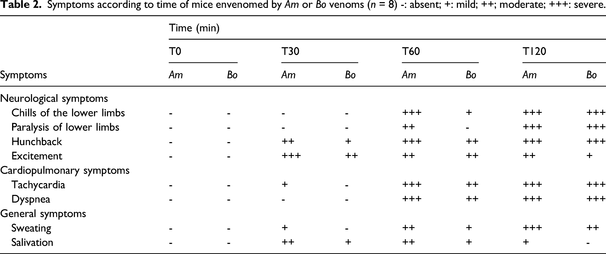
Symptoms according to time of mice envenomed by Am or Bo venoms (n = 8) -: absent; +: mild; ++; moderate; +++: severe.
Also, it should be mentioned that despite the taxonomic difference between the Am and Bo scorpions, there is a high homology of the toxic effects of their venoms and their antigenic properties. However, the venom Am led to toxic effects more consequent than the Bo venom.
Alterations disorders
The histological examination was performed for the following organs: brain, lung, heart, liver, and kidneys after an experimental envenomation of mice by the IP route, with the sLD of Am venom (245 μg/kg body weight) and Bo venom (665 μg/kg body weight). Compared to the control group, significant structural alterations in the histological sections were noted at 30, 60, and 120 min post-injection. Interestingly, all the studied organs were affected and the tissue damages generated by the Am venom were more important than those of the Bo venom (Figure 2). Histological sections of the mouse organs after 30, 60 and 120 min of experimental envenomation by Am and Bo venoms (x40) Bv: Blood vessel; Nf: Nerve fibers; Cg: Glial cell; N: Neurone; Hr: Hemorrhage; Vd: Vasodilation; Av: Alveole; Ase: Alveolar space enlargement; Ad: Alveolar destruction; Inf: Inflammatory cell infiltration; Mf: myofobrils; Dmf: Degeneration of the myofibrils; Hp: Hepatocyte; Sn: Sinusoid; Cv: Centrilobular vein; Tg: Turgescent; Esn: Sinusoidal dilation; Cr: Caryolysis; Nc: Necrosis; Pc: Pycnosis; Cng: Congestion; Tl: Tubular lumen; Gm: Glomerulus; Bc: Bowman’s capsule; Gd: Glomerular disorganization; Elt: Enlargement of tubular lumen (n = 8).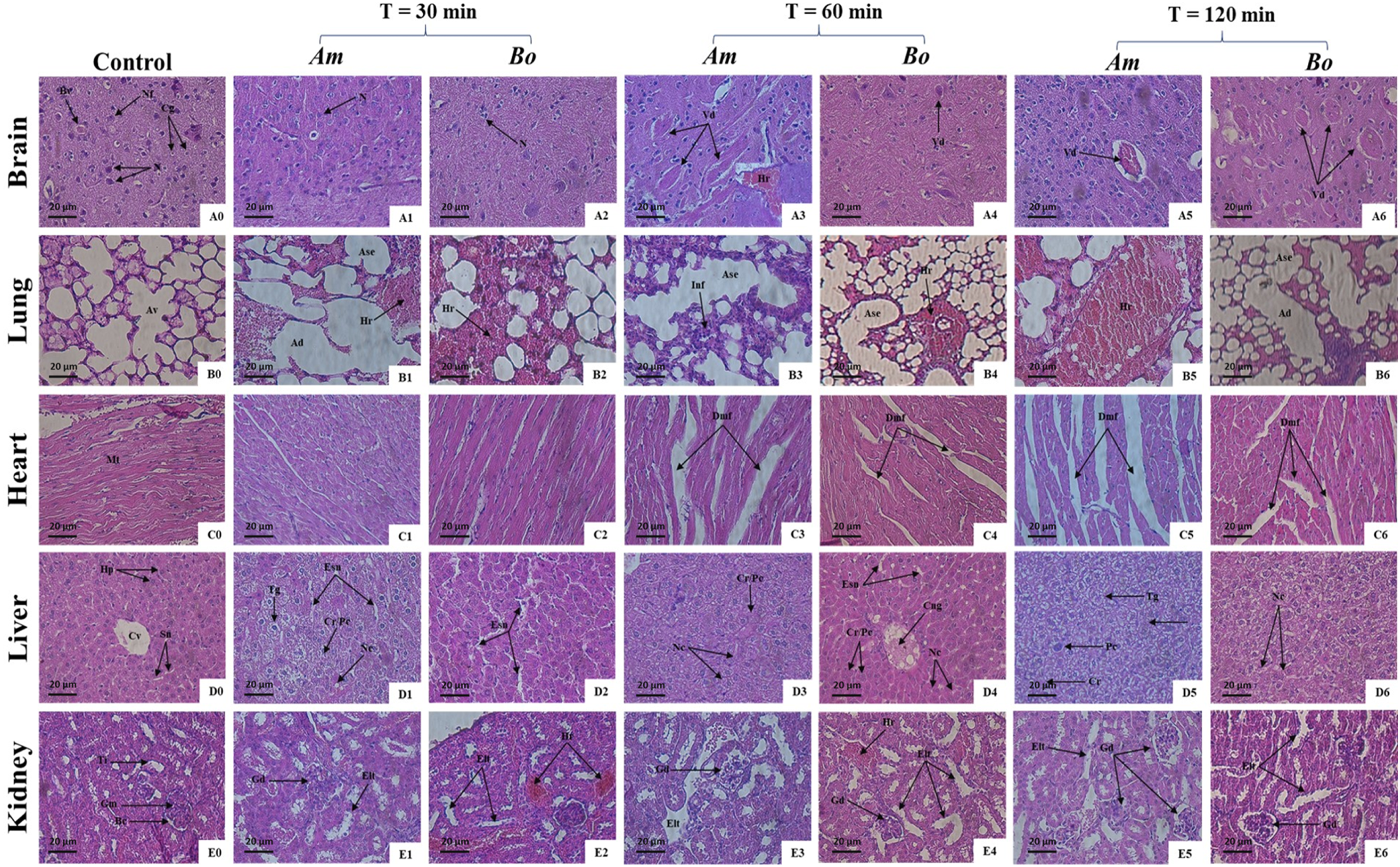
Macroscopically, after dissection of mice envenomed by sub-lethal doses of Am and Bo venoms, the organs (brain, lung, and kidney) presented red spots and we noted an increase in liver size (hepatomegaly). The Am scorpion venom appears to be more potent in terms of red spots and hepatomegaly than Bo scorpion venom.
Histopathological studies of the brain
The brain tissue of control mice appeared normal without any histological damage. However, the histological evaluation of the cerebral cortex of envenomed mice with the sub-lethal dose of Am or Bo venoms revealed no histological lesions in brain at 30 min. Though, excessive vasodilation at 60 and 120 min was found in both groups (Figure 3), while the hemorrhagic foci were registered in mice only at 60 min after administration of Am venom (Figure 3(A3)). Histological sections of the brain after 30, 60 and 120 min of the experimental envenomation by Am or Bo venoms (x40) (n = 8). Av: Alveole; Ase: Alveolar space enlargement; Hr: Hemorrhage; Ad: Alveolar destruction; Inf: Inflammatory cell infiltration.
Histopathological studies of the lungs
Histological examination of the lungs tissue of control mice appeared normal, while the examination of histological sections of the pulmonary tissue of envenomed mice by both venoms showed an important alveolar space enlargement and destruction associated to major hemorrhage with a diameter, in group traited with Am venom at 120 min, as large as the diameter in the other groups (Figures 4(B1–B6)). In addition, an important infiltration of inflammatory cells; macrophages were noted after 60 min of the injection by the Am venom (Figure 4(B3)). Histological sections of the lung after 30, 60 and 120 min of experimental envenomation by Am and Bo venoms (x40) (n = 8). Mf: myofibrils; Dmf: Degeneration of the myofibrils.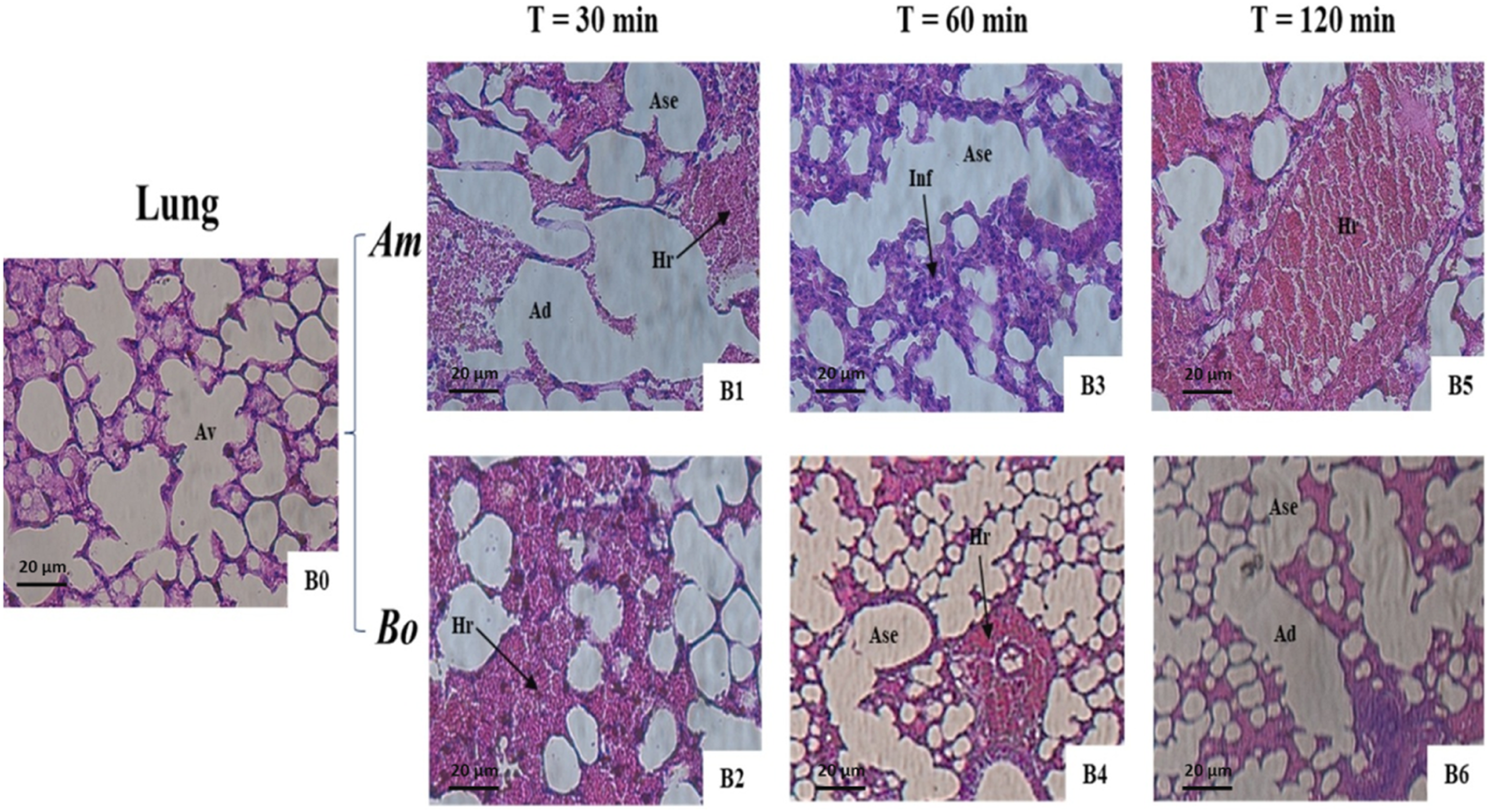
Histopathological studies of the heart
The heart tissue of the control group showed no visible lesion, while the heart tissue of envenomed mice revealed a degenerative myofibril since 60 min after envenomation for both venoms. However, the groups of mice sacrificed at 120 min showed massive deleterious degeneration (Figures 5(C5–C6)). The degree of damage due to venom from Am was more damaged than Bo venom. Histological sections of the heart after 30, 60 and 120 min of experimental envenomation by Am and Bo venoms (x40) (n = 8). Hp: Hepatocyte; Sn: Sinusoid; Cv: Centrilobular vein; Tg: Turgescent; Esn: Sinusoidal dilation; Cr: Caryolysis; Pc: Pycnosis; Nc: Necrosis, Cng: Congestion.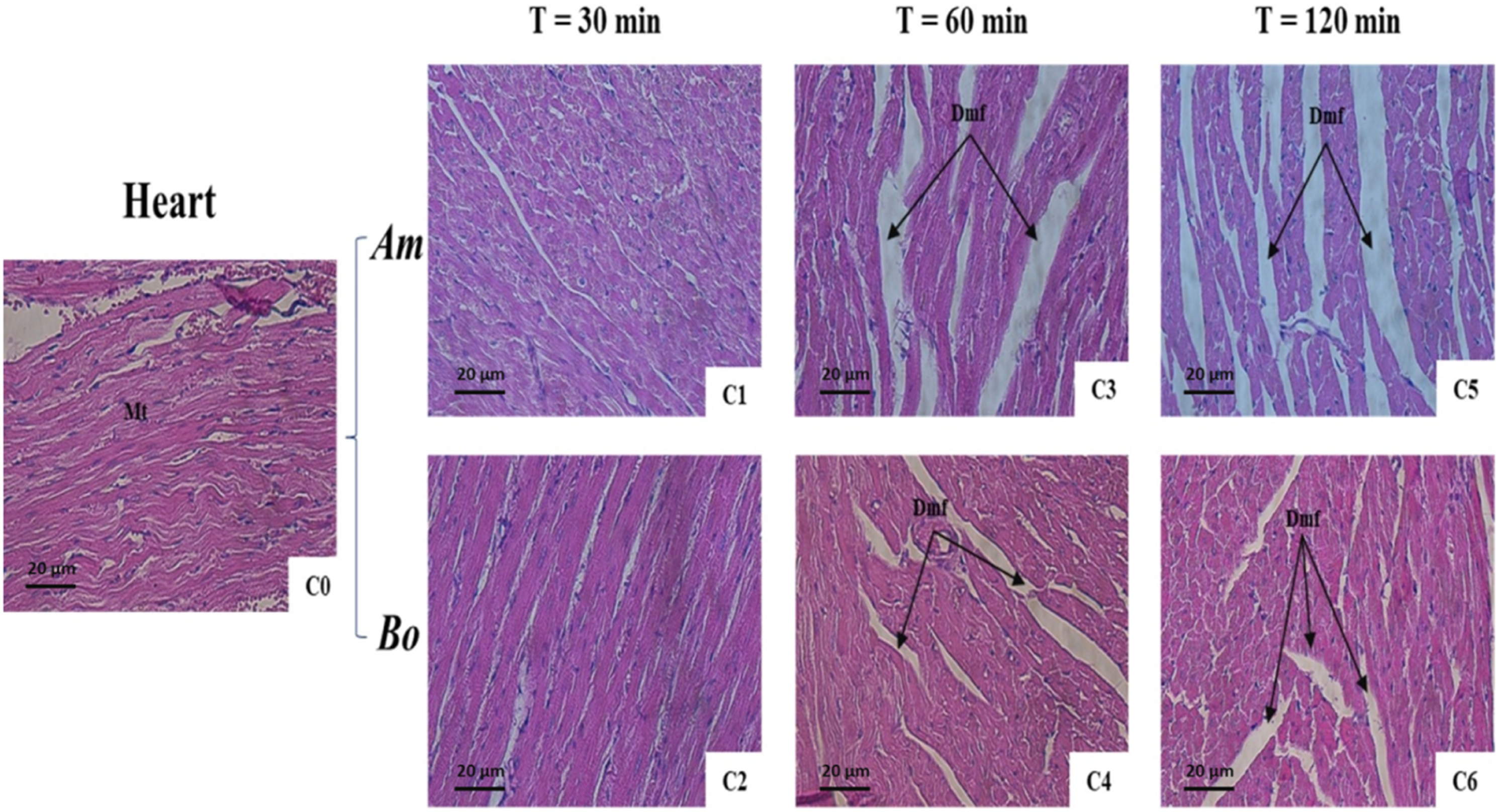
Histopathological studies of the liver
Control liver section showed a normal structure: centrilobular vein surrounded by hepatocytes with sinusoids. In the liver parenchyma, the injection of Am or Bo venoms induced dilation of the sinusoid spaces at 30 min for both groups and 60 min for Bo group. The nuclei of some cells were pycnotic or in caryolysis, while others were in necrosis mostly at 120 min (Figures 6(D5 and D6)). Also, an important focus of congestion was registered 60 min after envenomation in mice by the Bo venom (Figure 6(D4)). Histological sections of the liver after 30, 60 and 120 min of experimental envenomation by Am and Bo venoms (x40) (n = 8). Tl: Tubular lumen; Gm: Glomerulus; Bc: Bowman’s capsule; Gd: Glomerular disorganization; Elt: Enlargement of tubular lumen; Hr: Hemorrhage.
Histopathological studies of the kidney
The kidneys tissue of control mice showed normal glomerulus and tubular lumen. However, envenomed mice showed glomerular disorganization, Bowman’s capsule rupture, enlargement of the tubular lumen for both groups with different intensity. Hemorrhagic areas appeared at 30 and 60 min in groups envenomed by Bo venom (Figures 7(E2–E4)). These alterations were detected 30 min following envenomation and became progressively intense after 60 and 120 min post-injection. Histological sections of the kidney after 30, 60 and 120 min of experimental envenomation by Am and Bo venoms (x40) (n = 8). Tl: Tubular lumen; Gm: Glomerulus; Bc: Bowman’s capsule; Gd: Glomerular disorganization; Elt: Enlargement of tubular lumen; Hr: Hemorrhage.
Discussion
Scorpionism can lead to major clinical manifestations associated with multi-organ failure characterized by structural damage. In the present study, we examined the pathophysiological alterations of the most dangerous scorpions in the Moroccan, namely Am and Bo.
These toxic effects were maintained at 30, 60 and 120 min following the envenoming of Swiss mice by a sub-lethal dose of both venoms. The results of protein quantification showed that the Am venom is richer in protein (153.25 ± 3.22 mg/mL) than the Bo venom (117 ± 2.65 mg/mL). The toxic activity of Am venom is significantly more accentuate compared to Bo venom. Moreover, the LD50 toxicity values of collected Am and Bo scorpion venoms were 300 ± 25 and 875 ± 20 μg/kg body weight, respectively, this distribution is related to the low molecular weight (<10 kDa) of the toxins, which favors a quick entry of toxins into the blood circulation. Our results are in agreement with those reported previously, 22 where the LD50 was 290 and 855 μg/kg body weight for Am and Bo, respectively, using IP injection in mice. Daoudi et al. 23 reported that the Am venom had a higher lethal potency (180 μg/kg body weight) compared to Bo venom (310 μg/kg body weight). The Am venom showed an almost similar toxicity to that of the Androctonus australis hector venom (335 μg/kg body weight) via IP route. 22 The sLD of Tityus obscurus and Tityus serrulatus scorpions revealed a lesser lethal potential (3.13 and 0.99 mg/kg body weight, respectively) compared to Am and Bo venoms, 24 same for Mesobuthus eupeus venom with a LD50 equal to 6.95 mg/kg body weight. 25
After sLD intraperitoneal administration of the Am and Bo venoms, the mice exhibited the symptoms of envenomation, including neurological (chills, paralysis, hunchback and excitement), cardiopulmonary (tachycardia and dyspnea) and general symptoms (sweating and salivation). The classification of signs and symptoms of scorpion envenomation had been revised. 26 Then three classes are proposed; Class I includes a local manifestation (erythema, hyperesthesia, itching, necrosis, paresthesia, pain, swelling and tingling), class II consists of minor manifestations (convulsion, hypothermia, hyperthermia, miosis and mydriasis). The class III corresponds to severe manifestations that can be life-threatening (hypotension, dyspnea, pulmonary edema and paralysis). According to this classification, the observed symptoms after Am or Bo envenomation in our study was grouped in three classes; class I represented by local symptoms type hunchback and chills. The symptoms of class II included sweating and excitement. While the cardiopulmonary (tachycardia and dyspnea) and paralysis of lower limbs are registered in class III without life-threatening. In previous studies, Ozkan et al. 27 showed that Iurus dufoureius asiaticus venom was responsible for salivation, humpback, paralysis, excitability, weakness and coma. Ait Laaradia et al. 28 revealed severe symptoms by the Morrocan Buthus lienhardi scorpion venom represented by dyspnea, hypersalivation, chewing, mouth rubbing, rotating behavior, convulsions and a little scream, which were all dose-dependent. The Tityus bahiensis venom (2.5 mg/kg) also induced moderate manifestations of increased respiratory and salivary frequency, increased sensitivity and vocalization to touch, as well as rigidity in hind limbs. 29 The reported clinical effects are the consequence of nervous stimulation with increased release of mediators. Salivation is related to stimulation of bronchial mucus glands.30,31 The tachycardia effect is due to the fixation of toxins on beta-adrenergic receptors. 32 A mydriasis, as an other symptoms of envenomation, is induced by the massive release of catecholamine (such as norepinephrine and epinephrine) and stimulation of alpha receptor in dilator pupillary muscles. 33
Simultaneously, the histological examination of brain, lung, heart, liver, and kidney tissues in envenomed mice by sLD of Am and Bo venoms, showed several structural damages. The histopathological modifications were revealed at 30 min after envenomation at the level of lungs, liver, and kidney parenchyma, while no histological lesions were detected in brain and heart. The rapid apparition of structural disorganization was attributed to the fast diffusion kinetics of scorpion venom from the blood compartment to the tissue. 34 The nature of the target organ is also involved. However, richly vascularized organs, such as lungs and liver, are rapidly affected by scorpion venom compared to brain, heart and kidney tissues. 35 El Hafny et al. 36 revealed that after 30 min of subcutaneous injection of Am venom (sLD), a maximal concentration of venom was noted in the serum. De Oliveira-Mendes et al. 37 had reported by radioactivity test in mice, the quick absorption of Tityus serrulatus scorpion venom at the level of the bloodstream (maximum at 30 min), and then decreases to reach the tissues. The venom may reach each organ at different times depending on the intraspecific composition variety and abundance of toxins in each sample of the venom.
In the brain, Am and Bo scorpion venoms generated vasodilation features since 60 min of envenomation, with moderate hemorrhagic foci by the effect of the Am venom only at 60 min. Another study conducted by Malih et al. 38 showed neuronal darkness, cellular atrophy and atypia, associated with edema and hemorrhages as a result of envenomation by sLD of the Androctonus mauretanicus mauretanicus scorpion, whereas, the toxic fraction of Am venom expressed a neuronal constriction, congestion, vasodilation and edema following intra-cerebroventricular (ICV) administration of a sLD. 39 Experimentally, Ladjel-Mendil et al. 14 observed similar damages (edema, hemorrhage, necrosis…) with ICV injection of sLD of purified Kaliotoxin from Androctonus australis hector scorpion venom.
The histopathological alterations of the lung parenchyma had increased since 30 min after Am and Bo envenomation, a remarkable increase of alteration was noted progressively in rupture of the alveolar structure and the hemorrhagic area dimeter. However, the pathological effects of Am venom by ICV route revealed edemas in alveolar lumen and bronchial, congestion, several hemorrhages, a large deposition of fibrin and infiltration of inflammatory cells. 39 In parallel, the study of Malih et al. 38 evoked an alveolar space enlargement, hemorrhages, and edema after 60 min of experimental envenomation by Androctonus mauretanicus mauretanicus venom, a mononuclear cell infiltration is also noted by the effect of Am venom’s toxic fractions.
According to the histopathological studies on cardiac muscles tissue, a degeneration of myofibrils started at 60 min following the envenomation by both venoms. However, at T = 120 min, the heart muscle showed massive deleterious degeneration. Comparing the degree of damage on each cardiac muscle tissue sample due to venom from Am or Bo, the groups of mice treated with Am venom reveled the high level of histological damage in myofibrils than Bo venom. This result is in line with the study of Ait Laaradia et al. 40 noticed the focal fragmentation of myocardial fibers and hemorrhage 3 h following envenomation by Buthus lienhardi scorpion. A similar observation of myocardial fiber’s focal fragmentation was detected in mice envenomed 3h with a LD50 of B. occitanus venom. 41 Yet the animals treated with Androctonus australis hector scorpion venom presented the degeneration of myocardiocytes, accumulation of polymorphonuclear, granulocytes, macrophages and lymphocytes; associated with edema, acute hemorrhages and necrosis in the myocardium ventricle. 15
The histological screening of the liver of envenomed by both venoms showed a sinusoidal dilation, a remarkable destruction of hepatocytes (caryolysis, psycnosis and necrosis) in addition to a congestion and turgescence in mice treated by the Bo and Am venoms, respectively. The high hepatic toxic effects of the Am venom were also confirmed by intra-cerebroventricular inoculation of toxic fraction and then noted a sinusoidal dilation, turgescence and hypertrophy of nucleus or bi-nucleating, 39 whereas, the Tityus trivittatus scorpion venom causes structural impairments consisting of sinusoidal congestion, hepatocyte necrosis, and vacuolization. 42
The kidney seems to be another target of the Am and Bo venoms as highlighted by the analysis of the section which manifested by disorganized tubules, glomerulus and the destruction of Bowman’s space. In addition to remarkable hemorrhagic foci at 30 and 60 min only by the effect of Bo venom. The toxicity of Hemiscorpius lepturus scorpion venom to renal parenchyma induced signs of the damaged tubule, renal glomerulus 43 besides tubular inflammatory cell infiltration and hemorrhage by injection of Androctonus australis hector scorpion venom. 44
The neurological symptoms induced by the Am and Bo venoms are mostly related to nervous structures. These symptoms are observed after 60 min of envenomation, which were positively correlated to the time where severe damages in the brain tissue were detected. The same thing was observed for cardiopulmonary symptoms, where the alterations in the heart tissue were observed 60 min after envenomation.
Most importantly, this study highlighted the correlation between the intensity of signs appeared and the level of histopathological alterations observed at the same time following venom inoculation. However, the Am venom is highly toxic compared to the Bo venom according to the activity of venom, the rapidity and severity of symptoms and the histological lesions in mice organs.
Herein, we reported the severity of pathophysiological alterations induced by the most dangerous scorpions in Morocco; Am and Bo at the level of the main organs (brain, lungs, heart, liver and kidney). Also, we proved that the neurological, cardiopulmonary and the others symptoms detected reflect the alteration of tissue and therefore the functions of organs. Interestingly, this study correlated for the first time the kinetic of symptoms apparition, at the same interval post-injection of Am and Bo venoms, to the kinetic of tissular damages. This will help the physicians to focus on the pathophysiological process of scorpion venom to conceive a targeted and more effective treatment plan.
Footnotes
Acknowledgments
We acknowledge the team from the animal unit of the Pasteur Institute of Morocco in Tit Mellil and all participants who contributed to this work.
Declaration of Conflicting Interests
The authors declared no potential conflicts of interest with respect to the research, authorship, and/or publication of this article.
Funding
The authors received no financial support for the research, authorship, and/or publication of this article.