
Abstract
Melanoma a common skin tumor induced by excessive hyperplasia of abnormal melanocyte. Circular RNAs (circRNAs) play critical roles in various diseases and presented as the prognostic markers of melanoma. The present study was designed to confirm the effect of circ 0001591 on human melanoma cell growth and to elucidate its mechanism. Patient with melanoma was obtained from Shaanxi Provincial People’s Hospital. Cell viability of A2058 cell was detected by MTT assay. The expression of circ 0001591 in serum of patients with melanoma was increased. Up-regulation of circ 0001591 promoted cell growth and cell invasion, and reduced apoptotic rate of melanoma. Down-regulation of circ 0001591 reduced cell growth and cell invasion, and promoted apoptotic rate of melanoma.
Up-regulation of circ 0001591 induced PI3 K and p-Akt protein expressions in melanoma through induction of ROCK1 by suppression of miR-431-5p. Over-expression of circ 0001591 suppressed PI3 K and p-Akt protein expressions via suppression of ROCK1 in melanoma by induction of miR-431-5p. MiR-431-5p reduced the effects of circ 0001591 down-regulation on cell growth of melanoma through PI3K/AKT signal pathway. ROCK1 reduced the effects of circ 0001591 on cell growth of melanoma through PI3K/AKT signal pathway. Our findings demonstrated that circ 0001591 inhibits the progression of human melanoma through ROCK1/PI3K/AKT signal pathway by targeting ROCK1 by miR-431-5p.
Introduction
Melanoma, also called malignant melanoma, is a common skin tumor induced by excessive hyperplasia of abnormal melanocyte. 1 China is a low-incidence area of melanoma. However, the morbidity of malignant melanoma shows an increasing trend in recent years. 1 Currently, no potent agent is available for treating melanoma due to its early metastasis, rapid progression, poor prognosis and high mortality. 2 With the continuous development of anti-cancer traditional Chinese medicine (TCM) components, some active TCM ingredients have attained satisfying efficacy in clinical antitumor practice. 3,4 This has also provided a direction for developing therapeutic agents for melanoma.
PI3 K is a class of specific kinase for catalyzing phosphatidyl inositol lipid. It is a heterodimer constituted by a P110 catalytic subunit and a P85 regulatory subunit. 5 It possesses protein kinase activity and lipid kinase activity. Akt is a serine/threonine protein kinase, which is the key effector molecule of the PI3 K downstream. 5 It can be activated through phosphorylation. Thus, it can promote tumor growth and invasion through many pathways. 6 They include inactivating multiple apoptosis effector molecules, regulating cell cycle, activating the telomerase activity, and promoting angiogenesis and migration related programs. PI3K/Akt signaling pathway is originally activated through binding multiple cytokine receptors on cell membrane surface (including tyrosine kinase receptors, non-tyrosine kinase receptors, and insulin receptors) with extracellular growth factors. 7 PI3 K can subsequently translocate into the cell membrane to bind with the receptor casein kinase (such as EGFR) or connexin to be activated. 7 In tumor cells, the PI3K/Akt signaling pathway frequently shows pathological and composed activation. Research indicates that, the PI3K/Akt signaling pathway is in composed activation status in over 70% melanomas. 7
The Rho family is a small molecule G protein discovered in recent years. Its family member RhoC is involved in vital biological processes such as cell signal transduction and actin skeleton regulation (8). An increasing number of data suggest that, RhoC is closely correlated with the acquisition of tumor migration, invasion and metastasis phenotypes. 8 It is considered as the molecule switch of tumor metastasis and has received extensive attention in recent years. Rho kinase is a major effector molecule of Rho family, which can exert its function through ROCK. 9 Studies on the invasion and metastasis phenotypes of melanoma suggest that RhoC is closely associated with melanoma. 8,9 This has thereby ushered in the research upsurge regarding the relationship between RhoC and malignant tumor metastasis. 1/5 melanoma patients have developed metastasis at the time of diagnosis. 8 The high metastasis rate of melanoma renders it the ideal model for research on tumor metastasis. Existing studies find that the effect of RhoC is achieved through ROCK, one of its important effector proteins. ROCK is a metastasis-related serine/threonine protein kinase that is the most extensively studied one at present. It contributes to transforming the downstream target protein and phosphorylating the myosin light chain. 10 As a result, it can participate in cell aggregation, adhesion and fibrous contraction. 11 Cancer cells transfected with ROCK can produce the invasion characteristics independent of the serum. 11
Circular RNAs (circRNAs) is a kind of ncRNA with limited protein coding capacity. 12 Circular RNAs (circRNAs) play critical roles in various diseases and presented as the prognostic markers of Melanoma. 12,13 miRNA is a class of small non-coding RNA molecule about 22 nucleotides in length. It can regulate gene expression at post-transcription level and plays a key regulatory role in cell. 9 Research indicates that miRNA has extensive roles, which can serve as a oncogene as well as a tumor suppressor gene. 14 It can regulate the expression of multiple genes. 15 Thus, it plays a vital role in biological processes such as cell growth, proliferation, differentiation and apoptosis. Anticancer treatment aiming at improving disease condition and enhancing the cure rate with miRNA as the research direction has been gradually applied. 11 Particularly, the miRNA targeted therapy has the advantages that miRNA can serve as the effector to participate in multiple pathways, including cell differentiation, proliferation and survival. 14 The aim of the current study was to confirm the effect and elucidate the mechanism of microRNA 448 on human melanoma cell growth.
Materials and methods
Clinical samples
Patient with melanoma (n = 53) were obtained from Shaanxi Provincial People’s Hospital. Written informed consent was obtained from each patient, and the research protocols were approved by the Ethics Committee of the Shaanxi Provincial People’s Hospital. Serum samples of patient with melanoma and normal healthy volunteers were collected and saved at −80°C. Overall survival (OS) and disease free survival (DFS) were executed using telephone at every 3 months.
Quantitative RT-PCR
The extracted RNA from the serum samples and cells was treated with Trizol reagent (Invitrogen, Carlsbad, Calif, USA). MiRNA were inverse transcribed into cDNA by using PrimeScript RT Reagent Kit (Takara, Dalian, China). The real-time PCR were carried out on ABI 7500 Fast Real-Time PCR system (Applied Biosystems, USA) by using SYBRGreen PCR Kit (4389986, Takara, Dalian, China). These reactions were incubated at 95°C for 5 min, followed by 40 cycles of 95°C for 30 s, 60°C for 40 s, 72°C for 30 s. The relative expression of genes was counted by means of the 2-ΔΔCt relative quantification method. Circ 0001591 forward, 5’-CCGCTGCGGTGGAATGAGTG-3′; Circ 0001591 forward, 5’-TTCTTTCTCGTCGCCTCTTTTT-3′; miR-431-5p forward, 5′-ACGCGTGTCTTGCAGGCCGT-3′; miR-431-5p reverse, 5′-ATCCAGTGCAGGGTCCGAGG-3′; U6 forward, 5′-CTCGCTTCGGCAGCACA-3′; U6 reverse, 5′-AACGCTTCACGAATTTGCGT-3′.
Cell culture and transfection
A2058 cell obtained from Shanghai Institutes for Biological Sciences Cell Resource Center and were cultured in high glucose Dulbecco’s Modified Eagle’s Medium (DMEM, Gibco, Carlsbad, CA, USA) supplemented with 10% fetal bovine serum (Sigma Chemical Co.® (St. Louis, MO, USA). All the cells were incubated at 37°C in a humidified incubator with 5% CO2.
Circ 0001591, anti-circ 0001591, ROCK1 plasmid (CCCTCTCAGCCCCCTCGCC and TTCTCTGATGATCAGTAATG) and negative mimics were transfected into cell using Lipofectamine 2000 (Thermo Fisher Scientific, Waltham, MA, USA) according to the manufacturer’s protocol. Next, after transfection of 4 h, PI3 K inhibitor (LY294002, 10 nM, MedChemExpress) was added into cell.
MTT assay
Cell viability was detected by MTT assay. Cell was added by MTT for 4 h at 37°C. the culture was removed and 200 µL of DMSO was added to each well. The absorbance was evaluated at 492 nm using a microplate reader (Bio-Rad Laboratories Inc., Hercules, CA, USA).
Cell invasion assay
Cells were plated on the upper Transwell chambers (Costar; Corning Incorporated, Corning, NY, USA) at 24-well Transwell culture chamber and 500 ml of complete medium supplemented with 10% fetal bovine serum (Sigma Chemical Co.® (St. Louis, MO, USA) was added to the lower compartment. After 48 h of incubation, cells were fixed with methanol for 1 h and traversed cells on the lower side of the filter were stained with crystal violet and counted.
Lactate dehydrogenase (LDH) activity assay and caspase-3/9 activity assay
LDH activity levels were measured using LDH activity kit and the absorbance was evaluated at 492 nm using a microplate reader (Bio-Rad Laboratories Inc., Hercules, CA, USA).
Caspase-3/9 activity levels were measured using LDH activity kit and the absorbance was evaluated at 405 nm using a microplate reader (Bio-Rad Laboratories Inc., Hercules, CA, USA).
Apoptosis assay
Cells were washed in phosphatebuffered saline (PBS) and fixed in 70% ethanol at 4°C for 30 min. Cell was stained in PI/FITC-Annexin V in the presence of 50 μg/ml RNase A (Sigma-Aldrich) for 30 min at room temperature in the dark. Apoptotic rate was done by using a FACS can (Beckman Coulter, Fullerton, CA, USA) and analyzed using Flowjo 7.6.1 (FlowJo, LLC,).
Luciferase reporter assay
ROCK1 plasmids were synthesized and cloned into luciferase reporter plasmid (pMIR-REPORT) (Promega Corporation, Fitchburg, WI, USA). ROCK1 plasmids and circ 0001591 mimics were co-transfected into cell using Lipofectamine 2000 (Thermo Fisher Scientifc, Waltham, MA, USA) according to the manufacturer’s protocol. 48 h after transfection, activities of frefly and renilla luciferase were measured using the Dual Luciferase Assay (Promega Corporation) according to the manufacturer’s instruction.
Western blot assay
Total proteins were lysed with RIPA buffer and the concentrations of Proteins were measured by BCA Protein Assay Kit. Equal amounts of protein samples (50 μg) were separated by 10 percent SDS-PAGE and then transferred into PVDF membranes. Membranes were blocked with 5% non-fat in milk for 1 h at 37°C and incubated with ROCK1 (Abcam, Burlingame, CA, USA), PI3 K (Abcam, Burlingame, CA, USA), p-Akt (Abcam, Burlingame, CA, USA) and GAPDH (Abcam, Burlingame, CA, USA) at 4°C over-night. Membranes were washed with TBST for 20 min, incubated with anti-rabbit, and anti-mouse horseradish peroxidase (HRP)-conjugated secondary antibodies (Sigma Aldrich Co.) and visualized by enhanced chemiluminescence (ECL) reagent. Densitometric analysis was performed with Image-ProPlus 6.0 software (Media Cybernetics, Inc., Rockville, MD, USA).
Immunofluorescence assay
Cell after was washed with PBS and fixed with 4% paraformaldehyde for 15 min at room temperature. Cell was incubated with 0.2% TrisX100 in TBST for 15 min at room temperature and incubated with 5% BSA in TBST for 1 h at room temperature. Cell was incubated with ROCK1 at 4°C overnight and stained with 555- anti-rabbit secondary antibody for 1 h at 37°C. Cell stained with DAPI assay for 15 min at darkness and imaged by fluorescence microscopy (Nikon Eclipse TE3000-U, Japan).
Statistical analyses
All statistical analyses were performed using SPSS software 17.0 (n = 3). Data were presented as mean ± standard deviation (SD). One-way ANOVA was chosen for comparing between multiple groups, and Student’s t-test was used for comparing between two groups. P value was less than 0.05, which was considered statistically significant.
Results
The expression of circ 0001591 and ROCK1 in patients with melanoma
We first determined that the expression of circ 0001591 in serum of patients with melanoma was increased, compared with that in normal group (Figure 1A-1B). Overall survival (OS) and disease free survival (DFS) in patients with high expression of circ 0001591 were lower compared to those with low expression of circ 0001591 (Figure 1C-1D). These results showed that circ 0001591 might play a role in melanoma.

The expression of circ 0001591 and ROCK1 in patients with melanoma. The expression of circ 0001591 (A and B), ROCK1 expression (C and D) in patients with melanoma, ROCK1 expression (E, immunohistochemical), OS and DFS (F and G). Normal, para-carcinoma tissue; Melanoma, patient with melanoma group. ##p < 0.01 compared with patient with melanoma group.
Circ 0001591 regulated cell progression of human melanoma
Then, we explored the functional roles of circ 0001591 in cell progression of human melanoma. Anti-circ 0001591 mimics reduced the expression of anti-circ 0001591 in vitro model of melanoma, compared with negative mimics group (Figure 2A). Down-regulation of circ 0001591 reduced cell growth and cell progression of human melanoma, compared with negative mimics group (Figure 2B-2D). Down-regulation of circ 0001591 promoted apoptotic rate, and increased LDH activity levels in vitro model of melanoma, compared with negative mimics group (Figure 2E-2 J). Afterward, we analyzed the function of circ 0001591 in cell progression of human melanoma. As showed in Figure 3A, circ 0001591 expression was increased by circ 0001591 mimics, compared with negative mimics group. Then, over-expression of circ 0001591 promoted cell growth and cell progression of human melanoma, compared with negative mimics group (Figure 3B-3D). As showed in Figure 3E-3 J, over-expression of circ 0001591 reduced apoptotic rate, and inhibited LDH activity levels in vitro model of melanoma, compared with negative mimics group.
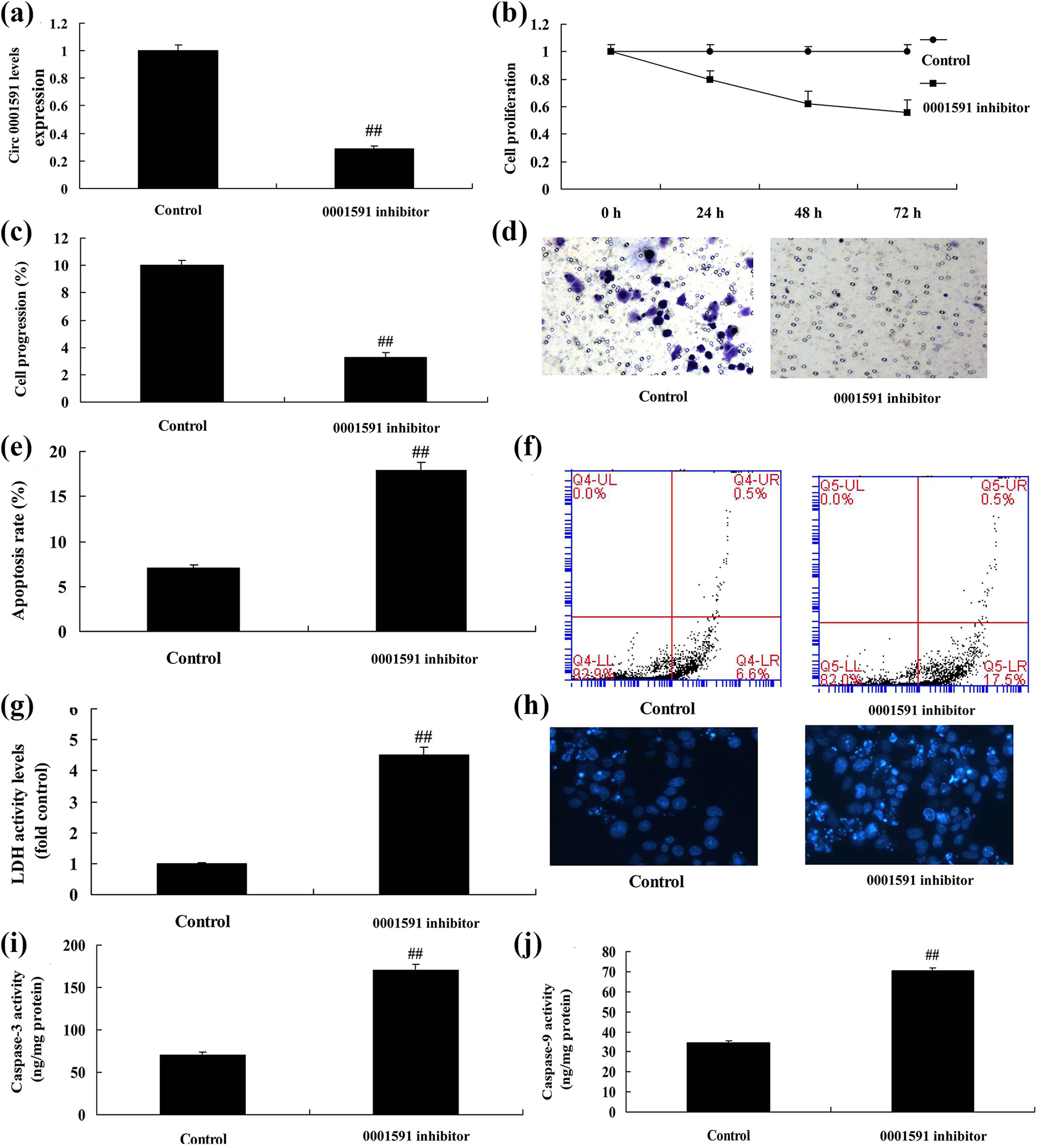
Down-regulation of circ 0001591 regulates cell progression of human melanoma. Circ 0001591 expression (A), cell growth (B), cell transfer (C and D), apoptosis rate (E and F), LDH activity levels (G), DAPI (H), caspase-3 and caspase-9 activity levels (I and J). Control, control negative mimics; 0001591 inhibitor, down-regulation of circ 0001591. ##p < 0.01 compared with control negative group.
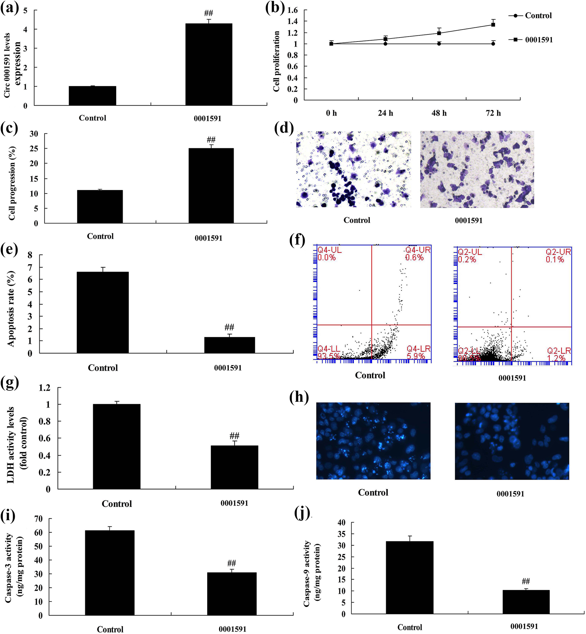
Circ 0001591 regulates cell progression of human melanoma. Circ 0001591 expression (A), cell growth (B), cell transfer (C and D), apoptosis rate (E and F), LDH activity levels (G), DAPI (H), caspase-3 and caspase-9 activity levels (I and J). Control, control negative mimics; 0001591, over-expression of circ 0001591; ##p < 0.01 compared with control negative group.
Circ 0001591 regulates miR-431-5p in human melanoma
To investigate the mechanism of circ 0001591 on melanoma, we used gene chip analyzed the changes of signal pathway. First, miR-431-5p expression was reduced in patients with melanoma, compared with normal group (Figure 4A). Meanwhile, in vitro model, circ 0001591 inhibited miR-431-5p expression, compared with negative group (Figure 4B). Then, this study analyzed these results and found that circ 0001591 maybe target spot miR-431-5p in melanoma (Figure 4C). Circ 0001591 3′UTR binding site is miR-431-5p and circ 0001591 reduced luciferase reporter activity levels, compared with negative group (Figure 4D-4E). Over-expression of circ 0001591 inhibited miR-431-5p expression, down-regulation of circ 0001591 increased miR-431-5p expression in vitro model, in comparison to negative group (Figure 4F-4G).

Circ 0001591 regulates miR-431-5p in human melanoma. Gene chip (A) in patients with melanoma (A) and vitro model of melanoma (B), Analysis result (C), Circ 0001591 3′UTR binding site is miR-431-5p (D), luciferase reporter activity levels (E), miR-431-5p by over-expression of circ 0001591 (F) and down-regulation of circ 0001591 (G). Control, control negative mimics; 0001591, over-expression of circ 0001591; 0001591 inhibitor, down-regulation of circ 0001591. ##p < 0.01 compared with control negative group.
Circ 0001591 regulates miR-431-5p by targeting ROCK1
To investigate the mechanism of circ 0001591/miR-431-5p on melanoma, we used gene chip analyzed the changes of signal pathway. As showed in Figure 5A, miR-431-5p suppressed the expression of PI3 K, and reduced ROCK1 expression. Meanwhile, signal pathway of melanoma, miR-431-5p and apoptosis showed that miR-431-5p regulates apoptosis by targeting ROCK1 in melanoma. Human ROCK1 3′UTR binding site is miR-431-5p and miR-431-5p reduced luciferase reporter activity levels, compared with negative group (Figure 5C-5D). Then, network signaling path showed that ROCK1/ PI3K/AKT signal pathway maybe participate in the function of miR-431-5p on apoptosis of melanoma (Figure 5E). If showed that over-expression of miR-431-5p reduced ROCK1 expression, compared with negative group (Figure 6A). In addition, over-expression of miR-431-5p suppressed the expression of PI3 K and p-Akt protein, and inhibited ROCK1 protein expression, compared with negative mimics group (Figure 6B-6E). Over-expression of circ 0001591 induced the expression of PI3 K and p-Akt protein, and promoted protein ROCK1 expression, compared with negative mimics group (Figure 6F-6I). These results suggested that circ 0001591 regulated PI3K/AKT signal pathway by targeting ROCK1.

Circ 0001591 regulates miR-431-5p by targeting ROCK1. Gene chip (A), signal pathway of melanoma (B), ROCK1 3′UTR binding site is circ 0001591 (C), luciferase reporter activity levels (D), network signaling path (E). Control, control negative mimics; miR-431-5p, up-regulation of miR-431-5p. ##p < 0.01 compared with control negative group.
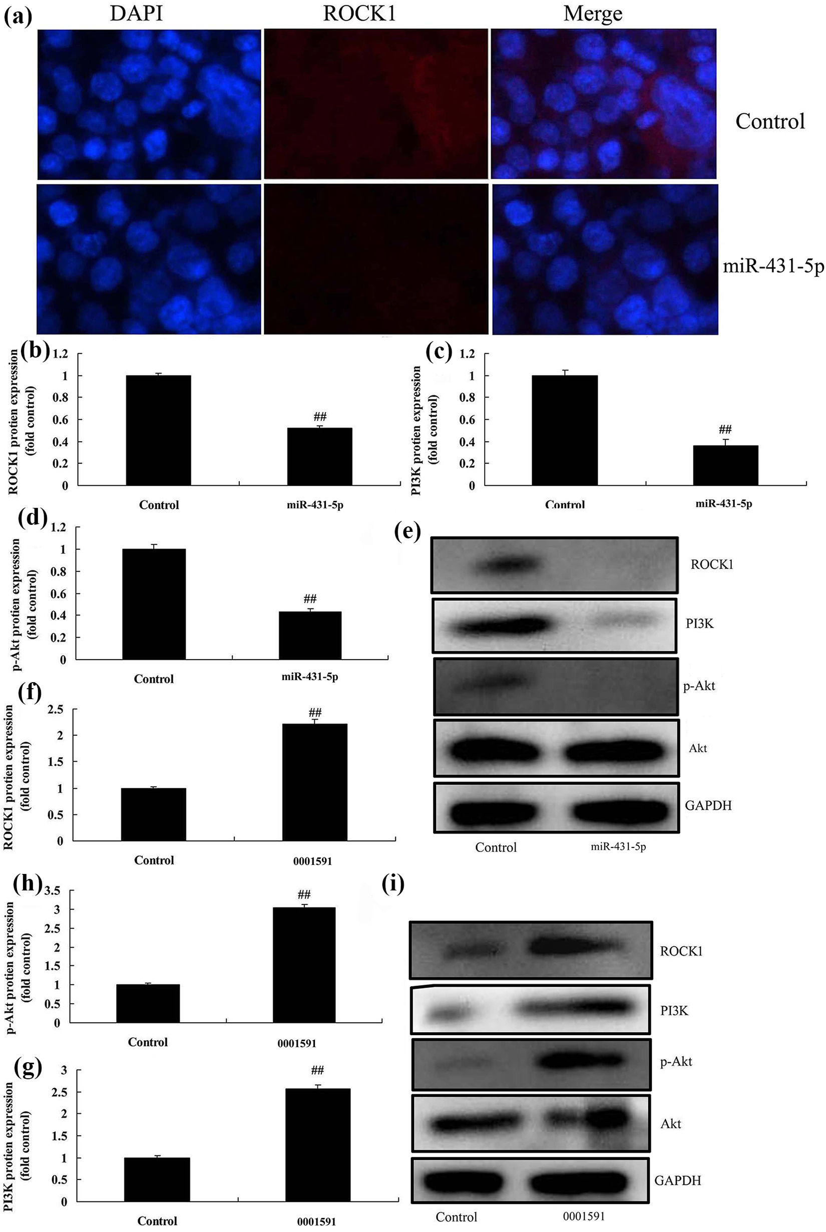
Circ 0001591 regulates miR-431-5p by targeting ROCK1/ PI3K/ p-Akt. ROCK1 expression by IF (A), ROCK1, PI3 K, p-Akt protein expression by statistical analysis (B, C and D) and western blot analysis (E) by down-regulation of circ 0001591; ROCK1, PI3 K, p-Akt protein expression by statistical analysis (F, G and H) and western blot analysis (I) by over-expression of circ 0001591. Control, control negative mimics; anti-346, down-regulation of circ 0001591; miR-346, over-expression of circ 0001591. ##p < 0.01 compared with control negative group.
Circ 0001591 regulates cell progression of human melanoma in vivo model
This study explored the effects of circ 0001591 on cell progression of human melanoma in vivo model. Over-expression of circ 0001591 promoted tumor volume, inhibited caspase-3/9 activity levels, and induced ROCK1, PI3 K and p-Akt protein expressions in vivo model, compared with negative group (Figure 7A-7 H).

Circ 0001591 regulates cell progression of human melanoma in vivo model. Tumor volume (A and B), caspase-3 and caspase-9 activity levels (C and D), ROCK1, PI3 K, p-Akt protein expression by statistical analysis (E, F and G) and western blot analysis (H). Control, control negative mimics; miR-346, over-expression of circ 0001591. ##p < 0.01 compared with control negative group.
MiR-431-5p reduced the effects of circ 0001591 down-regulation on cell growth of melanoma through ROCK1/PI3K/AKT signal pathway. Next, miR-431-5p expression was increased in miR-431-5p mimics and circ 0001591 group, compared with circ 0001591 group (Figure 8A). Over-expression of miR-431-5p reduced that of PI3 K, p-Akt in vitro model of melanoma by circ 0001591, in comparison to circ 0001591 group (Figure 8B-8E). However, over-expression of miR-431-5p reduced the effects of circ 0001591 on cell growth and cell progression, and apoptotic rate and LDH activity levels of melanoma, in comparison to circ 0001591 group (Figure 8F-8 N). Therefore, circ 0001591/ miR-431-5p induced cell apoptosis of melanoma by regulating PI3K/AKT signaling pathway via targeting ROCK1.
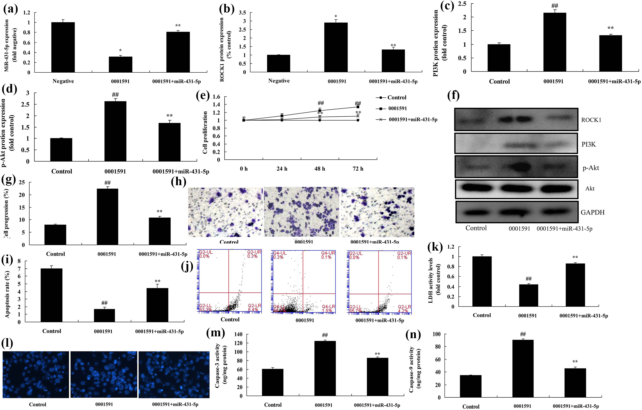
MiR-431-5p reduced the effects of circ 0001591 down-regulation on cell growth of melanoma through ROCK1/PI3K/AKT signal pathway. MiR-431-5p expression (A), ROCK1, PI3 K, p-Akt protein expression by statistical analysis (B, C and D) and western blot analysis (E), cell growth (F), cell transfer (G and H), apoptosis rate (I and J), LDH activity levels (K), DAPI (L), caspase-3 and caspase-9 activity levels (M and N). Control, control negative mimics; circ 0001591, over-expression of circ 0001591; circ 0001591 + miR-431-5p, over-expression of circ 0001591 and over-expression of circ 0001591. ##p < 0.01 compared with control negative group; **p < 0.01 compared with control negative group.
ROCK1 reduced the effects of circ 0001591 on cell growth of melanoma through PI3K/AKT signal pathway
Finally, we investigated the functional roles of ROCK1 in the effects of circ 0001591 on cell growth of melanoma. As showed in Figure 9A-9D, ROCK1 promoted the protein expression of ROCK1, PI3 K, p-Akt protein expression in vitro model of melanoma by anti-circ 0001591, in comparison with anti-circ 0001591 group. Then, we found that the activation of ROCK1 weakened the effects of circ 0001591 on cell growth and cell progression, and apoptotic rate and LDH activity levels of melanoma, in comparison with circ 0001591 group (Figure 9E-9 M).
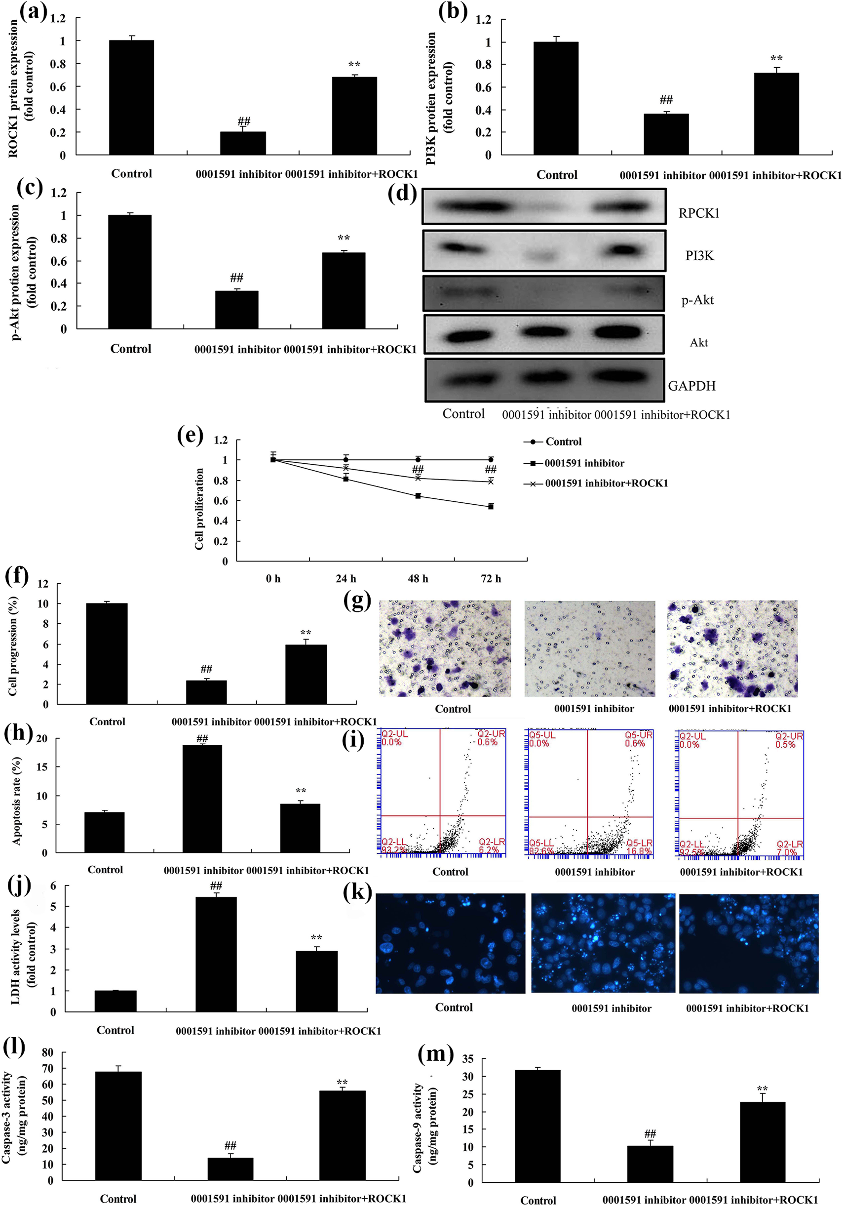
ROCK1 reduced the effects of circ 0001591 on cell growth of melanoma through PI3K/AKT signal pathway. ROCK1, PI3 K, p-Akt protein expression by statistical analysis (A, B and C) and western blot analysis (D), cell growth (E), cell transfer (F and G), apoptosis rate (H and I), LDH activity levels (J), DAPI (K), caspase-3 and caspase-9 activity levels (L and M). Control, control negative mimics; circ 0001591 inhibitor, down-regulation of circ 0001591; circ 0001591 inhibitor + ROCK1, ROCK1 and down-regulation of circ 0001591. ##p < 0.01 compared with control negative group; **p < 0.01 compared with control negative group.
Discussion
Malignant melanoma is a malignant melanocyte tumor mainly occurring in skin. It is characterized by high malignant grade, high metastasis, dismal prognosis and high mortality. 3 Currently, its etiology has not been completely illustrated yet. Research finds that multiple risk factors are related to the genesis and development of malignant melanoma. 3 They include the surrounding environment, sensitivity to sunlight, family history of melanoma, and poor structure. 2 Moreover, the genesis and development of malignant melanoma is also closely correlated with genetic variation. 16 miRNA can regulate the expression of target gene to repress the proliferation, invasion and metastasis of malignant melanoma. Some TCM components can also exert the antitumor effect through participating the signal transduction mechanism. 6 Consequently, understanding the relationship between miRNA and malignant melanoma, and TCM targeted therapy is of positive significance to its prevention. 14 Therefore, we found that the expression of circ 0001591 in patients with melanoma was increased, OS and DFS of circ 0001591 expression high were lower than those of circ 0001591 expression low. Xu et al. showed that miR-431 suppresses proliferation and metastasis of lung cancer. 17
The PI3K/Akt signal transduction pathway is constituted by PI3 K and Akt proteases. 18 The activation of the PI3K/Akt signal transduction pathway can suppress apoptosis induced by multiple stimulations and promote the cell cycle process. 18 Thereby, it can promote tumor cell survival and proliferation, and participate in angiogenesis. 19 It plays a vital role in the genesis, development and resistance of malignant tumor. 18 Besides, it is also involved in tumor invasion and metastasis. Research indicates that the PI3K/Akt pathway plays a critical role in regulating tumor cell proliferation, adhesion, survival, migration and invasion. 19 The results of the present study verified circ 0001591 regulates ROCK1/PI3K/AKT signal pathway by targeting miR-431-5p. Tanaka et al. reported that down-regulation of microRNA-431 inhibits viability of medulloblastoma and glioblastoma cells via PI3K/ Akt pathway. 20
ROCK is a serine/threonine kinase in the Rho downstream. There are two subtypes of ROCK protein, including ROCK1 and ROCK2. 21 The amino acid sequence identity in kinase region is as high as 92%. 21 They possess multiple common downstream target proteins. It is known that the activated Rho/ROCK pathway can regulate the activity of its downstream target proteins (such as myosin phosphatase 1 (MYPT1, MLCP) and LIM kinase. As a result, it will result in cytoskeleton remodeling and actin-myosin contraction. Moreover, it can promote the formation of cell stress fiber and focal adhesion. Meanwhile, it plays a key role in regulating cell morphological change, cell motion and cell adhesion. Abnormal expression and activation of the Rho/ROCK signal has been discovered in numerous tumors. They include liver cancer, bladder cancer, lung cancer and metastasis of melanoma. These results indicated that ROCK1 reduced the effects of anti-circ 0001591 on cell growth of melanoma through PI3K/AKT signal pathway. MiR-431-5p regulates apoptosis by targeting ROCK1 in melanoma. Tanaka et al. suggested that miR-431-5p regulates inflammatory in Relapsing-remitting multiple sclerosis (RRMS) via PI3K/Akt signaling. 20
The present study investigated the expression of circ 0001591 in patients with melanoma was increased. In vitro or vivo experiments showed that circ 0001591 promoted the progression of human melanoma through ROCK1/PI3K/AKT signal pathway by targeting miR-431-5p (Figure 10), suggesting that circ 0001591 may function as tumor motivator in melanoma. These findings implied that circ 0001591 might be applied as potential therapeutic target for melanoma.
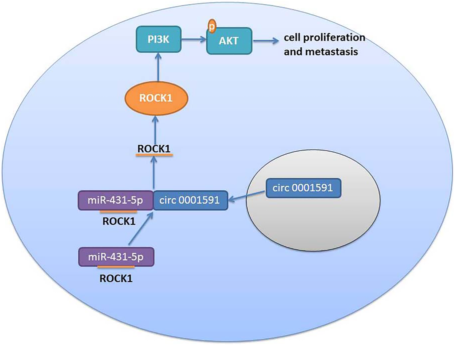
Circular RNA has circ 0001591 promoted cell proliferation and metastasis of human melanoma via ROCK1/PI3K/AKT by targeting miR-431-5p.
Supplemental material
Supplemental Material, complete_western_blot_image - Circular RNA has circ 0001591 promoted cell proliferation and metastasis of human melanoma via ROCK1/PI3K/AKT by targeting miR-431-5p
Supplemental Material, complete_western_blot_image for Circular RNA has circ 0001591 promoted cell proliferation and metastasis of human melanoma via ROCK1/PI3K/AKT by targeting miR-431-5p by Dong Yin, Guo Wei, Fan Yang and Xiaoyan Sun in Human & Experimental Toxicology
Supplemental material
Supplemental Material, PIK3 - Circular RNA has circ 0001591 promoted cell proliferation and metastasis of human melanoma via ROCK1/PI3K/AKT by targeting miR-431-5p
Supplemental Material, PIK3 for Circular RNA has circ 0001591 promoted cell proliferation and metastasis of human melanoma via ROCK1/PI3K/AKT by targeting miR-431-5p by Dong Yin, Guo Wei, Fan Yang and Xiaoyan Sun in Human & Experimental Toxicology
Supplemental material
Supplemental Material, ROCK1 - Circular RNA has circ 0001591 promoted cell proliferation and metastasis of human melanoma via ROCK1/PI3K/AKT by targeting miR-431-5p
Supplemental Material, ROCK1 for Circular RNA has circ 0001591 promoted cell proliferation and metastasis of human melanoma via ROCK1/PI3K/AKT by targeting miR-431-5p by Dong Yin, Guo Wei, Fan Yang and Xiaoyan Sun in Human & Experimental Toxicology
Footnotes
Author contributions
Dong Yin carried out the guarantor of integrity of the entire study, study concepts, study design, definition of intellectual content, clinical studies, data analysis, manuscript preparation, manuscript editing and manuscript review; Guo Wei and Fan Yang were dedicated to the literature research, experimental studies and data acquisition; Xiaoyan Sun was involved in the statistical analysis. All authors have read and approved this article.
Declaration of conflicting interests
The author(s) declared no potential conflicts of interest with respect to the research, authorship, and/or publication of this article.
Funding
The author(s) received no financial support for the research, authorship, and/or publication of this article.
Supplemental material
Supplemental material for this article is available online
References
Supplementary Material
Please find the following supplemental material available below.
For Open Access articles published under a Creative Commons License, all supplemental material carries the same license as the article it is associated with.
For non-Open Access articles published, all supplemental material carries a non-exclusive license, and permission requests for re-use of supplemental material or any part of supplemental material shall be sent directly to the copyright owner as specified in the copyright notice associated with the article.