
Abstract
Icariin, a major component of Epimedium species, was evaluated using isoproterenol (ISO)-induced cardiotoxicity in Wistar rats. Rats were treated with icariin at the doses of 1, 5, and 10 mg kg−1 orally for 15 days. Afterward, rats were administered with ISO (85 mg kg−1, subcutaneous) on 14th and 15th day to produce cardiac injury. Sildenafil (0.7 mg kg−1, intraperitoneal) was used as a positive reference to compare the effects of icariin. ISO-treated rats showed significant changes in hemodynamic parameters. Elevated levels of cardiac troponin T, nitric oxide, and tumor necrosis factor-alpha in serum, positive expression of nuclear factor-kappa B (NF-κB) and inducible nitric oxide synthase in cardiac tissue, and a decrease in serum level of interleukin-10, manifested inflammation and associated cardiac injury. However, pretreatment with icariin and sildenafil significantly prevented the hemodynamic fall and showed improved contractile and lusitropic states. Furthermore, pretreatment groups also showed a reversal of other toxicity markers to normal. Additionally, pretreatment with icariin and sildenafil significantly increased the myocardial cyclic guanosine monophosphate (cGMP) levels. Our results thus indicated the potential role of icariin in the restoration of the ISO-induced cardiac toxicity and restored membrane integrity through modulation of cGMP and NF-κB signaling.
Introduction
Cardiovascular diseases (CVDs) are the foremost cause of preventable deaths and disabilities globally. 1 CVDs mortality rate has declined in the past decades, but the premature deaths with CVDs in third world countries are still at an alarming level. Approximately, the 80% of global burden of CVDs occurs in lower and middle-income countries. 2 Myocardial infarction is the most common presentation of CVDs which accounts for the maximum number of mortalities. 3 Health-care providers are continually researching to develop novel strategies for management and treatment of CVDs, but the results are still far from optimum. 4
Isoproterenol (ISO) hydrochloride, a synthetic catecholamine, and β-adrenergic agonist cause severe oxidative stress in the myocardium resulting in infarct-like necrotic damage in rodents. 5 ISO generates reactive oxygen species (ROS) leading to lipid peroxidation and membrane permeability alterations. 6 Excess ROS production also leads to the necrosis of cardiac tissue and apoptosis. 7 In addition to generation of oxidative stress, ISO administration shows direct β-adrenergic effects such as tachycardia and hypertension. 8 Altogether, the pathophysiological changes produced by ISO in rat’s heart are comparable to those observed in human myocardial infarction.
Recently, phosphodiesterase-5 (PDE-5) inhibition has emerged as a novel treatment approach for CVDs. 9 Various preclinical and clinical studies showed the cardioprotective role of sildenafil and other PDE-5 inhibitors. 10 –12 A clinical study showed that sildenafil was able to prevent the development of heart failure in preserved ejection fraction patients. 13 Pre- and postconditioning of heart, 14 suppression of oxidative stress, and the opening of potassium channels 15 are the various mechanisms involving in the cardioprotection by PDE-5 inhibition.
Icariin, a flavonoid from Herba Epimedii, and a mild PDE-5 inhibitor, is widely used in traditional Chinese medicine for the treatment of coronary heart diseases, impotence, and osteoporosis. 16 Preclinical studies also supported the pleiotropic effects of icariin as it influence a vast range of molecular targets like phosphodiesterases, tumor growth factor-β, mitogen-activated protein kinase, peroxisome proliferator-activated receptor, nitric oxide synthase (NOS), sirtuin, and others. 17 Previous studies have shown the importance of icariin in cardiovascular disorders 18 and erectile dysfunction, 19 but the mechanism involved in cardioprotection by icariin is still unexplored.
On the purview of the above facts, we hypothesized that icariin could be a promising candidate to be examined as a novel approach for the management of myocardial infarction. Since it is a mild PDE-5 inhibitor, we have used sildenafil as a positive standard to compare the effects of icariin. Hence, this study was designed to assess the cardioprotective effects of icariin in ISO-challenged Wistar rats.
Materials and methods
Animals
All the experimental procedures involving the use of experimental animals were approved by the Institutional Animal Ethics Committee (protocol number 1042/2014, approval date: March 3, 2014), Hamdard University (India) by the Committee for the Purpose of Control and Supervision of Experiments on Animals guidelines. Albino male rats of Wistar strain having body weight 200–250 g were procured from the Central Animal House Facility, Jamia Hamdard, New Delhi (India). The animals were housed in polypropylene cages for 1 week to adopt the standard conditions (12 h light/12-hdark cycle; temperature, 23 ± 2°C; relative humidity 60 ± 5%). Animals were provided free access to the standard diet pellet and water ad libitum.
Drugs and reagents
Icariin (purity > 97% by high-performance liquid chromatography (HPLC)) and ISO (purity > 98%) were purchased from Cayman Chemicals (Michigan, USA) and TCI (New Delhi, India), respectively. The enzyme linked immunosorbent assay kits for cardiac troponin T (cTnT) and cyclic guanosine monophosphate (cGMP) were obtained from KinesisDx (California, USA) and Arbor Assays (Michigan, USA), respectively. Cytometric bead array multiplex kit for tumor necrosis factor-alpha (TNF-α) and interleukin-10 (IL-10) were procured from BD Biosciences (San Diego, California, USA). The antibodies used for immunohistochemistry were brought from Santa Cruz Biotechnology (California, USA) (nuclear factor kappa B (NF-κB) p65 antibody, NOS antibody). Other chemicals used in the experiment were of analytical grade. HPLC grade water was used for all biochemical analysis.
Experimental design
Animals were treated with the three doses of icariin and one dose of sildenafil chosen by earlier work done on Wistar strain of rats in erectile dysfunction and myocardial infarction, respectively.
15,19
Icariin was dissolved in 0.1% solution of dimethylsulfoxide (DMSO) in phosphate buffer solution. Sildenafil and ISO were dissolved in the required amount of normal saline. Further, rats were randomly divided into eight groups (n = 6) and treated as per the following details.
Vehicle control: Received vehicle (0.1% DMSO in phosphate buffer solution) for 15 days, plus 0.1 ml saline, s.c. on 14th and 15th day of the treatment.
ISO treated: Received vehicle for 15 days, plus ISO (85 mg kg−1 body weight, s.c.) on 14th and 15th day of the treatment.
Icariin per se: Received icariin (10 mg kg−1, oral) for 15 days.
Sildenafil per se: Received sildenafil (0.7 mg kg−1, i.p.) for 15 days.
Icariin 1 + ISO: Received icariin (1 mg kg−1 body weight, orally) for 15 days, plus ISO (85 mg kg−1 body weight, s.c.) on 14th and 15th day of the treatment.
Icariin 5 + ISO: Received icariin (5 mg kg−1 body weight, orally) for 15 days, plus ISO (85 mg kg−1 body weight, s.c.) on 14th and 15th day of the treatment.
Icariin 10 + ISO: Received icariin (10 mg kg−1 body weight, orally) for 15 days, plus ISO (85 mg kg−1 body weight, s.c.) on 14th and 15th day of the treatment.
Sildenafil + ISO: Received sildenafil (0.7 mg kg−1 body weight, i.p.) for 15 days, plus ISO (85 mg kg−1 body weight, s.c.) on 14th and 15th day of the treatment.
At the end of 15 days treatment, hemodynamic measurement was done. Blood was collected and allowed to clot to collect serum. Rats were killed to excise out the heart which was then soaked between filter papers to remove excess fluid. The heart to body weight ratio was then calculated.
Hemodynamic measurement
After 24 h of the last dose, animals were weighed and anesthetized with urethane (1 g kg−1, i.p.). The left ventricular pressure (LVP) and maximum and minimum rates of developed left ventricular pressure LV (dP/dT max) and LV (dP/dT min) were recorded in situ using a micromanometer-tipped catheter (Millar Instruments, Texas, USA). Left ventricular end diastolic pressure (LVEDP) and cardiac contractility index were calculated offline from the LVP data (Power Lab system-4/35; AD Instruments, Australia). After completion of hemodynamic studies, blood sample was collected. Serum was subsequently separated by centrifugation and stored at −80 ± 5°C for biochemical assays. Hearts were removed, washed in ice-cold normal saline, soaked by tissue paper, and then weighed. A small piece of heart sample was preserved in formalin solution (10%) for immunohistochemistry. Remaining sections of the heart were kept in liquid nitrogen for biochemical estimations.
Estimation of nitric oxide
The accumulation of nitrite in the myocardial tissue is an indicator of the production of nitric oxide (NO). Colorimetric assay with Griess reagent (0.1% N-(1-naphthyl) ethylenediamine dihydrochloride, 1% sulfanilamide, and 2.5% phosphoric acid) was used to estimate NO. Equal volumes of Griess reagent and supernatant were mixed, incubated for 10 min at room temperature, and recorded absorbance at 540 nm. The concentration of nitrite in the supernatant was determined from a sodium nitrite standard curve.
Estimation of cGMP and cTnT
The cGMP level in myocardial tissue and cTnT level in serum were quantified using commercially available ELISA kit by following the manufacturer’s instructions.
Measurement of TNF-α and IL-10
Levels of IL-10 and TNF-α cytokines in the serum were estimated using cytometric bead array kit (BD Biosciences, San Diego, California, USA). Serum samples from all the experimental groups were collected and processed for analysis of cytokines using a BD LSR II flow cytometer as per the manufacturer’s instructions. FCAP Array Software (BD v.3.0.) was used to generate standard curves for both cytokines and concentration of each cytokine in pg ml−1 was calculated.
Immunohistochemistry of myocardial tissue
Paraffin sections of the hearts were deparaffinized with xylene and acetone for a fixed time interval. Sections were rehydrated with a graded series of ethanol followed by washing under running double distilled water. These sections were subjected to antigen retrieval using citrate buffer (pH 6). Sections were cooled at room temperature for 10 min, incubated in 4% hydrogen peroxide for 15 min to remove the background staining, and then washed for three times using Tris-buffer solution. Further, the sections were incubated at 4°C with the corresponding primary antibodies (NOS and NF-κB 65 antibody, Santacruz Biotechnology, California, USA). Sections were rinsed with buffer and incubated for 1 h with the peroxidase-conjugated secondary antibody. The reaction was visualized using diaminobenzidine solution. Photographs were taken with a camera-enabled with the Meiji microscope at different resolutions. Fiji (Image J) Software, Maryland, USA was used for the semiquantification of the protein expression by reciprocal intensity method. The range of pixel intensities of images was in between 0 to 250. Values 0 and 250 indicate the darkest and the lightest shade of image color, respectively. 20
Data presentation and statistical analysis
Data are presented as mean ± standard error of the mean for six rats. Statistical comparisons of all results were performed using Prism software package Version 7 (GraphPad Software Inc., San Diego, California, USA). One-way analysis of variance followed by Tukey’s test was performed to observe and identify significant differences among various groups. Differences in values were considered statistically significant at p < 0.05.
Results
Gravimetric parameters
Table 1 presents the changes in body weight, heart weight, and heart weight to body weight ratio in various groups. After ISO administration, heart weight of ISO-treated group significantly increased in comparison to the vehicle control group (p < 0.001). The body weight of all the treatment groups has shown nonsignificant alterations. The heart weight to body weight ratio in ISO-treated group increased significantly in comparison to the vehicle control group (p < 0.05). Pretreatment with icariin (10 mg kg−1) significantly prevented the pathogenic changes in heart weight and heart weight to body weight ratio (p < 0.001 and p < 0.05, vs. ISO-treated group) which was comparable to sildenafil. Icariin at 5 mg kg−1 dose significantly prevented the alteration in heart weight (p < 0.01) but failed to prevent the changes in heart weight to body weight ratio. Icariin at 1 mg kg−1 dose has not shown any significant prevention (p > 0.05). Icariin per se and sildenafil per se groups showed nonsignificant changes (p > 0.05) as compared to ISO-treated group.
Effect of icariin and sildenafil on the gravimetric parameters.a

ISO: isoproterenol; SEM: standard error of the mean; ANOVA: analysis of variance.
a Data are expressed as mean ± SEM (n = 6 animals per group). Significance was determined by one-way ANOVA followed by Tukey’s multiple comparison test.
b p < 0.01 significant versus vehicle control.
c p < 0.05 significant versus vehicle control.
d p < 0.01 significant versus ISO treated.
e p < 0.001 significant versus ISO treated.
f p < 0.05 significant versus ISO treated.
Hemodynamic parameters
ISO administration stimulated the deleterious changes in rat hemodynamics among various groups. These changes were characterized by fall in LVP (Table 2), increased LVEDP (Table 2), decreased cardiac contractility index (Table 2), and decreased contractile and lusitropic state (Figure 1) of heart. The toxic group showed significant fall in LVP and cardiac contractility index (p < 0.001 each) and increase in LVEDP as compared to the vehicle control group (p < 0.001). Pretreatment with icariin at 5 and 10 mg kg−1 substantially prevented the fall in LVP (p < 0.01 and p < 0.001, respectively) as compared to the ISO-treated group. Pretreatment with icariin (5 and 10 mg kg−1) also prevented the increase in LVEDP (p < 0.001 each) and fall in cardiac contractility index (p < 0.05 and p < 0.001, respectively) as compared to the ISO-treated group. Pretreatment with sildenafil successfully prevented all the harmful changes observed in ISO-treated group (p < 0.001). However, the lower dose of icariin (1 mg kg−1) was found ineffective in the prevention of hemodynamic changes (p > 0.05). Per se groups showed nonsignificant changes as compared to the vehicle control group (p > 0.05).

Effects of icariin and sildenafil on myocardial contractility. (a) dp/dT max and (b) dp/dT min. Values are mean ± SEM for six rats in each group. ***p < 0.001, versus vehicle control group. # p < 0.05, ## p< 0.01, ### p < 0.001, versus ISO-treated group. SEM: standard error of the mean; ISO: isoproterenol.
Effect of icariin and sildenafil on the LVP, LVEDP, and cardiac contractility index.a

LVP: left ventricular pressure; LVEDP: left ventricular end diastolic pressure; SEM: standard error of the mean; ISO: isoproterenol; ANOVA: analysis of variance.
a Data are expressed as mean ± SEM (n = 6 animals per group). Significance was determined by one-way ANOVA followed by Tukey’s multiple comparison test.
b p < 0.01 significant versus vehicle control.
c p < 0.01 significant versus ISO treated.
d p < 0.001 significant versus ISO treated.
e p < 0.05 significant versus ISO treated.
Figure 1 shows the changes in LV (dP/dT max) and LV (dP/dT min) in various groups. Treatment with ISO significantly decreased the contractility and relaxation of left ventricle (p < 0.001). Pretreatment with icariin (10 mg kg−1 day−1) and sildenafil (0.7 mg kg−1) reversed the changes in LV (dP/dT max) (p < 0.001 each) and LV (dP/dT min) (p < 0.05 and p < 0.01, respectively) when compared with the ISO-treated group. Icariin at 1 mg kg−1 dose showed nonsignificant changes on contractility and lusitropic state of ISO exposed rats (p > 0.05). Per se groups showed nonsignificant changes as compared to the vehicle control group (p > 0.05).
Effect of icariin on NO, cGMP, and cTnT levels
Table 3 presents the changes in nitrite levels, cGMP levels, and cTnT levels in various groups after ISO administration. Treatment with ISO considerably increased the level of nitrite and cTnT when compared with the vehicle control group (p < 0.001 each), whereas cGMP in the ISO-treated group remained unaltered as compared to the vehicle control group (p > 0.05). Pretreatment with icariin (5 and 10 mg kg−1) significantly decreased the nitrite and cTnT levels (p < 0.001 each) and increased the cGMP levels (p < 0.001 each) as compared to the ISO treatment group. Icariin at 1 mg kg−1 dose significantly decreased the nitrite levels (p < 0.01) but showed nonsignificant changes in cGMP and cTnT levels (p > 0.05). Pretreatment with sildenafil significantly decreased the nitrite level (p < 0.001) and cTnT level (p < 0.001) and increased the cGMP level (p < 0.001) as compared to the ISO-treated groups.
Effect of icariin and sildenafil on the levels of nitrite, cGMP, and cTnT.a
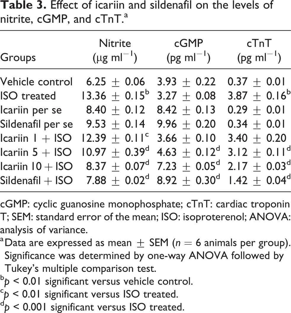
cGMP: cyclic guanosine monophosphate; cTnT: cardiac troponin T; SEM: standard error of the mean; ISO: isoproterenol; ANOVA: analysis of variance.
a Data are expressed as mean ± SEM (n = 6 animals per group). Significance was determined by one-way ANOVA followed by Tukey’s multiple comparison test.
b p < 0.01 significant versus vehicle control.
c p < 0.01 significant versus ISO treated.
d p < 0.001 significant versus ISO treated.
Effects of icariin on serum IL-10 and TNF-α levels
Figure 2 shows the changes in serum level of IL-10 and TNF-α in various groups. Treatment with ISO significantly increased the level of TNF-α (p < 0.001) and significantly decreased the level of IL-10 as compared to the vehicle control group (p < 0.001 each). Pretreatment with icariin (5 and 10 mg kg−1) significantly prevented the declining levels of IL-10 (p < 0.001 at each dose vs. ISO-treated group). On the other hand, pretreatment with icariin at 5 and 10 mg kg−1 doses substantially decreased the elevated TNF-α levels (p < 0.001 for each dose vs. ISO-treated groups). Similarly, pretreatment with sildenafil normalized the altered cytokine levels when compared with the ISO-treated group (p < 0.001). The lower dose of icariin, however, showed nonsignificant changes in comparison to the ISO-treated group (p > 0.05).
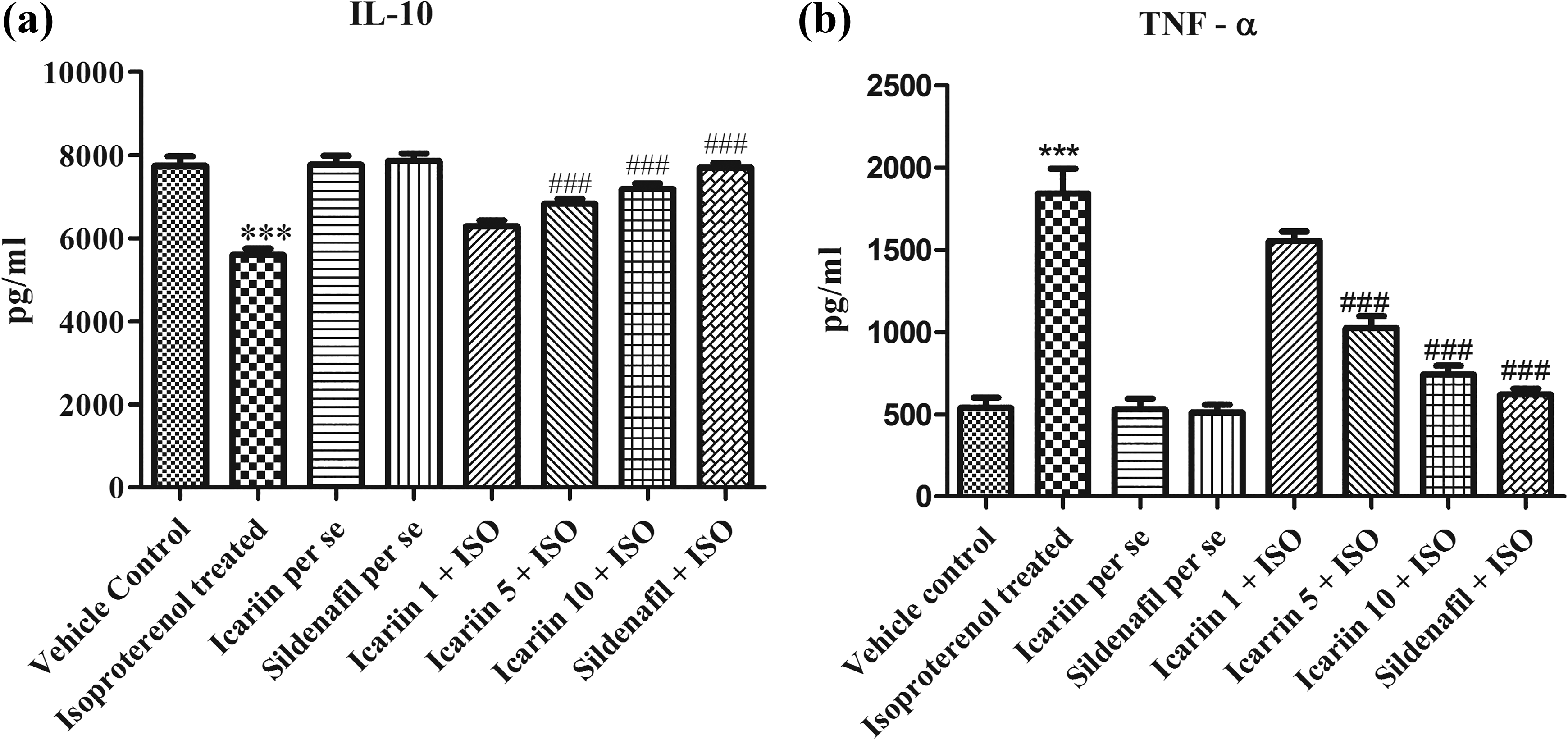
Effect of icariin and sildenafil on serum levels of IL-10 and TNF-α. (a) IL-10 and (b) TNF-α. Values are mean ± SEM for six rats in each group. ***p < 0.001, versus vehicle control group. ### p < 0.001, versus ISO-treated group. IL-10: interleukin-10; TNF-α: tumor necrosis factor-alpha; SEM: standard error of the mean; ISO: isoproterenol.
Effects of icariin on the expression of NF-κB and inducible NOS in various groups
Figure 3(a) to (h) and 3(i) to (p) shows alteration in expression of NF-κB and inducible nitric oxide synthase (iNOS) in various groups. ISO-treated groups showed overexpression of both NF-κB and iNOS in myocardial tissue. On the contrary, pretreatment with icariin (5 and 10 mg kg−1) and sildenafil decreased the elevated expression of iNOS and NF-κB, whereas the lower dose of icariin showed insignificant effects on their expression.
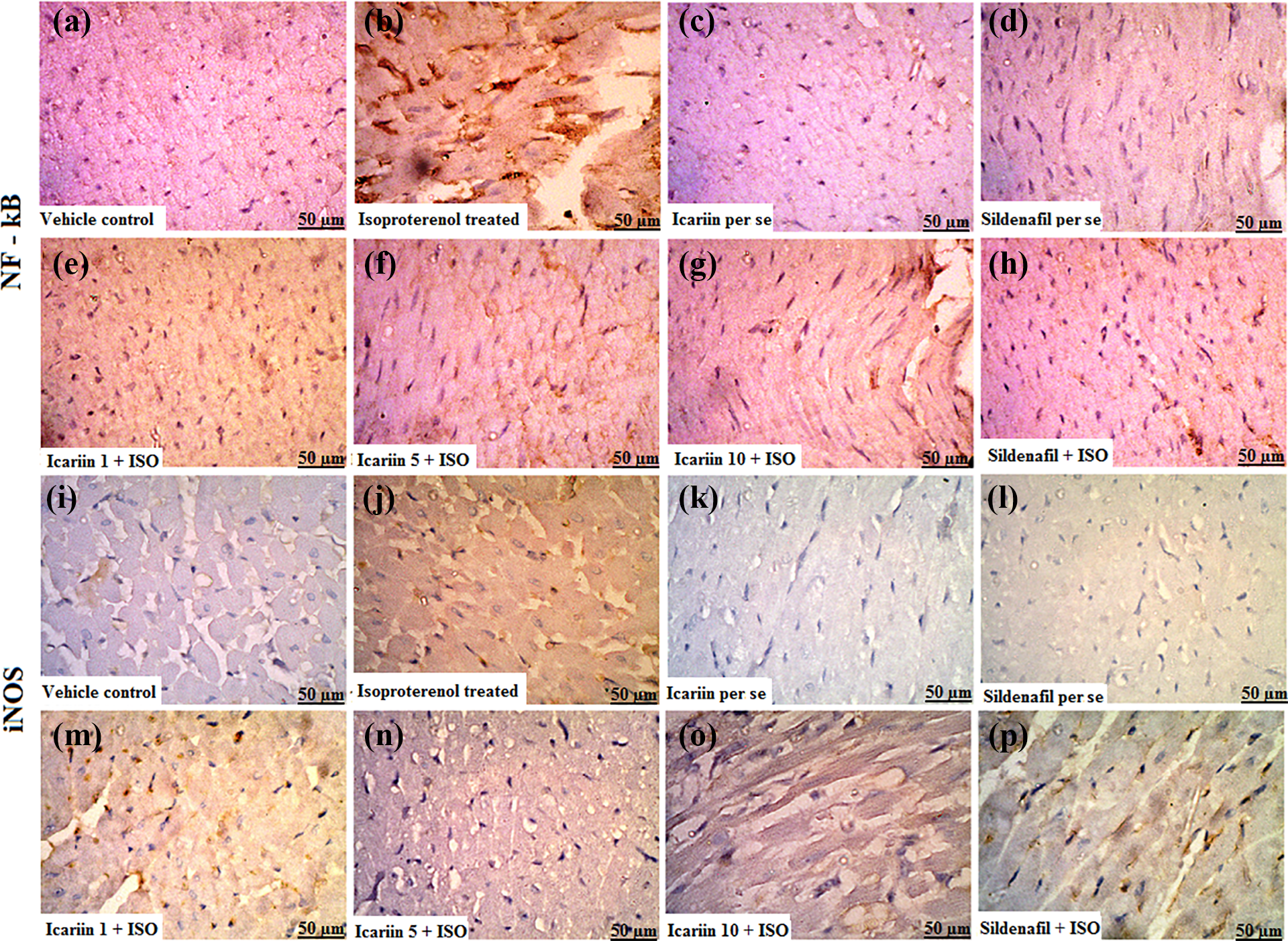
(a)–(h) Photomicrographs showing effect of icariin and sildenafil on expression of NF-κB in various groups. (g)–(l) Photomicrographs showing effects of icariin and sildenafil icariin on expression of iNOS in various groups. Photomicrographs were taken by Meiji fluorescent microscope at ×40. NF-κB: nuclear factor-kappa B; iNOS: inducible nitric oxide synthase.
Figure 4 shows the semiquantification of the expression of NF-κB and iNOS in myocardial tissue. The ISO-treated group showed significant expression of NF-κB (p < 0.001) and iNOS (p < 0.001) as compared to the vehicle control group. Pretreatment with icariin (5 mg kg−1, p < 0.01 and 10 mg kg−1, p < 0.001) and sildenafil (p < 0.001) significantly prevented the elevated expression of NF-κB in myocardial tissue. Further, pretreatment with icariin (10 mg kg−1, p < 0.001) and sildenafil (p < 0.001) significantly prevented the elevated expression of iNOS in myocardial tissue. However, icariin at 5 mg kg−1 showed nonsignificant prevention in comparison to the ISO-treated group (p > 0.05). Icariin at 1 mg kg−1 dose failed to prevent the increase in expression of iNOS and NF-κB.

Semiquantitative estimation of expression of NF-κB and iNOS in various groups. (a) NF-κB and (b) iNOS. Values are mean ± SEM for six rats in each group. ***p < 0.001, versus vehicle control group. ## p< 0.01, ###p < 0.001, versus ISO-treated group. NF-κB: nuclear factor-kappa B; iNOS: inducible nitric oxide synthase; SEM: standard error of the mean; ISO: isoproterenol.
Discussion
ISO (at supramaximal dose) has been reported to cause oxidative stress and nitrative stress and induces expression of iNOS/NF-κB which leads to the generation of various cytokines responsible for inflammation and apoptosis. 21 Thus, measuring the level of NO, cytokines (TNF-α and IL-10), and expression of iNOS and NF-κB provides sufficient background for establishing their role in inflammation and cardiac injury.
It has been found that ISO stimulates iNOS for the bulk production of NO which interacts with superoxide radicals and produces peroxynitrite, a highly cytotoxic molecule, that cause considerable cardiac damage via activation of NF-κB. NF-κB further upregulates pro-inflammatory genes leading to increased production of TNF-α (proinflammatory cytokine) and decreased production of an anti-inflammatory molecule-like IL-10. 22 –24
In our study, keeping the above inflammatory cascade in mind, we have tried to evaluate iNOS, NF-κB, TNF-α, and IL-10 to ascertain their probable role in cardiac damage induced by ISO. We found a rise in the level of NO, TNF-α, iNOS, and NF-κB expression and a decrease in anti-inflammatory molecule IL-10 which strongly indicated the involvement of nitrative stress due to NO production induced by iNOS and activation of NF-κB in cardiac inflammation and damage. Icariin, however, very well reversed this condition toward the normal supposedly by suppressing the expression of iNOS/NF-κB and the associated inflammatory inducers of the cascade, which was also reported in previous investigations. 25,26 Thus, icariin acted as a cardioprotective molecule by counteracting the effects of ISO.
To understand the other mechanisms of icariin-mediated protection in ISO-induced cardiac injury, we assessed the level of cGMP in the heart tissue as it is a known vasodilator. cGMP is an intracellular secondary messenger that mediates multiple cellular functions and morphological processes in the heart via activation of cGMP-dependent protein kinases and opening of mitochondrial KATP channel (adenosine triphosphate-sensitive potassium channels) that restores ATP and calcium level in myocytes. 27 PDE-5 enzymes that cause hydrolysis of cGMP if inhibited will cause a rise in cGMP level which will add to cardioprotection. 14 The results of our studies showed that icariin significantly increased the cGMP levels and is mainly attributed to its PDE-5 inhibiting property.
Apart from the assessment of the above parameters, we also estimated gravimetric, hemodynamic, and membrane damage markers which indicated cardiotoxicity by ISO and protection by our drug, icariin. In the gravimetric evaluation, we have taken heart weight to body weight ratio and found a significant increase in the ratio between vehicle control and ISO-treated group (p < 0.05). These findings are in agreement with multiple studies demonstrating increased heart weight in ISO-treated rats. 28,29 The change in heart weight is probably attributed to the development of edema in intramuscular spaces, infiltration of inflammatory cells, fibrosis, and extensive necrotic changes. 30 High dose of icariin significantly prevented this alteration in comparison to the ISO-treated group. Icariin-induced elevation in myocardial cGMP levels is probably responsible for the prevention of changes in heart weight and its ratio to body weight. 31
The impairment of rat’s heart hemodynamics and its restoration in ISO toxicity also substantiated our findings. The results showed a fall in left ventricular systolic pressure (LVP) and further, a consecutive decline in inotropic (dP/dT max) and lusitropic (dP/dT min) state of the heart. The significant increase in LVEDP and fall in LV [dP/dT max]/P were also observed which indicated the elevated preload and decreasing cardiac output. Our observations comply with various previous studies that showed diminished hemodynamics after ISO administration. 32,33 Pretreatment with icariin significantly reduced these injurious effects which go well with the previous reports of PDE-5 inhibitors showing improvement in cardiac dynamics. 34,35
One of the consequences of ISO toxicity is the damage of myocardium that results in leakage of cTnT in serum. 36 Our results showed a considerable increase in serum cTnT level which indicated severe myocardial damage. Administration of icariin successfully restored this myocardial damage as indicated by the reduction in serum cTnT level which may be attributed to an increase in cGMP level. 37
Thus, it is evident from this study that the administration of ISO (85 mg kg−1) caused cardiotoxicity which was considerably reverted by icariin at the doses of 5 and 10 mg kg−1. Icariin at the lower dose (1 mg kg−1), however, did not show any significant protection.
Our drug icariin has very well reversed the toxic effects of ISO by modulating cGMP levels and suppressing inflammatory signaling. The limitation of our study is, however, that we have not studied the direct effect of icariin on β-receptor in terms of its effect on the expression or its role as a β-receptor antagonist/partial agonist. In order to elucidate the mechanism involved in cardioprotection by icariin involving β-receptor, in vitro interaction of icariin directly with the receptor using cell line can be investigated in future.
Conclusion
Taken together, we can conclude that increase in the myocardial cGMP level and suppression of NF-κB signaling has a significant role in cardiac protection. Our drug, icariin successfully increased the cGMP level by inhibiting PDE-5 and suppressed the activation of NF-κB. Beside this icariin also reverted other detrimental effects of ISO on the heart and behaved as a cardioprotective agent.
Footnotes
Acknowledgment
The authors are thankful to U.G.C. for providing RFSMS-BSR fellowship to the first author and grateful to Professor Md. Fahim, HIMSR, Jamia Hamdard for providing the facilities to perform hemodynamic assessments.
Declaration of conflicting interests
The author(s) declared no potential conflicts of interest with respect to the research, authorship, and/or publication of this article.
Funding
The author(s) received no financial support for the research, authorship, and/or publication of this article.