
Abstract
Liver is a precious organ to maintain body life. Hepatotoxicity is a worldwide health problem that is still a challenge for research. Although countless pharmaceutical drugs and herbal compounds were screened for their hepatoprotective effects, the death from hepatotoxicity is increasing. Thus, there is continuous necessity of searching for the hepatoprotective effect of commonly used drugs. Accordingly, our aim was to examine a hepatoprotective potential for the antihypertensive drug, verapamil, and searching for new insights underlie its protective mechanism. Four groups of adult male rats were randomly arranged as controls, thioacetamide (TAA) hepatotoxic, and TAA + verapamil treated. Serum liver enzyme, hepatic antioxidant, lipid peroxidation, and inflammatory parameters were assessed. Gene relative expression for heme oxygenase-1 (HO-1), nuclear factor-erythroid 2-related factor 2 (Nrf2), phosphoinositide 3-kinase (PI3K), and serine/threonine-specific protein kinase (Akt) were quantified in hepatic tissue. TAA caused hepatic injury evident both histopathologically and biochemically by a decrease in all gene expressions. Verapamil alleviated the injury via its antioxidant and anti-inflammatory effects that were suggested to be via upregulation of the previous gene expressions. In conclusion, the calcium channel blocker, verapamil, that is used widely as antihypertensive exhibits a valuable hepatoprotective effect. The protection partially rests on activation of Nrf2/HO-1 and PI3K/Akt pathways.
Introduction
Organ toxicity becomes an upgrading problem with increasing usage of chemical substances and exposure to xenobiotics in the surrounding environment. The liver is the most vulnerable organ for toxicity owing to its anatomical site and function. 1 The study of hepatotoxicity, in particular, becomes a greater focus of research in order to protect this precious organ.
Thioacetamide (TAA) is a sulfur compound that enters in different industries as leather and paper. Historically, TAA was used as a fungicide, but it was rapidly banned due to its toxicity to the liver. Since that, TAA has served as a popular model to study liver toxicity, fibrosis, and cancer. 2 TAA offers a hepatotoxicant model that resembles human with prolonged injury and high regenerative capacity. TAA is a great trigger of oxidant damage and release of inflammatory mediators by its hepatic metabolites. 3
The cornerstone of liver toxicity is the inflammatory squeal of free oxidants. Mitochondrial dysfunction results from the intracellular oxidant stress in hepatocytes leading to oncotic necrosis rather than apoptosis. 4 Liver, as a machine of metabolism, is the first organ that faces most chemicals and xenobiotics in order to detoxify them. The liver is consequently vulnerable to the hazardous and the harmful effect of such agents. Despite liver has a valued antioxidant machinery system, repeated exposure to toxicants may incapacitate its antioxidant mechanisms, resulting in toxicity. 1
Calcium, the famous second intracellular messenger, regulates various vital cellular processes and is involved in diverse pathological processes as the major cardiovascular complications. 5 It was proved that calcium influx inside cells acts as a spark for releasing the inflammatory mediators under certain conditions. Calcium ions can activate nitric oxide synthase with the elaboration of nitric oxide (NO) and phospholipase A2 with the elaboration of prostaglandins, leukotrienes, and thromboxanes. Accordingly, the inhibitors of calcium current play a role in damping inflammation. 6 Calcium channel blockers are members of drugs that not only regress inflammation, but also exhibit antioxidant properties that enable them to diminish tissue injury. 7,8 Among calcium channel blockers, verapamil, in addition to its anti-inflammatory effect, exerts a unique protective effect on liver and neurons that was demonstrated to be calcium current blocking independent. 9 –11 These data encourage us to examine the hepatoprotective effect of verapamil on TAA toxicity and explore the mechanistic strategies upon toxicity.
Materials and methods
Chemicals and primers
Verapamil and TAA powders were purchased from Sigma, St. Louis, MO, USA. A quantitative real-time polymerase chain reaction (RT-PCR) kit and the primers for heme oxygenase-1 (HO-1), nuclear factor-erythroid 2-related factor 2 (Nrf2), phosphoinositide 3-kinase (PI3K), and Akt were obtained from Thermo Scientific, Fremont, CA, USA. Other compounds used were acquired commercially.
Animals
Adult male albino rats about 200 ± 20 g weight were obtained from the National Research Center, El-Giza, Egypt. Twenty-eight rats were arranged in standard plastic cages under conditioned situations and left 1 week for acclimatization. They were arranged in four groups (seven rats); normal control, verapamil control, TAA hepatotoxic group, and TAA + verapamil treated. Both verapamil control and TAA + verapamil groups received verapamil at the lowest dose 5 mg/kg dissolved in distilled water orally for 7 days. Then TAA was dissolved in normal saline and injected i.p. as a single dose of 150 mg/kg in the seventh day of the experiment for both TAA hepatotoxic rats and TAA + verapamil rats. Controls received the same vehicle i.p. and orally. Doses of verapamil and TAA were chosen according to our pilot experiment and other referenced studies, respectively. 12,13 The experimental procedures were approved by the Local Ethics Committee of the Faculty of Medicine, Minia University, Egypt (approval no: 77_7/2018), and in accordance with the Guide for the Care and Use of Laboratory Animals of the National Research Council. 14
Sample collection and storage
Animals were allowed to feed and drink ad libitum for 48 h after TAA injection to allow TAA metabolism and toxicity, except the last fasting 12 h. Rats were then anesthetized and euthanized to collect blood samples and dissect liver tissue. Blood was centrifuged to obtain clear sera that were kept at 20°C to assess serum parameters. Liver of each rat was divided into three parts; one for RNA extraction, other for homogenization, and the last part was kept in 10% formalin for histopathological examination.
Biochemical analysis
Serum parameters
Assessment of liver enzymes (alanine aminotransferase (ALT) and aspartate aminotransferase (AST)), in addition to total bilirubin in the collected sera was achieved by colorimetric kits (Biodiagnostic, Egypt).
Hepatic homogenate parameters
Portions of liver were homogenized in phosphate buffer (pH 7.4) and then centrifuged to obtain supernatant for assessment of different biological parameters.
The lipid peroxidation product, malondialdehyde (MDA), was measured colorimetrically at 535 nm wavelength after its reaction with thiobarbituric acid. The method was described in detail by Buege and Aust. 15 Hepatic tissue concentration of the antioxidant, reduced glutathione (GSH), was measured through its reaction with 5,5′-dithiobis-(2-nitrobenzoic acid) with the consequence of a yellow compound detected at 412 nm. 16 Assessment of superoxide dismutase (SOD) activity was based on inhibition of pyrogallol autoxidation by SOD. The color produced was monitored in the spectrophotometer at 420 nm. 17 NO end products, in the form of nitrite and nitrate, were determined chemically by a method of Ridnour et al. 18 Briefly, nitrate was reduced into nitrite by copperized cadmium and then total nitrite was estimated after adding Griess reaction, and the formation of colored chromophore was measured at 540 nm.
Real-time reverse transcription polymerase chain reaction
Hepatic tissue was analyzed for the relative gene expressions of HO-1, Nrf2, PI3K, and Akt using RT-PCR. The extraction of RNA from hepatic tissue was performed by RiboZol RNA extraction reagent (AMRESCO, Solon, USA), as directed by the manufacturer’s instructions. Performance of RT-PCR was done using a Thermo Scientific one-step kit, with 50 ng RNA template per reaction in 25 μL reaction volume containing 70 nM of specific primers in the RT-PCR Detection System (Kapa Biosystems, Wilmington, MA, USA). Analysis of the SYBR Green data (Thermo Fisher Scientific; Fremont, CA,USA) was performed and quantified to glyceraldehyde-3-phosphate dehydrogenase (GAPDH) as a reference gene. The sequences of the used primers were shown in Table 1. The expression level of genes was calculated and mounted relative to control, where control samples were set at a value of 1. 19
Primers of genes.
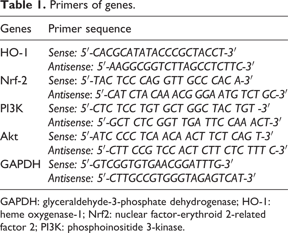
GAPDH: glyceraldehyde-3-phosphate dehydrogenase; HO-1: heme oxygenase-1; Nrf2: nuclear factor-erythroid 2-related factor 2; PI3K: phosphoinositide 3-kinase.
Histopathological examination of hepatic tissue
Hepatic tissue was removed from formalin and embedded in paraffin to prepare 5 µm sections for staining with hematoxylin–eosin (H&E). Slides were examined under a light microscope by an expert pathologist blind to the experiment. The severity of liver injury was scored according to modified HAI grading necroinflammatory score, which was based on four findings; periportal hepatitis, confluent necrosis, apoptosis, and portal inflammation. The final score was calculated by the summation of the four categories of necroinflammation per rat. 20
Statistical analysis
The results were investigated by GraphPad Prism version 5 while they expressed as means ± SEM. A one-way repeated measures analysis of variance, followed by Tukey’s test, was applied. Differences with a p value <0.05 will be considered significant.
Results
Liver enzymes and bilirubin
Serum ALT, AST, and bilirubin were used traditionally as indicators of liver injury. Rats injected by TAA displayed a significant rise in their levels as compared to control rats. Liver enzymes increased more than three times than control, while total bilirubin increased nearly 20-fold than control. Verapamil administration reduced their levels significantly (Table 2).
Effect of verapamil on serum levels of liver enzymes (ALT and AST) and bilirubin in TAA hepatotoxicity in rats.a

TAA: thioacetamide; ALT: alanine aminotransferase; AST: aspartate aminotransferase.
a Parameters were expressed as means ± SEM (n = 6–7).
bcSignificantly different (at p < 0.05) from the normal control group and the TAA group, respectively.
Effect of verapamil on oxidative stress parameters
Table 3 illustrated obviously the deleterious effect of TAA on hepatic antioxidant defenses together with significant elevation of lipid peroxidation product, MDA in hepatic tissue. As shown in the table, verapamil by increasing significantly the hepatic SOD activity and GSH concentrations could restore the antioxidant status of the liver. Verapamil significantly dropped the elevated malondialdehyde to their normal values. The hepatic tissue content of NO is observed to be increased significantly as compared to control rats (Table 3). The normal hepatic NO content was obtained after verapamil treatment as compared to the hepatotoxic group.
Effect of verapamil on hepatic MDA, SOD, GSH, and NO in TAA hepatotoxicity in rats.a

TAA: thioacetamide; MDA: malondialdehyde; SOD: superoxide dismutase; GSH: glutathione reduced; NO: nitric oxide.
a Parameters were expressed as means ± SEM (n = 6–7).
bcSignificantly different (at p < 0.05) from the normal control group and the TAA group, respectively.
Effect of verapamil on Nrf2/HO-1 and PI3k/Akt mRNA expression
The relative expression of HO-1, Nrf2, PI3K, and Akt mRNAs in hepatic tissue demonstrated that TAA caused a significant decrease in their expression in comparison to the control groups (Figure 1). Verapamil treatment resulted in upregulation of their expression as compared to the TAA group as well as the control group.

Relative gene expressions of mRNA for HO-1, Nrf2, PI3K, and Akt in hepatic tissue. Parameters were expressed as means ± SEM (n = 6–7). TAA: thioacetamide; HO-1: heme oxygenase-1; mRNA: Messenger RNA; Nrf2: nuclear factor-erythroid 2-related factor 2; PI3K: phosphoinositide 3-kinase; GAPDH: glyceraldehyde-3-phosphate dehydrogenase. abSignificantly different (at p < 0.05) from the normal control group and the TAA group, respectively.
Effect on liver histopathological pictures
Figure 2(a) displays that TAA administration in rats caused hemorrhage, inflammatory cellular infiltration, and disorganized hepatocytes. These pathological changes were significantly disappeared in rats treated by verapamil. The verapamil control group showed normal values for all parameters as normal control.

A photomicrograph of hepatic tissue stained with hematoxylin and eosin at 400x magnification: control groups showed a normal architecture. TAA hepatotoxic group showed a marked distortion of architecture, periportal lymphocytic infiltration (black arrow), areas of hemorrhage (yellow arrow), and necrosis (red arrow). TAA + verapamil–treated group showed restoration of hepatic architecture with less parenchymal injury. Parameters were expressed as means ± SEM (n = 6–7). TAA: thioacetamide; CTRL: control. abSignificantly different (at p < 0.05) from the normal control group and the TAA group, respectively.
Discussion
The present study presents a new insight into other molecular mechanisms for verapamil hepatoprotection in the most popular model of hepatotoxicity, TAA. TAA is a potent centrilobular hepatotoxicant, which acts only after its biotransformation by the liver into TAA sulfoxide and thioacetamide-S, S-dioxide. These reactive metabolites are highly toxic to hepatocyte by acting as modifiers for lipid and proteins. 21
In accordance, our study revealed an elevation of hepatic injury markers together with the disruption of hepatic lobules that indicated damaged hepatic cells in TAA rats. The results of the present study in line with others demonstrated the ability of TAA to induce hepatic oxidative damage evident by lipid peroxidation and deterioration of antioxidant defense mechanisms in the liver. 21,22 However, the centrilobular necrosis evoked by TAA was related to elaboration of NO secondary to activation of inducible nitric oxide synthase (iNOS) in the liver by TAA. 23 Accordingly, we found an elevation of NO in TAA toxic rats indicating the role of inflammation in the hepatotoxicity. It was known that iNOS depends on calcium ions for activation 6 ; consequently, the calcium channel blocker verapamil was able to prevent the elaboration of hepatic NO induced by TAA. The anti-inflammatory effect of verapamil was clearly studied in different models of inflammation. 9,24 Moreover, different explanations were assumed for its anti-inflammatory property that depend on its ability to inhibit cytokine productions and recruitment of macrophages. 24,25
Searching in depth for other potential mechanisms, we found that different genes were upregulated with verapamil treatment after TAA toxicity. PI3K is a member of cellular kinase family that produces the intracellular second messenger phospholipid PIP3 through downstream activation of other signaling molecules as Akt. 26 PI3K/Akt pathway implicated recently in diverse cellular function as cell differentiation and inflammation. 27 Despite the role of PI3K/Akt in inflammation is controversial, 26 our findings in line with others suggested the inhibitory effect of this pathway on inflammation in the liver. 28 –30 Verapamil achieved decreased NO content together with upregulation of PI3K/Akt gene expression in hepatic tissue.
Verapamil succeeded in restoring the normal pattern of hepatic tissue together with improving the hepatic antioxidant status. The antioxidant effect of verapamil was revealed by several studies in the cardiovascular system and kidney 8,31 ; however, the present study focused on the mechanistic value for its upregulation to the antioxidant genes HO-1 and Nrf2. Our findings are greatly strengthened by Lee et al. who based the protective role of verapamil in acetaminophen cytotoxicity on its induction for Nrf2/HO-1 pathway. 32 Another in vitro study explored the ability of verapamil to translocate Nrf2 nuclearly in hepatocytes to induce HO-1, which resulted in reduction of oxidative stress and cellular injury. 33 Based on our results and the previous knowledge, we suggested that verapamil has an esteemed molecular effect beyond its blocking of calcium channel.
Conclusion
The present study displayed a perfect preventive effect for the antihypertensive, verapamil on hepatotoxicity of TAA through its antioxidant and anti-inflammatory effects. The latter emerging effects were suggested to be mediated via upregulation of PI3K/Akt and Nrf2/HO-1 pathways.
Footnotes
Acknowledgement
The authors thank Dr. Hanaa Hasanein and Dr. Nagwa Zenhom for their precious help in histopathology and RT-PCR, respectively.
Declaration of conflicting interests
The author(s) declared no potential conflicts of interest with respect to the research, authorship, and/or publication of this article.
Funding
The author(s) received no financial support for the research, authorship, and/or publication of this article.