
Abstract
Organophosphorus flame retardants (OPFRs) are a group of chemicals widely used in various everyday use products. Tris(2-chloroethyl)phosphate (TCEP) and tris(1-chloro-2-propyl)phosphate (TCPP) are one of the commonly used chemicals belonging to this group. Due to the need of limitation of the use of polybrominated diphenyl ethers (PBDEs) as retardants, the share of the compounds tested in our experiments in chemicals production systematically increases. There is limited information about the influence of halogenated OPFRs on living cells, especially on the immune system cells. That is why the aim of this study was to assess the impact of TCEP and TCPP on viability and morphological alterations of human peripheral blood mononuclear cells (PBMCs). The cells were incubated with selected flame retardants in the concentrations ranging from 0.001 to 1 mM for 24 h. It was found that TCEP at 1 mM and TCPP at 0.5 mM decreased viability of PBMCs, while only TCPP induced morphological alterations in the incubated cells. The results of our experiments suggest that TCPP is more cytotoxic than TCEP, which can be explained by the presence of methyl groups in the molecule of this compound. Similar to other studies, our data also suggest that OPFRs are suitable replacements for PBDEs.
Introduction
Flame retardants (FRs) have been introduced into the industry in order to keep up high flammability standards of various products. The widespread use of these compounds has been noted in technological processes, in which final products such as polymers, synthetic resins, varnishes, dyes, adhesives, electronics, plastics, furniture and many others are used. 1 Among the compounds that delay or inhibit combustion processes, organophosphorus flame retardants (OPFRs) have high importance. Recently, due to the significant reduction of use of polybrominated diphenyl ethers (PBDEs), 2 the utilization of OPFRs has risen significantly.
Totally, world consumption of FRs is estimated to will reach 2.8 milion ton in 2018. 3 The share of phosphate flame retardants in FRs production in Europe is estimated at 20%, which corresponds to approximately 295,000 ton of these synthetic compounds produced per year. 2 Europe Flame Retardants Association 4 indicated that annual production of organophosphate esters increased in China up to 15% and reached 70,000 ton in 2007.
Despite the fact that FRs are used as the components of various materials and liquids, they are not chemically bonded with basic compounds, for example, polymers forming these products. Consequently, FRs relatively easily migrate from materials by leaching or abrasion during their use or recycling and thus they enter the environment. 5,6 OPFRs have been determined in detectable concentration in water, 7,8 air and dust, 9 –13 sediments, 14,15 soil, 16 biota 17 –20 and humans, particularly in urine, 21 –23 blood 20,24 and breast milk. 25,26 The exposure to OPFRs can occur through alimentary tract (with food and drinking water) and by inhalation of polluted air. 1,26
Literature data have indicated that OPFRs cause adverse effects in laboratory animals. It has been found that these compounds exhibit carcinogenic, allergic and neurotoxic potential and induce infertility and cardiovascular disorders. 27 –29 One of the most commonly used chlorinated OPFRs is tris(2-chloroethyl)phosphate (TCEP) and tris(1-chloro2-propyl)phosphate (TCPP) (Figure 1).

Chemical structures of TCEP and TCPP. TCEP: tris(2-chloroethyl)phosphate; TCPP: tris(1-chloro-2-propyl)phosphate.
TCEP is an FR with annual world production estimated from 500 to 5000 ton. 30 This compound exists in several commercial preparations namely Disflamoll TCA, Antiblaze 100, Fyrol CEF or Celluflex CEF. It has been found that TCEP reveals carcinogenic and neurotoxic properties in laboratory animals. 31,32 The study of Van de Eede et al. 33 found that mean daily intake (by inhalation) of this retardant by adults for Flemish population was 0.10 ng/kg/day. Poma et al. 34 found TCEP in many market foodstuffs, for example, in cereals (0.25 ng/g), fat/oils (1 ng/g) and vegetables (0.43 ng/g).
Annual world production of TCPP in 1997 was approximately 40,000 ton. 32 Actually, TCPP is known under such trade names as Levagard PP, Fyrol PCF or Tolgard TMCP, and its main application is the reduction of flammability of polyurethane foams used in buildings.
Similar to TCEP, TCPP has been found in cereals (1.23 ng/g), pastries (0.81 ng/g) and fats/oils (0.75 ng/g). 34 Although the estimated human daily intakes of TCEP and TCPP are below the reference doses (22 ng/kg/day for TCEP and 80 ng/kg/day for TCPP), 35 the production of this group of FRs dramatically increases, which is due to restriction of use PBDEs.
There are limited data about toxicity of OPFRs, particularly in relation to the immune system cells. To the best of our knowledge, there is no experimental data about the effect of TCEP and TCPP on peripheral blood mononuclear cells (PBMCs). Taking above into consideration, we have decided to evaluate the effect of these two aliphatic chlorinated FRs on this cell type.
Materials and methods
Chemicals
Tris(2-chloroethyl)phosphate (TCEP) and tris(1-chloro-2-propyl)phosphate (TCPP) were purchased from Sigma, Germany. Because of the limited water solubility in water, tested compounds were dissolved in dimethyl sulphoxide (DMSO) with the final concentration of this solvent in a sample equal to 0.4%. To evaluate the effect of DMSO on PBMCs, we have compared changes in viability and morphology changes in negative controls with DMSO or phosphate buffered saline (PBS). No statistically significant differences between compared samples (data not shown) were observed, which means that concentration of DMSO used in our experiments was non-toxic to PBMCs. Furthermore, previous studies have shown that the above DMSO was non-toxic to PBMCs.
36
Gradient used for PBMCs isolation (lymphocyte separation medium (LSM)) and Roswell Park Memorial Institute (RPMI 1640) medium with
Methods
Cell isolation and treatment
PBMCs used in these experiments were isolated from a leucocyte-buffy coat collected from blood of healthy, non-smoking volunteers (aged 18–55), who showed no signs of infection. The leucocyte-buffy coat was taken from Blood Bank in Łódź, Poland. For each of the parameters studied, PBMCs from three blood donors (three leucocytes-buffy coats) were taken.
The cells were isolated on LSM (1.077 g/cm
3
). Then, the cells were centrifuged at 600 × g for 30 min at 20°C. Fraction of PBMCs was collected and resuspended in erythrocyte lysis buffer (150 mM ammonium chloride, 10 mM sodium bicarbonate, 1 mM ethylenediaminetetraacetic acid, pH 7.4) and incubated for 5 min at 20°C. After incubation, PBS was added immediately, and the samples were again subjected to centrifugation at 200 × g for 15 min at 20°C. The supernatant was removed, the cells were washed twice with RPMI with
The final concentrations range of each studied retardant was from 0.001 to 1 mM, which corresponds to the level of these compounds determined in the environment. PBMCs were incubated with TCEP and TCPP for 24 h to analyse cells viability, changes in cell morphology and Hoechst 33342/PI staining assay. The incubation was performed at 37°C in 5% CO2 atmosphere in the total darkness.
Cell viability (necrotic changes)
In order to assess viability of PBMCs exposed to TCEP and TCPP, the assay with calcein-AM and PI was conducted. Calcein-AM is a hydrophobic, non-fluorescent acetoxymethyl derivate of calcein, which easily penetrates cell membrane. Esterases present in cell membrane and intracellular environment of living cells hydrolyse calcein-AM to fluorescent form (calcein). This form is hydrophilic, positively charged product, which (if cell membrane is intact) is retained in the cytoplasm. Calcein has an excitation maximum (λ ex) of 494 nm and emission maximum (λ em) of 517 nm. Opposite properties exhibit PI, which is cell membrane-impermeable fluorescent dye. PI penetrates into the cells with disintegrated membrane and binds to DNA of necrotic cells through intercalation, which dramatically increases fluorescence of this dye. Maximum of complexed PI fluorescence excitation (λ ex) and emission (λ em) is 535 and 617 nm, respectively.
PBMCs were incubated with TCPP and TCEP in the final concentrations ranging from 0.001 to 1 mM for 24 h at 37°C (in the total darkness). Then, the samples were centrifuged at 200 × g for 5 min at 4°C, the supernatant was removed and the cells were resuspended with RPMI with
Morphological changes of PBMCs
Cell size
Using flow cytometry, changes in cell morphology can be determined. For this purpose, scattered light (FCS, forward light scatter) on linearly flowing PBMCs was focused. The received data were in the form of diagram, which described value of light scatter versus cells number. In the next step, obtained data were analysed by a commonly used, standard computer programme (WinMDI 2.8). Value of forward scatter characteristics (FSC) parameter was expressed as a percent of control value.
Before the measurement, PBMCs had been exposed to TCPP and TCEP for 24 h at 37°C (in total darkness). Measurement of FSC parameter was performed using a flow cytometer (LSR II, Becton-Dickinson), and FCM gate on PBMCs had been established for data acquisition. The data were obtained for a total of 10,000 events per sample.
Cell granulation
Due to optical properties of intracellular components, it is also possible to assess the changes in cells granularity using flow cytometry. It has been assumed that this quantity is correlated with changes in side scatter characteristics (SSC) parameter.
Cells were incubated with or without TCEP and TCPP for 24 h at 37°C (in the total darkness). Then, value of laser beam side scatter by granular components of the cells was measured using a flow cytometer (LSR II, Becton-Dickinson). The obtained data were elaborated and showed in analogical ways as in case of FSC parameter.
Hoechst 33342 and PI staining
Morphological changes in PBMCs exposed to TCEP and TCPP were also conducted by double staining with Hoechst 33342 and PI using fluorescence microscopy. After 24-h PBMCs incubation with tested retardants at 37°C (in the total darkness), the cells were centrifuged at 200 × g for 3 min at 4°C. After centrifugation, the supernatant was decanted and the cells were suspended in 0.5 ml PBS. Then, the mixture of 1 µl Hoechst 33342 and 1 µl PI (both at the final concentration of 1 mg/ml) was added to the cell suspension, which were incubated for 1 min at 37°C in the total darkness. In the next step, the cells were analysed by fluorescence microscopy (Olympus IX70, Japan) at 400× magnification. Due to morphological and staining characteristics, the cells were classified as viable (blue fluorescence), apoptotic (bright blue fluorescence) and necrotic (red fluorescence). 37 The most representative areas for photography documentation were chosen.
Statistical analysis
Cell viability, FSC and SSC parameters were expressed as mean ± standard deviation (SD) of three experimental repetitions; an experimental point was a mean value of at least three replications. The differences between multiple groups were evaluated using one-way analysis of variance followed by Tukey posteriori test. p Values less than 0.05 were considered to be of statistical significance. Data were analysed using STATISTICA software (StatSoft, Inc., Tulsa, Oklahoma, USA).
Results
Cell viability
To evaluate changes in PBMCs viability exposed to selected OPFRs, the assay with calcein-AM and PI was used. Performed analyses showed that tested compounds decreased PBMCs viability, but significant difference was only at their highest concentration. Statistical significant changes were observed from the concentration of 0.5 and 1 mM for TCPP and TCEP, respectively (Figure 2). At 1 mM, TCPP decreased PBMCs viability up to 47.7%, while TCEP up to 76.9% (versus control 83.5%).

Changes in human PBMCs viability after 24-h incubation with TCEP or TCPP in the concentrations ranging from 0.001 to 1 mM. The analysis was conducted by flow cytometry using fluorescent stains: calcein-AM and propidium iodide. Mean ± SD obtained from three experiments (three blood donors). Statistically significantly different from negative control (*p < 0.05). PBMCs: peripheral blood mononuclear cells; TCEP: tris(2-chloroethyl)phosphate; TCPP: tris(1-chloro-2-propyl)phosphate.
Cell size
It was found that TCEP and TCPP altered FSC parameter in PBMCs, which indicated that these compounds affected cell size. It was also noticed that tested OPFRs showed different effect on the FSC parameter in PBMCs. TCEP decreased cells size, but these changes were not statistically significant. TCPP more strongly decreased analysed parameter; statistically significant changes were observed in cells exposed to TCEP at 0.25 mM (Figure 3).

Changes in human PBMCs size expressed as changes in FSC parameter after 24-h incubation with TCEP and TCPP in the concentrations ranging from 0.001 to 1 mM. Mean ± SD obtained from three experiments. Statistically significantly different from negative control (*p < 0.05). PBMCs: peripheral blood mononuclear cells; TCEP: tris(2-chloroethyl)phosphate; TCPP: tris(1-chloro-2-propyl)phosphate; FSC: forward scatter characteristics. SD: standard deviation.
Cell granulation
TCEP and TCPP altered SSC value in tested cells, which showed that they changed PBMCs granulation.
Among the OPFRs analysed, TCEP to a lesser extent caused alteration in PBMCs granulation. It was found that this compound, similar to FSC analysis, did not cause statistically significant changes in SSC parameter even at its highest concentration of 1 mM. In contrast to TCEP, TCPP from the concentration of 0.25 mM induced substantial changes in SSC parameter (Figure 4).

Changes in human PBMCs (alterations to SSC parameters) after 24-h incubation with TCEP and TCPP in the concentrations ranging from 0.001 to 1 mM. Mean ± SD obtained from three experiments. Statistically significantly different from negative control (*p < 0.05). PBMCs: peripheral blood mononuclear cells; TCEP: tris(2-chloroethyl)phosphate; TCPP: tris(1-chloro-2-propyl)phosphate; SSC: side scatter characteristics.
Hoechst 33342 and PI staining
Morphological and necrotic changes were also assessed by double staining Hoechst 33342/PI. Both markers have the ability to bind with DNA through intercalation, but PI, in contrast to Hoechst 33342, does not have the ability to penetrate membrane of living cells. PI stained necrotic cells with organized chromatin structure and emitted red fluorescence, while cells stained with Hoechst 33342 were viable cells with organized chromatin and emitted blue fluorescence. 38,39 Necrotic effect was observed in cells treated by TCEP and TCPP at 0.5 and 1 mM, respectively (Figure 5), while stronger necrotic effect was noticed in PBMCs treated with TCPP (Figure 5(b)).
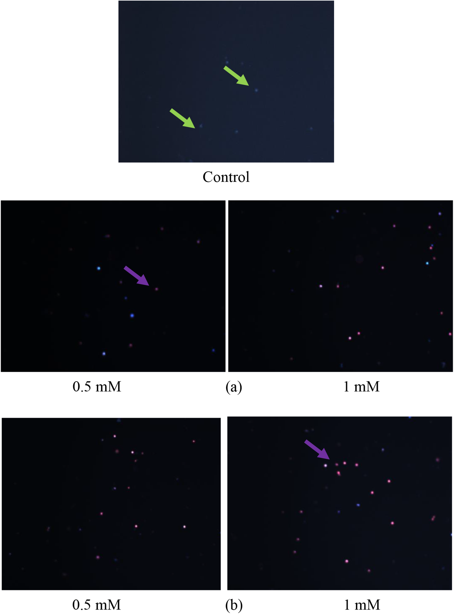
Representative images of morphological changes (as changes in cell membrane integrity) in human PBMCs after 24-h incubation with TCEP (a) and TCPP (b) in the concentrations 0.5 and 1 mM after double staining with Hoechst 33342/PI: viable cells with preserved cell membrane integrity and organized chromatin – blue fluorescence (green arrows), necrotic cells with damaged cell membrane and organized chromatin – red fluorescence (violet arrows).
Discussion
It is widely accepted that organic, chlorinated FRs exhibit atmospheric stability and, as a result, they can be transferred to long distances from the source. 40 Despite the frequent detectable presence of these compounds in the environment, there is limited information about toxicity mechanism of these chemicals, especially in the immune system cells. In the light of the growing demand for FRs with the phosphate group (OPFRs), extended studies on the toxicity of these compounds to living organisms should be carried out.
The purpose of this study was to evaluate necrotic and morphological changes in human PBMCs exposed to selected OPFRs. We have analysed influence of two commonly used organophosphorus-chlorinated FRs, TCEP and TCPP (Figure 1).
It was shown that both OPFRs analysed decreased PBMCs viability, while TCPP induced stronger necrotic changes in this parameter (Figure 2). Interestingly, statistically significant changes in cell viability were observed only at the highest concentrations of the compound studied, that is, at 0.5 and 1 mM of TCEP and TCPP, respectively. The above data indicate a relatively low toxicity of these two OPFRs, and particularly TCEP. Similar results were obtained by Föllmann and Wober 41 who found that TCPP and TCEP at above 1 mM induced moderate toxicity in V79 cells (hamster fibroblasts). Moreover, Crump et al. 42 revealed that TCPP, even at 300 µM, was not cytotoxic for neuronal embryos cells (CEN) and hepatocytes embryos cells (CEH). Previous works of Jarema et al., 43 Dishaw et al. 44 and McGee et al. 45 have shown that TCEP caused lower developmental lesions in comparison to other OPFRs.
In this study, we have reported that TCPP to a greater extent than TCEP reduced cell viability and induced changes in cells size and granulation (Figure 3). Stronger changes in tested parameters which were observed in PBMCs exposed to TCPP may be associated with different chemical properties of these compounds.
TCEP and TCPP contain highly electronegative chlorine atoms, which may partly be responsible for cytotoxic effect of these compounds in the cells studied. It was found that pentachlorophenol (PCP) induced the strongest morphological changes and ATP depletion in human lymphocytes when compared to other tested phenols with smaller number of chlorines. 46 Author explained that PCP reached the cells and caused their swelling, which was correlated with ATP depletion. Interestingly, Dishaw et al. 47 in the study of neurodifferentiation of PC12 observed that TCEP and TCPP, despite lower number of substituents than tris(1,3-dichloro-2-propyl)phosphate (TDCPP) and tris(2,3-dibromopropyl)phosphate (TDBPP), showed significant effects. Researchers supposed that major impact on phenotypic fate of PC12 cells depended on halogen substitution patterns in OPFRS, but not on the molecular size of that group of chemicals.
It is noteworthy that TCPP is structurally similar to another commonly used compound, chlorpyrifos, the chemical used as an insecticide for which numerous studies have shown neurotoxic and endocrine disrupting potential. Furthermore, it should be mentioned that toxic potential of many substances is related to their metabolism in living cells. It was found that TDCPP, aliphatic OPFRs, is transformed in living organisms to bis(1,3-dichloro-2-propyl)phosphate (BDCPP). BDCPP is characterized by higher stability than parent substance, and thus many of harmful effects observed for TDCPP may be in fact due to the formation of its metabolite (Chu et al. 48 ).
Mechanism of toxicity of OPFRs may be also correlated with K o/w values of these compounds. For the TCPP and TCEP tested in our experiment, values of K o/w are 2.55 and 1.44, respectively, what might suggest that these chemicals have relatively strong bioaccumulation properties (Van der Veen and de Boer, 2012). 1 Similar conclusions in relation to above-described OPFRs were proposed by Howard and Muir 49,50 who listed TCEP and TCPP as potentially persistent and capable of bioaccumulation. We hypothesized that alterations in PBMCs size and granulation observed in our experiments (Figures 3 and 4) can partly be the effect of interaction of TCPP and TCEP residues with cell membrane, which may be additionally due to above-described K o/w values. Morphological alterations induced in tested cells can thus be the result of membrane damage, loss of its integrity and finally lysis and reduction of cell volume through its shrinking. It has been proven that chlorinated derivatives such as trichlorophenol and perfluorooctanesulphonate induced changes in Vero cells membrane components such as proteins and phospholipids. 51 Authors of the above study suggested that changes in membrane induced by these compounds led to alterations in rigid structure and cell shape. Similar to our findings, Chen et al. 52 observed morphological alterations in TM3 Leydig cells after 24-h exposure to high doses of triphenyl phosphate (TPP) and TCEP.
Another factor that may be associated with increased toxicity of TCPP is the presence of three methyl group attached to oxygen atoms in the molecule of this compound (Figure 1). Methyl groups change chemical properties of the molecule depending on the electrons and stereochemistry. Branching of the carbon chain with methyl groups mostly results in increased toxicity of the compound. It is known that introduction of the methyl group into aromatic ring results in an increase in its toxic potential. In some studies, it has been demonstrated that presence of methyl residue in phenols contributes to haemoglobin oxidation. 53,54 Authors also suggested that different toxicity of aromatic compounds depended on the type of attached substituent. Moreover, these researchers revealed that phenols with methyl groups caused mainly oxidation of haemoglobin, while phenols with chlorine substituents more strongly contributed to lipid peroxidation.
Referring to the above, it is worth noticing that toxicity of TCEP and TCPP, although relatively small to PBMCs, may be associated with generation of reactive oxygen species (ROS) and oxidative stress in cells. Chen et al. 55 found that after 35 days of oral TCEP administration to mice, there was a functional impairment of antioxidant enzymes like catalase, superoxide dismutase and glutathione S-transferase (GST) in Sertoli cells of the studied animals. In turn, Li et al. 56 noted that TCPP significantly increased ROS production in human L02 cells. Similar results were obtained by Arukwe et al. 57 who using polymerase chain reaction analysis showed that the exposure of juvenile salmon to TCEP and other OPFRs, that is, tris(2-butoxyethyl) phosphate, increased the expression of glutathione peroxidase and GST genes at environmental dosages in liver and brain cells of the studied species. Moreover, it was observed that the tested compounds increased lipid peroxidation in liver cells, the process considered to be one of the major markers of oxidative stress occurring in living organisms.
Despite numerous studies determining the mechanism of OPFRs toxicity, action of these chemicals is still unclear and further research studies are needed.
In conclusion, it was noted for the first time that TCEP and TCPP decreased PBMCs viability only at concentration that is attributed to poisoning subacute with these compounds. It was found that organophosphorus-chlorinated FRs studied altered PBMCs size and granulation. Between the two tested compounds, stronger toxicity was noted for TCPP. Our results suggest that OPFRs may be appropriate substitutes of brominated flame retardants (BFRs), and these results may be a suitable base for further research for understanding toxicity of organophosphorus retardants.
Footnotes
Acknowledgements
The investigation was supported by a statutory research admitted for Department of Molecular Genetics and Department of Biophysics of Environmental Pollution.
Declaration of Conflicting Interests
The author(s) declared no potential conflicts of interest with respect to the research, authorship and/or publication of this article.
Funding
The author(s) disclosed receipt of the following financial support for the research, authorship and/or publication of this article: This work was partially financed by Student Grant obtained from Rector of the University of Lodz.