
Abstract
Effects on postnatal development of Swiss albino mice exposed to nickel (Ni2+) ions as nickel chloride haxahydrate (NiCl2·6H2O) during the gestation periods were evaluated in this study. Administration of Ni to pregnant females by gavage (46.125, 92.25, and 184.5 mg Ni/kg body weight (b.w.)) at doses below median lethal dose during 0–5 (preimplantation period), 6–13 (organogenetic period), and 14–18 days (fetal period) postconception. The dams were allowed to deliver and raise their pups. Significant (p < 0.05) decrease in litter size was observed after 184.5 mg Ni/kg b.w. during the three gestation periods particularly from preimplantation period as compared to organogenetic and fetal periods in comparison with the control group. Exposure to 184.5 mg Ni/kg b.w. during fetal period revealed higher mortality as compared to organogenetic period. Exposure to 184.5 mg Ni/kg b.w. increased the eye, limb, and tail anomalies during organogenetic period. Gestation index from preimplantation period was low at all the doses. Live birth index decreased during preimplantation and organogenetic periods after 184.5 mg Ni/kg b.w. The viability and weanling of pups decreased during all periods after 92.25 and 184.5 mg Ni/kg b.w. doses. A dose-dependent highly significant (p < 0.01) decrease in the body weight of offspring from day 0 to 6 weeks of age at all the doses during different gestation periods were observed. Maximum body weight decrease was observed in offspring from organogenetic period. This study concludes that young ones are vulnerable during different gestational and lactation period which indicates that Ni ingested by mothers constitutes a great threat to the progeny.
Keywords
Introduction
Environmental pollution particularly from industrialization can cause inevitable problems such as pollution of air, water, and soil. Both the quality and quantity of water are severely threatened by industrial sewage. Nickel (Ni) is very commonly found in surface and ground water, mostly originating from industrial activities and anthropogenic discharges. It also enters the water bodies through atmospheric deposition 1 and exists as hexahydrate in natural waters with pH < 9. 2 Humans absorb about 40 times as much Ni from water under fasting conditions. 3 After 10 min of flushing in the morning, Ni concentration in screened household drinking water were significantly decreased from 10.79 μg/L to 7.23 μg/L. 4
Exposure to Ni can produce a spectrum of adverse reproductive effects. 5 –7 A case study of occupational exposure of Russian women to Ni hydrometallurgy refinery plant resulted in congenital malformations in live-born infants, higher incidences of spontaneous and threatening abortions, and cardiovascular and musculoskeletal defects. 8 –12 Keeping in view the impact of Ni upon human health, the present study was planned to evaluate the effects of nickel chloride hexahydrate (NiCl2·6H2O) to pregnant Swiss albino mice on postnatal development during all the three gestation periods (i.e. preimplantation, organogenetic, and fetal periods, respectively).
Materials and methods
Experimental design
Swiss albino mice were procured from the Indian Veterinary Research Institute, Bareilly, Uttar Pradesh, India, and maintained on standard mice feed (Aashirwad Limited, Chandigarh, India) and provided tap water ad libitum. Males and females were housed for mating in the ratio of 3:1. Females were examined for vaginal plugs, and the day on which vaginal plug was observed was counted as day 0 of pregnancy. They were separated and their gestational days were recorded. NiCl2·6H2O obtained from Hi-Media (97.0%) Laboratories Pvt Ltd (Mumbai, Maharashtra, India) was used. The dose of Ni was chosen by calculating median lethal dose (LD50), and they were selected below LD50, that is, 369 mg Ni/kg body weight (b.w.). The pregnant females were assorted into 4 groups of 15 mice each. Group I were given tap water and served as a control, whereas groups II, III, and IV were given 46.125, 92.25, and 184.5 mg Ni/kg b.w. as NiCl2·6H2O. Each group were administered by gavage during 0–5 days (preimplantation period), 6–13 days (organogenetic period), and 14–18 days (fetal period) postconception, respectively. Experiments were approved by the Departmental Animal Ethics Committee, Department of Zoology, University of Rajasthan, Jaipur, Rajasthan, India.
The pregnant females of groups I–IV were allowed to reach term, deliver normally, and raise their pups. The litter size and sex ratio of neonates were recorded. The offsprings were examined for morphological anomalies, also the eye opening, pinna detachment, hair appearance, vaginal opening, and testes descent. The weights of these offsprings were recorded weekly up to the age of 6 weeks to determine their growth.
The following indices were calculated. 13
Viability index: viability index was calculated on postnatal day (PND) 4, 7, and 14 of age.

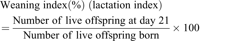
Live birth index
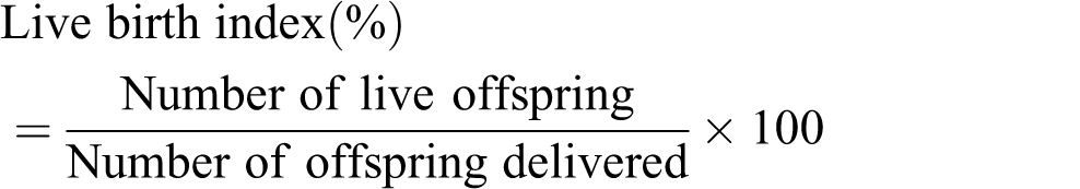
Weaning index
Gestation index

Statistical analysis
Data were evaluated in the Microsoft Office Excel 2003 sheet, and the significance of data was determined either using one-way analysis of variance or using one-way Mann–Whitney U test. Differences were considered to be significant (p < 0.05) and highly significant (p < 0.01).
Results
The average number of litter size per dam during preimplantation period (46.125 mg Ni/kg b.w.) was equal to that of the control group, although significant (p < 0.05) decrease after 92.25 and 184.5 mg Ni/kg b.w. exposure was observed when compared with their respective control groups. Exposure during the organogenetic and fetal periods caused nonsignificant decrease (after 46.125 and 92.25 mg Ni/kg b.w. dose levels), while significant (p < 0.05) decrease was observed (after 184.5 mg Ni/kg b.w.) when compared with their respective control groups (Table 1).
Influence on the postnatal development of mouse offspring exposed to Ni2+ as NiCl2·6H2O during gestational periods.

Ni2+: nickel ion; NiCl2·6H2O: nickel chloride hexahydrate; b.w.: body weight.
a p < 0.05: significant using one-way Mann–Whitney U test.
During preimplantation period, no mortality was observed after 46.125 and 92.25 mg Ni/kg b.w. dose levels. However, at 184.5 mg Ni/kg b.w. dose, 11.76% of pup mortality was occurred. Most of the deaths occurred within the first week of age. Exposure during organogenetic and fetal periods (46.125 mg Ni/kg b.w.) did not cause any pup mortality. However, after 92.25 and 184.5 mg Ni/kg b.w. dosage, the mortality was found to be 9.52 and 18.75%, respectively, during organogenetic period, whereas during fetal period it was 11.11 and 22.22%, respectively (Table 1).
The percentage of two sexes, that is, male and female was not altered in the preimplantation, organogenetic, and fetal periods at all the three dose levels. No change in the time of pinna detachment, eye opening, hair appearance, vaginal opening as well as testes descent was observed during different gestation periods at all the three dose levels (Table 1).
The eyes, limbs, and tail were found to be normal in surviving animals at all the three dose levels during preimplantation and fetal periods respectively. However, during organogenetic period after 92.25 and 184.5 mg Ni/kg b.w. dose levels, 5.55 and 6.25% animals showed unilateral microphthalmia (Figure 1(c)). Also, 18.75% total limb anomalies in the offspring were noticed after 184.5 mg Ni/kg b.w. administration. Few animals showed left hind limb without phalanges (6.25%), whereas others revealed absence of left and right hind limbs (12.25%; Figure 1(a) and (b)). High incidences (25.00%) of tail anomalies in the offspring were evident after 184.5 mg Ni/kg b.w. dose. These included absence (6.25%) and short tail (18.75%; Figure 2) anomalies. X-Ray of short-tailed animals revealed fused caudal vertebrae (Figure 3; Table 2).

Morphological anomalies in the offsprings showing (a) left hind limb without phalanges (Ph), (b) absence of left and right hind limbs (L), and (c) unilateral (Mi) at the dose of 184.5 mg Ni/kg b.w. during organogenetic period. Ph: phalanges; Mi: microphthalmic; Ni: nickel; b.w.: body weight.

Photograph showing (a) absence and (b) short tail offsprings at the dose of 184.5 mg Ni/kg b.w. during organogenetic period. Ni: nickel; b.w.: body weight.

X-Ray showing fused Cv in offsprings at the dose of 184.5 mg Ni/kg b.w. during organogenetic period. Cv: caudal vertebrae; Ni: nickel; b.w.: body weight.
Morphological anomalies in mouse offspring exposed to Ni2+ ion as NiCl2·6H2O during organogenetic period.

Ni2+: nickel ion; NiCl2·6H2O: nickel chloride hexahydrate; b.w.: body weight.
The gestation index during preimplantation period was affected by 75% at all the three dose levels, while no effect was seen during organogenetic and fetal periods. The neonates at the time of birth were found to be normal after exposure to 46.125 and 92.25 mg Ni/kg b.w. during preimplantation, organogenetic, and fetal periods. However, after 184.5 mg Ni/kg b.w. dose, the percentage of neonates (at the time of birth) was 88.23 and 93.75% during preimplantation and organogenetic periods, respectively. The survivability of neonates and lactation by mother during preimplantation period was 88.23% at PND 4, 7, 14, and 21 after 184.5 mg Ni/kg b.w. dose while during organogenetic period were 94.44, 94.44, 88.88, and 88.88% at the dose level of 92.25 mg Ni/kg b.w.; these values declined to 87.50, 87.50, 81.25, and 81.25% at 184.5 mg Ni/kg b.w. dose at PND 4, 7, 14, and 21, respectively. On exposure during fetal period, the viability index of offspring after 92.25 mg Ni/kg b.w. was less effective than after 184.5 mg Ni/kg b.w. at PND 4, 7, and 14 (94.44%, 94.44, and 88.88% and 88.88, 83.33, and 83.33%, respectively). Weaning index at PND 21 after 92.25 mg Ni/kg b.w. exposure was 90.47% that dropped to 77.77% after 184.5 mg Ni/kg b.w. administration. It was found to be more sensitive in fetal period than the organogenetic period (Table 3).
Effect on different indices of mouse offspring exposed to Ni2+ (as NiCl2·6H2O) during three gestation periods.

Ni2+: nickel ion; NiCl2·6H2O: nickel chloride hexahydrate; b.w.: body weight.
Prenatal exposure to 46.125 mg Ni/kg b.w. dose during the preimplantation period showed nonsignificant decrease in body weight of offspring and significant (p < 0.05) decrease at first and sixth week of age after 92.25 mg Ni/kg b.w. dose. However, highly significant (p < 0.01) decrease at the time of birth, first and third week of age at 184.5 mg Ni/kg b.w. exposure was observed. During organogenetic period after 46.125 mg Ni/kg b.w. dose exhibited significant (p < 0.05) decrease in the body weight of offspring at day 1 to 2nd week of age and 4th week to 6th week of age, while highly significant (p < 0.01) decrease was observed at 3rd week of age. However, after 92.25 and 184.5 mg Ni/kg b.w. a steep decline in the body weight at postpartum was observed which was highly significant (p < 0.01) from day 1 till the last week of age when compared with the control. Exposure during fetal period after 46.125 mg Ni/kg b.w. dose showed nonsignificant decrease in the body weight of offspring. After 92.25 mg Ni/kg b.w. dose, the decrease was highly significant (p < 0.01) at day 1 and 3rd week of age, whereas significant (p < 0.05) decrease during 1, 2, 4, 5, and 6 weeks of age was observed. However, after 184.5 mg Ni/kg b.w. highly significant (p < 0.01) decrease from day 1 to 6 weeks of age was observed as compared to their respective control groups. Maximum decrease in body weight was observed in mice offspring from organogenetic period, whereas this decrease was low in fetal period followed by preimplantation period (Table 4).
Body weight (in grams) of mouse offspring exposed to Ni2+ as NiCl2·6H2O during three gestation periods (mean ± SEM).

Ni2+: nickel ion; NiCl2·6H2O: nickel chloride hexahydrate; b.w.: body weight; ANOVA: analysis of variance.
a p < 0.05: significant using one-way ANOVA.
b p < 0.01: highly significant using one-way ANOVA.
Discussion
A significant decrease in litter size was observed during the three gestation periods particularly from preimplantation period as compared to organogenetic and fetal periods. This could be related to strong resorption in the uteri leading to decline in the number of live pups. Reduced number of live pups was also reported by Schroeder and Mitchener 14 in a three generation study with Ni, NiCl2, and subsulfide. 15 During fetal period, the mortality was higher compared with the organogenetic period. Dead pups were also observed in the organogenetic period. This may be due to the fact that Ni probably crosses the placenta (direct cytotoxic effect) adversely influencing the development of the fetus leading to the fetal mortality. Soluble NiCl2 has been found in the cytoplasm and nucleus, 16 and it is hypothesized that Ni2+ ions compete with other ions to enter the cells through divalent metal transporter I. 17 The overwhelming amount of Ni2+ ions may replace the metal ions that are required for the structure and function of enzymes leading to their inactivation which would contribute to Ni-induced mortality. Further, the physiological disturbances must also have strongly contributed to the pup mortality before weaning. 5
No change in the time for the appearance of physiological landmarks, that is, pinna detachment, hair appearance, eye opening, vaginal opening, and testes descend at all the three dose levels during different gestation periods were observed in the present study. Similar results were obtained with arsenic in rats. 18,19
The occurrence of unilateral microphthalmic eyes in the present study during the period of organogenesis was observed in few fetuses after NiCl2 administration. Sunderman Jr et al. 20 noticed that the inhalation of nickel tetracarbonyl resulted in both unilateral and bilateral anophthalmic and microphthalmic pups. Hassoun and Dencker 21 suggested that the absence of harderian glands probably causes the eyes to sink deeply into the orbits resulting in narrowing the palpebral fissures leading to microphthalmic eyes.
Few pups during organogenetic period had limb deformities such as absence of limbs (amelia) and phalanges (aphalangia) at the high-dose level. Reduction in the length of digit bones and loss of the middle phalanges were noticed by Takahara et al. 22 This has been correlated to excessive apoptosis in the interdigital regions. In the developing phalanges, the condensed mesenchymal cells were gradually decreased and failed to form cartilage model that resulted in developmental failure of the phalanges. 22 Several authors 23 –25 have also reported limb deformities in mice and rats.
Abnormalities such as complete absence and short tail have been observed in the present study during organogenetic period. Exposure to various other metals like chromium in rats, 26 –28 lead in mice, 23 and cadmium in rats 29 have also caused tail anomalies. Our findings indicate that exposure during organogenetic period is most sensitive in producing tail anomalies caused either due to absence or reduced number of urorectal caudal vertebrae or due to direct death of the few presumptive tail cells. 30
The gestation/pregnancy index during preimplantation period was lower at all the three dose levels, whereas no effect was observed on exposure during organogenetic and fetal periods to any of the three dose levels. This is in agreement with the studies of Ni in mice 5 and cadmium in rats. 31 Leonard and Jacquet 32 reported that prenatal exposure to Ni could partially be due to the production of certain changes in the mitotic apparatus provoking cellular death at the critical stage of development leading to decreased rate in the gestation index. The percentage of live pups at birth decreased in preimplantation and organogenetic periods. Similar findings were noticed with mercury in mice 33 and arsenic in rats. 18,19 The survivability (PND 4, 7, and 14) and weanling (PND 21) of pups were decreased at all the three gestation periods. Similar findings were also obtained with Ni in rats, 5 mercury in mice, 33 and arsenic in rats. 18,19 Maximum body weight reduction at all the three dose levels was observed in mice offspring from organogenetic period. Parallel were the observations of several authors 15,34 –36 in rats. Generally, that the Ni2+ ions form complexes with a number of proteins raise the possibility that it may significantly alter protein conformation and change their function and cellular homeostasis 37 thus leading to decrease in weight of the offspring. Jacobsen et al. 38 showed that Ni is transported through the placenta and is excreted in milk. Other factors can also contribute to growth retardation such as decreased milk consumption as observed by Dostal et al. 39 who reported that after the exposure of NiCl2 not only the milk production decreased but also the milk quality changed which probably was not palatable to the offspring leading to reduced weight of the offspring. Maternal or fetal metabolic disturbances causing growth retardation of offspring has been reported in mice, rats, and rabbits. 40,41
Conclusion
Conclusively, the present investigation with Ni as NiCl2 6H2O exhibited significant decrease in litter size as well as body weight of the offspring, high incidences of mortality, increased rate of morphological anomalies in eyes, limb, and tail, and lower different indices. Thus, it may be concluded that Ni is hazardous and has toxicological consequences. The young ones are vulnerable during the different gestational as well as nursing period; also the toxicity is transferred in part to the next generation through breast milk after birth, which reflects that maternal Ni ingested by mothers constitutes a great threat to the progeny.
Footnotes
Acknowledgment
Authors thank the Head, Department of Zoology, University of Rajasthan, Jaipur, Rajasthan, India, for providing necessary facilities.
Conflict of interest
The authors report no conflict of interest. The authors alone are responsible for the content and writing of the article.
Funding
This research received no specific grant from any funding agency in the public, commercial, or not-for-profit sectors.