
Abstract
Galacto-oligosaccharide (GOS) is a naturally occurring prebiotic that beneficially affects the host by selectively stimulating growth and/or activity of one or a limited number of colon bacteria to improve host health. A novel GOS was administered by gavage to male and female Sprague Dawley rats at 0, 500, 1000, and 2000 mg/kg/day for 10 weeks. In males, administration of GOS was initiated prior to mating and continued for 91 days. Females received GOS beginning 2 weeks prior to mating through day 20 of lactation. Parents were observed daily, and body weight (BW) and feed consumption were measured. Vaginal smears, mating behavior, and observation of delivery/lactation were evaluated in parents. Effects on the reproductive function of parents including gonad function, estrous cycle, mating performance, fertility, delivery and lactation, and effects on the growth and development of pups were examined. No deaths occurred, and no general toxicological effects or abnormal reproductive functions were observed in any dose group. Pups were observed at birth and the following measurements were undertaken: BW, external differentiations, sensory functions, and reflex reactions during lactation and just prior to necropsy. No external malformations or differences in the number of pups, in the sex ratio, or BW at birth occurred in any dose group. Growth and development of pups were normal. The No Observed Effect Level for reproductive function of male and female parent animals and for the growth and development of their offspring was at least 2000 mg/kg/day.
Introduction
Galacto-oligosaccharides (GOS), also known as oligogalactosyllactose, oligogalactose, oligolactose, or transgalactooligosaccharides, are nondigestible ingredients that stimulate the growth of bacteria such as bifidobacteria and lactic acid bacteria in the colon. 1 –3 The increased activity of these bacteria results in a number of health-related benefits attributed directly or indirectly from the organic acids produced by fermentation. 4 –6 GOS is added to a number of commercial food products for both infants and adults.
GOS have been used as food ingredients in Japan and Europe for at least 30 years and recently has been added to the U.S. food supply, where its use is growing. Besides differences in the purity among commercially available products, there are differences in the oligosaccharide linkages due to different enzymes used in GOS production. The GOS, which is the subject of this toxicity study, is manufactured by a two-step enzymatic reaction from lactose. This GOS has been shown to be well tolerated by humans. 7 Its safety also has been demonstrated in a repeated dose 90-day toxicity study in rats and in juvenile rats (in manuscript) and by the absence of genotoxicity in the Ames assay, Escherichia coli WP2uvrA, cultured fibroblast cells from Chinese hamster lungs, in an in vivo micronucleus assay of mouse peripheral blood, and an in vivo comet assay in rats. 8
To our knowledge, since there is no published reproductive toxicity study with GOS, this study was conducted to examine the effects of GOS on the reproductive functions of male and female parental animals and on the growth and development of their offspring. Treatment started just prior to mating and lasted for 91 days in males, whereas for females, it began 2 weeks prior to mating and continued up to the day before the pups were weaned.
Methods
Compliance statement
This study was conducted under the following guidelines: (1) “OECD Principles of Good Laboratory Practice” OECD Council: November 26, 1997; (2) “OECD Guideline for Testing of Chemicals 415” OECD Council: May 26, 1983; (3) Law Concerning the Protection and Control of Animals” Law No. 105, October 1, 1973, Revised on June 2, 2006 as Law No. 50 “Standards Relating to the Care and Management of Laboratory Animals and Relief of Pain” Notification No. 88 of the Ministry of the Environment, Japan, April 28, 2006; and (4) “Guidelines for Proper Conduct of Animal Experiments” Science Council of Japan, June 1, 2006. The protocol was reviewed by the Institutional Animal Care and Use Committee and approved according to the Guidelines for Animal Studies of the testing facility.
Test article
The GOS (Yakult Honsha Co. Ltd, Tokyo, Japan) used in this study is a sugar manufactured by a two-step enzyme reaction of lactose with Sporobolomyces singularis (first step) and β-galactosidase derived from Kluyveromyces lactis (second step). The Brix sugar content of the GOS was 75%, and the sugar composition of the GOS used in this study was 56.9% GOS and 43.1% monosaccharides. Stability analysis showed that the test article (GOS) was stable at room temperature over the administration period of the study. The concentration of GOS in the test article was verified by high-performance liquid chromatography using both an exclusion type ion exchange column and a normal phase column (LC 10 Series, Shimadzu Corporation, Tokyo, Japan).
Preparation of dosing solutions
Distilled water suitable for injection (Japanese Pharmacopoeia; D.W., Lot No. 1A76 and 1B72; Otsuka Pharmaceutical Factory Inc., Japan) was used as the control vehicle. The appropriate amount of the test article (GOS) was weighed in a beaker and dissolved in distilled water to a specified volume. After mixing, the test solution was divided into 1-day aliquots and stored in brown glass bottles in a refrigerator until used. The proportion of each measured concentration to the theoretical value was between 93.99 and 100.44% and within the acceptable range (proportion of the GOS to the theoretical values: 100.0 ± 10.0%). Dosing solutions were stable for 8 days at refrigeration temperature and for 24 h at room temperature.
Experimental animals
A total of 228 Sprague Dawley specific pathogen-free (SPF) strain (Crl: CD (SD)] male (aged 4 weeks) and nulliparous female (aged 3 weeks) rats, obtained from Atsugi Breeding Center, Charles River Laboratories Japan Inc., Japan, were quarantined/acclimatized for 12 days for males and 68 days for females. During this period, animals were observed for general condition, such as external appearance, nutritional condition, and behavior once every day and weighed on the day of receipt, on day 3 after receipt, and at least once a week thereafter. Vaginal smears were collected from all females for 14 days before grouping.
In total, 96 males and 96 females (normal estrous cycles) were selected based on body weight (BW) on the day of grouping (day before start of dosing), and administration was started at 5 weeks of age for males and at 12 weeks of age for females. BWs were within the range of mean ± 20% (198–236 g for males and 237–327 g for females). Of these, 10 females with abnormal estrous cycle, 3 additional females, and 13 males were euthanized by carbon dioxide asphyxiation and excluded from the study.
Animals were individually identified by ear tags upon arrival. After grouping, each animal was assigned a four-digit number by the dose level and sex. All cages had labels that were color coded according to dose levels, indicating the study number, administration route, dose level, sex, animal number, ear tag number, scheduled date of necropsy (males), date of copulation (males and females), and date of delivery (females).
Animals were housed in an animal room in which the temperature was maintained at 22 ± 3°C, relative humidity at 50 ± 20%, ventilation at 10–15 changes per hour, and 12-h illumination per day (07:00–19:00 h). All animals were housed individually in bracket-type metal wire mesh cages (width (W) × diameter (D) × height (H): 254 × 350 × 170 mm3; Nihon Cage Co., Ltd, Japan) except during mating. During the period from day 17 of gestation to day 21 of lactation, females were housed in the unit of litter in plastic Econo cages (W × D × H: 340 × 400 × 185 mm3: CLEA Japan Inc., Japan) with bedding (white flakes: Charles River Laboratories Inc., Japan, Lot No. 2011-1). Animals were allowed free access to pelleted diet nitromethane fraction (radiation sterilized, Oriental Yeast Co. Ltd, Tokyo, Japan) via stainless steel feeders and to tap water (Gotemba City Water, Japan) via an automatic water supply system (premating and mating) or using water bottles (during gestation and lactation). Each lot of feed and bedding was analyzed for contaminants by Eurofins Scientific Analytic (Japan). The water supplied to the animals was analyzed by Shibaura Semtek Co. Ltd (Japan) on a quarterly basis in accordance with the Japanese Waterworks Law.
Animals were assigned to dosing groups by a combination of the block placement method and a random sampling method using a computer (requisite number of groups was made using the block placement method, and group numbers and individual numbers were assigned using the random sampling method). Each group consisted of 24 males and 24 females. Animals were dosed daily at 0, 500, 1000, and 2000 mg/kg/day by oral gavage using a flexible stomach sonde for 10 weeks before mating and 3 weeks after the start of mating for males and 2 weeks before mating, during mating, and gestation up to day 20 of lactation for females. The dose volume was 5 mL/kg of BW, calculated based on the animal’s most recently recorded BW. For delivering females, administration was carried out immediately after delivery. The animals in the control group received the vehicle (water for injection) alone in the same manner.
The following observations, measurements, and examinations were undertaken.
All parental animals were observed for the presence or absence of abnormalities in clinical signs such as external appearance, nutritional condition, posture, behavior, and excrement before, immediately after, and between 1 and 3 h after dosing of the day during the administration period, and on the day of necropsy. Periodical observation including handling of the animals was carried out once or twice a week except for lactating dams. Pregnant females were observed on days 0, 7, 14, and 20 of gestation.
Males were weighed on days 1, 8, 15, 22, 29, 36, 43, 50, 57, 64, 71, 78, 85, and 92 of administration. Females were weighed on days 1, 8, and 15 of administration, days 0, 4, 7, 11, 14, 17, and 20 of gestation, and days 0, 4, 7, 11, 14, 17, and 21 of lactation. For dams for which delivery was completed after administration, BW was measured after the end of delivery. Feed consumption from the previous day was measured on days 2, 8, 15, 22, 29, 36, 43, 50, 57, 64, and 71 of administration for males and on days 2, 8, and 15 of administration, days 1, 4, 7, 11, 14, 17, and 20 of gestation, and days 2, 4, 7, 11, 14, 17, and 21 of lactation for females.
Vaginal smears were collected from all females every day from the day following the start of administration to the day copulation was confirmed. Vaginal smears consisting of many cornified epithelial cells were classified as estrous period, and the number of estrouses and the number of days from the estrous to the next estrous (estrous cycle) were determined.
Males and females in the same dose group were co-housed overnight on a one-to-one basis (day 0), and copulation was confirmed either by vaginal plug or sperm in vaginal smears. The maximum mating period was 21 days. The gestation period was the number of days following successful copulation to the end of delivery in unit of 0.5 days. Pregnant females were observed twice a day from day 21 to day 25 of gestation for the absence/presence of delivery and completion of delivery, and for the presence/absence of abnormalities in the delivery (presence/absence of treatment on placenta/amnion).
Dams were allowed to nurse their own pups until day 21 after delivery and lactation condition (gathering pups, nesting, and lactation of dams) was observed.
On day 21 of lactation, necropsies were performed on all male and female parent animals following termination by exsanguination via the abdominal aorta under isoflurane anesthesia. Organs and tissues in the external, cephalic, thoracic, and abdominal regions were examined macroscopically. Dams that had total litter loss during the lactation period were killed by exsanguination and necropsied in the same manner but at the time, when there were no surviving pups in the observation before dose administration of the day. For dams, the number of implantation sites was counted at necropsy. Females for which delivery had not been confirmed by the morning of day 25 of gestation (1 female in the control group, 2 females in the 500 mg/kg group, 1 female in the 1000 mg/kg group, and 1 female in the 2000 mg/kg group) were killed by exsanguination via the abdominal aorta under isoflurane anesthesia and subjected to necropsy to confirm the presence/absence of gestation.
The pituitary, testes, epididymis, prostate, seminal vesicle (including coagulating gland), ovary, uterus, vagina, macroscopic lesions, and the part for individual identification (ear auricle with an ear tag) were removed from all parental animals, fixed, and preserved in phosphate-buffered 10% formalin. However, testes and epididymis were fixed in Bouin’s solution and preserved in phosphate-buffered 10% formalin. Thin sections were cut for all tissues except the ear auricle and stained with hematoxylin–eosin. Only tissues from males and females that did not copulate were examined microscopically because there were no effects from administration of the test article.
The number of live born and stillborn pups was counted on day 0 after birth and observed for the presence/absence of external abnormalities and sexed. Live born pups were weighed, while stillborn pups were fixed and preserved in Bouin’s solution.
Live born pups were observed for mortality once a day and nursed by a dam until day 21.
Live born pups were weighed on the same days as dams and mean BW was calculated for each male and female pup in each litter. The number of pups per litter was adjusted randomly to a total of eight pups, four males and four females, on day 4 after birth. Pups were examined for pinna detachment and weighed. If litter size were greater than eight, the extra pups were anesthetized with isoflurane and preserved in Bouin’s solution. If the litter size were less than eight, all pups were maintained. From day 11 after birth, pups were examined for incisor eruption (days 11 and 14 after birth) and eyelid opening (day 14 and 17 after birth). Two males and two females per litter, selected at random, were examined for righting reflex on day 10 and for air righting reflex, pupillary reflex, Preyer’s reflex, and pain reflex on day 20. On day 21, all pups were subjected to laparotomy under isoflurane anesthesia, killed by exsanguination via the abdominal aorta and examined for the presence/absence of abnormalities in the organs/tissues in the external, thoracic, and abdominal regions.
Statistical analysis
Using the following equations, the copulation index, insemination index, fertility index, delivery index, external differentiation index, and reflex indices were calculated for each group, and the length of gestation period, stillbirth index, external abnormality index, birth index, sex ratio on days 0 and 4 after birth, and viability index on days 4 and 21 after birth were calculated for each litter. Mean BW and righting reflex reaction time was calculated for male pups and female pups for each litter.
Copulation index (%) = (Number of animals copulated/Number of animals housed together) × 100
Insemination index (%) = (Number of males impregnated females/Number of males copulated) × 100
Fertility index (%) = (Number of pregnant females/Number of females copulated) × 100
Delivery index (%) = (Number of females that had live born pups/Number of pregnant females) × 100
Gestation period (days) = Number of days from day 0 of gestation to the day of delivery
Stillbirth index (%) = (Number of stillborn pups/Number of pups born) × 100
Index of external abnormalities (%) = (Number of pups with external abnormalities/Number of pups born) × 100
Birth index (%) = (Number of live born pups/Number of implantation sites) × 100
Sex ratio = Number of live born male pups/Number of live born pups
Viability index on day 4 after birth (%) = (Number of live pups on day 4 after birth/Number of live born pups) × 100
Viability index on day 21 after birth (%) = (Number of live pups on day 21 after birth/Number of live pups on day 4 after birth) × 100
Index of external differentiation (%) = (Number of pups with differentiation/Number of pups examined) × 100
Index of reflex (%) = (Number of pups that showed reflex/Number of pups observed) × 100
The difference between the control group and each dose group was analyzed for statistical significance as follows. For uncopulated females, only the data of copulation index were included in the statistical analysis. The following data were excluded from statistical analysis: BW of females during the mating period and BW of uncopulated females; BW on the day of necropsy of females that had not delivered by day 25 of gestation and dams that had total litter loss during the lactation period; and BW and feed consumption data during the gestation period of the animals that were judged to be not pregnant. For BW measurements (except for females during the mating period), BW gain (males: from days 1 to 71 of administration, females: from days 1 to 15 of administration, days 0 to 20 of gestation, and days 0 to 21 of lactation, and live born pups: days 0 to 21 after birth), feed consumption, number of estrouses observed, estrous cycle, number of days until copulation, length of the gestation period, number of implantation sites, number of live born pups, sex ratio and the reaction time for righting reflex, and the group mean with standard deviation were calculated and tested using Bartlett’s test for homogeneity of variance (level of significance: 0.01, bilateral). Then, comparison was carried out between groups by Dunnett’s test (levels of significance: 0.05 and 0.01, bilateral) for homogeneous data and by Steel’s test (levels of significance: 0.05 and 0.01, bilateral) for heterogeneous data. 9 –12
For the stillbirth index, index of external abnormalities, live birth index, and survival index, the group mean with standard deviation was calculated followed by Steel’s test (levels of significance: 0.05 and 0.01, bilateral). 11 The copulation index, insemination index, fertility index, delivery index, items in the observation of external differentiation, and indices of reflexes in air righting reflex, pupillary reflex, Preyer’s reflex, and pain reflex in the sensory functions and reflex responses were calculated from the number of copulated animals, number of male animals that impregnated females, number of pregnant females, number of females that delivered live born pups, number of pups that showed differentiations, and number of pups that showed normal reflexes; comparison between groups was carried out using χ 2 test with Yates’ continuity correction (levels of significance: 0.05 and 0.01, bilateral). If there were cells with expected frequency of not more than five, comparison between groups was carried out using Fisher’s direct probability method (levels of significance: 0.05 and 0.01, bilateral). 9
Results
No deaths occurred in any group and there were no abnormal clinical signs in any of the parental males or females. Decreased lactation behavior was observed in 1 dam in the 500 mg/kg group on day 1 of lactation (all pups died on day 2) and in 1 dam in the 2000 mg/kg group on days 1 and 2 of lactation (13/14 pups died). In addition, all pups died on day 2 of lactation in 1 dam in the control group. The deaths of these pups were incidental since the incidence of their occurrence was unrelated to the dose.
The results of BW and feed consumption are shown in Figures 1 and 2, and Figures 3 and 4, respectively. In females, a significantly low feed consumption was observed in the 2000 mg/kg group on days 1, 4, and 11 of gestation, but similar changes were not observed during premating or during lactation. Otherwise, there were no significant differences in feed consumption.
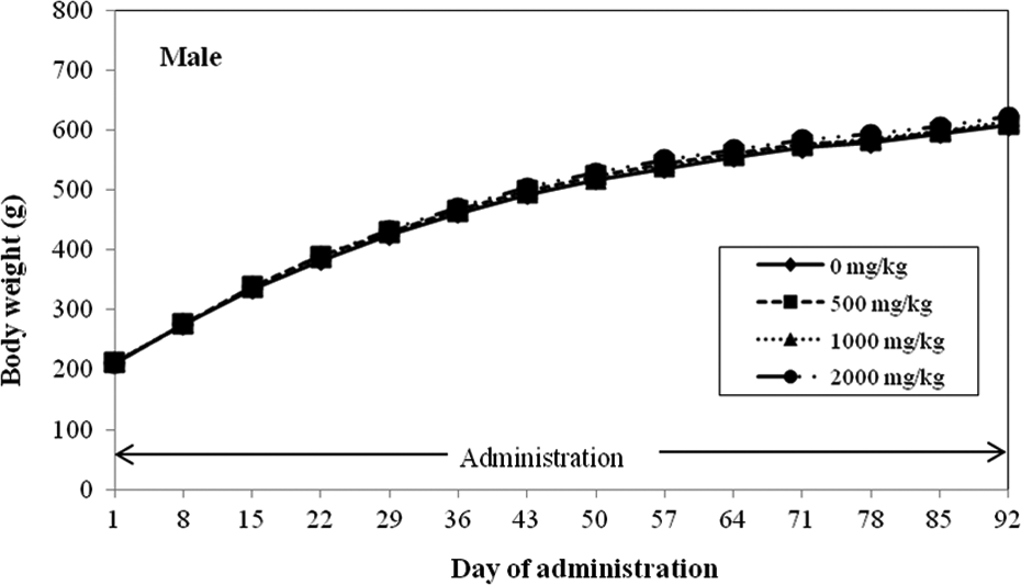
A one-generation reproduction toxicity study in rats treated orally with GOS. Body weight of male rats. GOS: galacto-oligosaccharide.

A one-generation reproduction toxicity study in rats treated orally with GOS. Body weight of female rats. GOS: galacto-oligosaccharide.
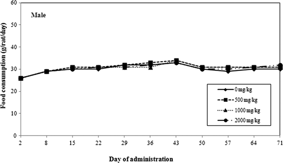
A one-generation reproduction toxicity study in rats treated orally with GOS. Feed consumption of male rats. GOS: galacto-oligosaccharide.

A one-generation reproduction toxicity study in rats treated orally with GOS. Feed consumption of female rats. GOS: galacto-oligosaccharide.
Most of the females showed 4-or 5-day estrous cycles, and there were no significant differences caused by GOS in the number of estrouses or mean number of days of the estrous cycle. There was a significantly low value in the number of estrouses in the 1000 mg/kg group. This change was due to the occurrence of a prolonged diestrous period of 10 days in 1 female, and it was thought to be a pseudopregnancy caused by stimulus of a swab. After recovery from the pseudopregnancy, this female showed a normal estrous cycle, normally copulated, became pregnant, and delivered a normal litter.
Most pairs had copulation within 5 days after the start of mating; there were no significant differences in the number of days until copulation, copulation index, insemination index, or fertility index between the control group and any test article group. Only a single pair in the control group and in the 500 mg/kg group did not copulate. The number of females that copulated but were not pregnant was 1, 2, 1, and 1 in the 0, 500, 1000, and 2000 mg/kg groups, respectively. The incidence was low, not dose related and within the historical background data of the test facility.
All females had normal delivery between days 21 and 23 of gestation, and there were no significant differences between the control group and any treatment group in the delivery index, length of the gestation period, number of implantation sites, stillbirth index, number of live born pups, or live birth index.
There were no gross pathological (all parental animals) or histological changes (only males and females that did not copulate) in any organs/tissues caused by the test material. There were no significant differences between control and any test article group in the number of male and female live born pups, sex ratio, or BW. There were no significant differences between the control group and any test article administration group in the viability index of live born pups on day 4 or 21 after birth. There were no significant differences between the control group and any test article group in BW from day 0 to 21 after birth or in BW gain during this period. A significantly low value in the differentiation ratio was observed in pinna detachment on day 4 after birth and for incisor eruption (first time) on day 11 after birth in the 1000 mg/kg group and in eyelid opening (first time) on day 14 after birth in the 1000 and 2000 mg/kg groups; however, these changes were incidental since they were all minimal changes, clearly dose unrelated, and differentiation in incisor eruption and eyelid opening was not observed in any animals at the second observation, and the changes were within the range of variations in the background data of the test facility. With the exception of delayed incisor eruption, eyelid opening, and pinna detachment (all of which were not dose related), there were no other external abnormalities.
There were no significant differences between the control group and any test article administration group in the reaction time for righting reflex, and all animals showed normal responses in air righting reflex, pupillary reflex, Preyer’s reflex, and pain reflex.
Discussion (revised and expanded)
GOS has been reported to have a number of beneficial effects in human, in part due to its fermentation in the colon resulting in an increase in the number of beneficial organisms including bifidobacteria and lactobacilli with a decrease in certain pathogens. 13 –16 In addition, systemic effects produced by GOS include positive changes in mineral balance (i.e., calcium, magnesium, and phosphorus), beneficial effects on plasma levels of cholesterol, increased production of soluble immunoglobulin A, and decreased incidence of atopic disease in at risk subjects. 6,7
The safety of this novel GOS has been demonstrated in a repeated dose 90-day toxicity study in rats and by the absence of genotoxicity in the bacterial reverse mutation assay (Ames test) in Salmonella typhimurium TA98, TA100, TA1535, and TA1537 strains and in E. coli WP2uvrA strain, in a chromosome aberration assay in cultured Chinese hamster lung (in nternational units) cells, in the micronucleus assay with mouse peripheral blood erythrocytes,16 and recently in a comet assay.
Because GOS has not been evaluated in a reproductive test, GOS was administered orally by gavage to groups of 24 male and 24 female Sprague Dawley strain SPF rats (Crl: CD (SD)) at 0, 500, 1000, and 2000 mg/kg/day for 10 weeks before the start of mating through mating up to the day before necropsy (administered for 91 days) to males and for 2 weeks before the start of mating through the mating, gestation, and up to day 20 of lactation (administration for 57 to 66 days) to females. Control animals received distilled water orally at the same volume and time as animals in the test groups. Both parent animals were observed for general condition prior to and during mating and females were observed during delivery and lactation. In addition, the following measurements or observations were made: BW and feed consumption, vaginal smears, necropsy, and histopathological examination (for uncopulated animals only). Gonad function, estrous cycle, mating performance, fertility, delivery and lactation, and effects on the growth and development of live born pups were measured to determine the potential effects of GOS on the reproductive function of male and female parental animals. General overall condition, sex, and BW were measured for live born pups at the time of birth. Observation for external differentiations and examination of sensory functions and reflex reactions during lactation and just prior to necropsy were performed.
No parental animals died in any group and there were no significant effects of the test article, GOS, on the general condition, BW, feed consumption or any of the other parameters evaluated. There were no abnormalities in the number of estrouses, mean duration of estrous cycle, number of days until copulation, copulation index, insemination index, number of implantation sites, or fertility index in any group. No untoward effects in the pituitaries or in the reproductive organs were examined during histopathological observation in the two pairs of animals that did not copulate (one pair each in the control and 500 mg/kg/day dose). There were no abnormalities in delivery conditions including delivery index, length of gestation, stillbirth index, number of live born pups, and live birth index. In addition, lactation behavior of females was normal.
Based on these results, administration of GOS at 2000 mg/kg/day had no general toxicological effect on male or female parental animals or on reproduction/development from premating or on copulation in males or females or implantation up to maintenance of pregnancy in females.
There were no effects from administration of the test article on the number of live born male or female pups, sex ratio, or external observations at the time of birth or on BW, survival, or external differentiation during lactation. In addition, since there were no effects from administration of GOS on the sensory function or reflex responses of live born pups, there were no abnormalities at the time of weaning; the No Observed Effect Level for GOS on the general condition and reproductive functions of parental animals and on the growth and development of the next generation was at least 2000 mg/kg/day under the condition of the present study. This study along with the other toxicological evaluations with a long history of use of GOS in a variety of foods throughout the world continue to support the safety of this prebiotic for use both in adult and infant foods.
Footnotes
Acknowledgment
The authors would like to acknowledge Ms Yoko Watanabe for her valuable technical advice.
Conflict of interest
The authors declared no conflicts of interest.
Funding
This research received no specific grant from any funding agency in the public, commercial, or not-for-profit sectors.