
Abstract
Rutin and quercetin were investigated for their effects on blood pressure and antioxidant defense system of rats fed with 8% sodium chloride-supplemented diet (high salt diet) for 6 weeks. Animals fed with high salt diet demonstrated an increase in systolic, diastolic, pulse, and mean arterial blood pressures (p < 0.05) as well as lipid peroxidation but decreases in the activities of antioxidant enzymes compared with control group. Groups post-treated with rutin and quercetin for 2 weeks showed significant reversals in the values of these indices compared with the group fed with only the high salt diet but not post-treated. The high salt diet also led to significant increase in serum glucose, urea, creatinine, triglycerides, low-density-lipoprotein, and total cholesterol concentrations. Treatment with rutin and quercetin ameliorated the effects of high salt diet on these biochemical indices. The reference standard, nifedipin was less effective than rutin and quercetin. The results of this study highlight the risk of high salt consumption on cardiovascular health and the potent antioxidant and antihypertensive property of rutin and quercetin.
Introduction
Hypertension, also known as high blood pressure is a silent killer. It is a major contributor to the morbidity and mortality arising from other diseases like heart failure, stroke, and diabetes. 1 The incidence of hypertension has been on a steady increase 2 and has been aggravated in less developed and underdeveloped societies due to factors like poverty, pressure of making ends meet, lack of regular medical checkup, and dietary indiscretion. 3 High salt intake through consumption of salted foods is still rampant in many traditional communities, where it is directly used in cooking or employed as a preservative for certain foods. 4 In urbanized and industrialized communities, high salt intake is also prevalent due to the increased consumption of processed foods.
Antihypertensive drugs like nifedipine and ramipril, though generally effective, often have side effects, 5 and the discovery of natural remedies without these side effects has been a subject of intensive research. Phytopharmaceuticals and other plant-based alternatives are becoming more and more popular, as many populations in both developed and developing nations increasingly embrace herbal medicine. 6 Rutin and quercetin are flavonoids that have been found to occur abundantly in plants. 7 The vasorelaxant and antihypertensive property of both compounds have been demonstrated. 8 Although there is information on the effect of rutin and quercetin in spontaneous hypertensive rats, there is dearth of information on their antihypertensive property against sodium chloride (NaCl)-induced hypertension. The present study has therefore been designed to evaluate the effect of rutin and quercetin on NaCl-induced hypertension in rats and to compare their efficacy with that of nifedipin.
Materials and methods
Chemicals
Epinephrine, thiobarbituric acid (TBA), trichloroacetic acid, glycine, hydrogen peroxide, quercetin, and rutin were obtained from Sigma-Aldrich (St Louis, Missouri, USA). All other reagents were of analytical grade and were obtained from standard suppliers.
Animal handling and treatment
Wistar albino rats (200–250 g), bred at the animal house of the Department of Physiology, Lagos University Teaching Hospital, Lagos, Nigeria, were used. They were all clinically healthy and maintained under standard laboratory conditions of temperature (27.0 ± 0.5°C), relative humidity (73.0 ± 15%), and 12 h darkness and 12 h light cycle for 2 weeks before inducing hypertension. Animals were handled and used in accordance with the international guide for the care and use of laboratory animals. 9 Animals were divided into nine groups. Group 1 (control) was fed with commercial diet and water ad libitum throughout the period of experiment and received an equivalent volume of distilled water in lieu of test substances. Group 2 was fed with a high salt diet (rat chow supplemented with 8% NaCl w/w) alone for 6 weeks after which commercial diet and distilled water were given for 2 weeks. Animals in group 3 were fed with high salt diet for 6 weeks, after which they were fed with commercial diet and administered with 1.67 mg/kg Nifedipine orally for 2 weeks. Animals in groups 4, 5, and 6 were fed with high salt diet for 6 weeks, after which they were fed with commercial diet and administered with graded doses of rutin (50, 100, and 150 mg/kg, respectively) for 2 weeks. Similarly, animals in groups 7, 8, and 9 were fed with high salt diet for 6 weeks, after which commercial diet and graded doses of quercetin (50, 100, and 150 mg/kg, respectively) were administered for 2 weeks. Rutin and quercetin were administered orally once daily for the 2 weeks.
Measurement of blood pressure
Blood pressure was measured according to the method of Adeboye et al.
10
After the period of treatment, rats were anaesthetized with 5 ml/kg urethane–chloralose (25% urethane and 1% chloralose). As soon as the animals reached the state of surgical anesthesia, each of them was placed on its back on a rat dissecting board. The trachea was exposed and cannulated. The carotid artery was located and carefully exposed, a bulldog clip was placed on the two ends of the artery and it was then cannulated towards the heart. The bulldog clip was removed and the cannula was pushed in further and then tied securely. Using a disposable syringe, the cannula was filled with heparinized saline (1% heparin in 9% NaCl) to prevent blood clotting. Heparinized saline was filled into a three-way tap and the cannula. The other end of the tap was connected to an isometric force transducer for blood pressure recording. After the stabilization period of 30 min, the blood pressure was recorded using a Glass polygraph Model 7D (calibration: 1 cm = 40 mmHg). The mean arterial blood pressure (MABP) was calculated using the formula

Biochemical estimations
Animals were killed by the overdose of anesthesia. A 10% homogenate was prepared and centrifuged at 5000g and then 15,000g to obtain supernatants used to assay for the extent of lipid peroxidation and the activities of antioxidant enzymes, respectively. Before killing the animal, blood was drawn from the carotid artery into plain and heparinized tubes that were then centrifuged at 5000g to obtain sera and plasma, which were used for biochemical estimations. Lipid (total cholesterol, triglyceride, high-density-lipoprotein (HDL), and low-density-lipoprotein (LDL)), glucose, creatinine, and urea levels were estimated in the sera, while nitric oxide (NO) level was estimated in the plasma.
Lipid peroxidation assay
TBA reactive species (TBARS) production in the liver was determined as described by Ohkawa et al. 11 This is based on the reaction of chromogenic substrate and malondialdehyde (MDA). The absorbance was measured against blank at 532 nm.
Assays for antioxidant enzymes
Superoxide dismutase (SOD) activity was evaluated according to the method described by Misra and Fridovich. 12 Catalase activity was measured by the method of Aebi. 13 Evaluation of glutathione peroxidase (GPx) activity was carried out according to Chance. 14
Estimation of NO level
Nitric oxide level was estimated as described by Green et al. 15 The principle of the assay is the reduction of nitrate by copper–cadmium alloy, followed by color development with Griess reagent (sulfanilamide and N-naphthylethylenediamine) in acidic medium.
Estimation of the levels of serum metabolites
Levels of all serum biochemical indices and total protein concentration were estimated with an automated analyzer (Hitachi 902, Roche Diagnostics, Boehringer-Mannheim, Germany) using kits supplied by Roche Diagnostics (Germany).
Statistical analysis
Values are expressed as mean ± SD or SEM. Statistical evaluation was performed by one-way analysis of variance followed by Duncan’s new multiple range test using SPSS 11.09 for windows. The significance level was set at p < 0.05.
Results
There was elevation in the blood pressure in the animal group fed with only the high salt diet as revealed with the significant increases (p < 0.05) in systolic (174.25 ± 9.89 mmHg), diastolic (129.75 ± 3.33 mmHg), MABP (145.00 ± 2.55 mmHg), pulse pressure (42.50 ± 11.12 mmHg), and heart rate (362.00 ± 18.65 bpm) compared with the control (83.00 ± 3.50; 69.00 ± 3.42; 75.40 ± 4.02; 19.50 ± 1.71; 414.50 ± 5.85 bpm, respectively) (Table 1). Groups post-treated with the flavonoids showed significant dose-dependent reductions in these indices.
Hemodynamic parameters of experimental animals.a

SYS: systolic blood pressure; DIA: diastolic blood pressure; PP: pulse pressure; MABP: mean arterial blood pressure; HR: heart rate.
aValues are expressed as mean ± SEM; n = 4. Values with the same superscript letter down a column are not significantly (p > 0.05) different.
Figure 1 showed that the high salt diet significantly (p < 0.05) increased the level of MDA in the liver of the NaCl-induced hypertensive group (45.18 ± 4.08 U/g tissue) as compared to control (17.57 ± 4.08 U/g tissue). Treatment with the flavonoids nullified the increase. Similarly, the activities of the enzymatic antioxidants such as catalase, SOD, and GPx were decreased in the NaCl-induced hypertensive group compared with control (p < 0.05). Treatment with the flavonoids also counteracted the effects seen in the induced group in a dose-dependent manner (Figures 2 to 4, respectively). The group fed with the high salt diet alone showed significant increases in serum creatinine (44.49 ± 1.10 µmol/l), urea (14.32 ± 0.69 mmol/l), and glucose (6.80 ± 0.73 mmol/l) levels compared with control (15.11 ± 0.71 µmol/l, 6.93 ± 0.89 mmol/l, and 0.43 ± 0.04 mmol/l, respectively). Treatment with the flavonoids brought about significant reductions in the creatinine, urea, and glucose levels compared with the group fed with the high salt diet alone (Table 2). Also, as depicted in Figure 5, the levels of serum triglycerides, LDL, and total cholesterol increased significantly (1.51 ± 0.22; 0.9 ± 0.04; 2.39 ± 0.21 mmol/l; p < 0.05) in the NaCl-induced hypertensive group compared with the control (0.33 ± 0.03; 0.18 ± 0.05; 1.37±1.00 mmol/l), while the level of HDL in the NaCl-induced hypertensive group decreased significantly (0.35 ± 0.13 mmol/l; p < 0.05) as compared to control (0.80 ± 0.04 mmol/l). However, treatment with the flavonoids reversed the effect of NaCl on the lipid profile.
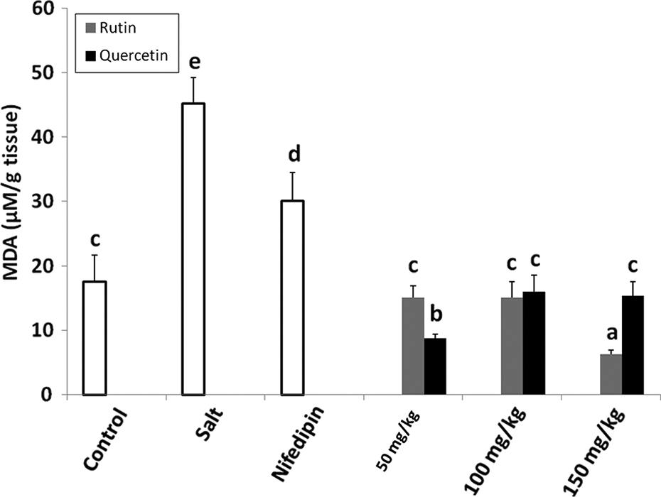
Effect of rutin and quercetin on the MDA concentration in salt-induced hypertensive rats. Values are mean ± SD; n = 6; Values with the same letters are not significantly different (p > 0.05). MDA: malondialdehyde.

Effect of rutin and quercetin on catalase activity in salt-induced hypertensive rats. Values are mean ± SD; n = 6; Values with the same letters are not significantly different (p > 0.05).
Serum biochemical indices in control and treated groups.a
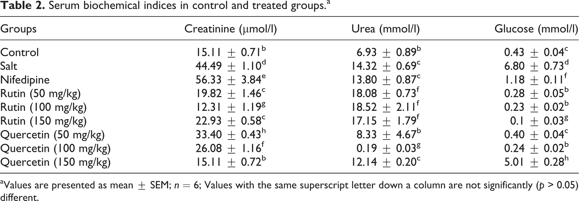
aValues are presented as mean ± SEM; n = 6; Values with the same superscript letter down a column are not significantly (p > 0.05) different.
Figure 6 indicates that the plasma NO concentration of the animals decreased significantly (p < 0.05) in the NaCl-induced hypertensive group (38.6 ± 5.13 µM) as compared to the control (95 ± 2.00 µM). Quercetin ameliorated the decrease only at 150 mg/kg (62.06 ± 1.87 µM), while rutin exerted dose- dependent increases in the concentration of plasma NO.
Discussion
The NaCl-induced elevation in the blood pressure (Table 1) is in agreement with earlier findings. 16 Quercetin has been reported by previous authors to lower blood pressure, 17 –20 which is in accord with the results obtained in this study. The antihypertensive effect of quercetin as demonstrated in this study may also explain, at least in part, why previous epidemiological reports show an inverse relationship between dietary flavonoid intake and heart disease risk. 21 –25 Experimental studies showed that polyphenolic compounds may reduce the arterial pressure in rats and enhance the vasorelaxant process. 26,27 Rutin and quercetin probably decreased the blood pressure by decreasing the heart rate, which is a major determinant of the cardiac output. 28
Feeding the high salt diet to rats caused increase in lipid peroxidation but reduction in the activities of SOD, catalase, and glutathione peroxidise (Figures 1 to 4), which could be due to oxidative stress. 29 –32 Salt-induced hypertension has been discovered to be associated with increased oxidative stress and renal expression of NADPH oxidase and decreased activity of SOD. 33 High salt diet may indirectly induce endothelial dysfunction through intermediate mechanisms that are associated with oxidative stress. 34,35 Nifedipine significantly increased the activity of catalase and GPx. Some cardiovascular drugs may also have antioxidant properties, as has been shown for some calcium channel blockers and adrenoceptor antagonists. 36
Oxidative stress appeared to be directly involved in the renal dysfunction observed in Dahl Salt-sensitive rats, 29 which could be the cause of elevation in the serum concentration of creatinine, urea, and glucose (Table 2). Creatinine and urea are markers used in evaluating kidney functions. 37 Impaired kidney function causes hardening of the arteries and may result in high blood pressure. 3,38,39 Also, persistent hyperglycemia causes diabetes mellitus, which is a high risk factor for secondary hypertension that might be responsible for the increase in the level of plasma glucose. Rutin has been reported as a potent advance glycation enzyme inhibitor. 40,41
High salt diet caused significant increase in serum concentrations of triglycerides, LDL, and total cholesterol and reduction in the serum concentration of HDL. This could promote atheroma development in arteries. 42 An inverse correlation between flavonoid intake and total plasma cholesterol concentrations has been reported. 43 There was decrease in the level of NO concentration in plasma in the high-salt group as compared with the control (Figure 6). Salt sensitive rats are deficient in NO. 44 In NO-deficient hypertension, the acceleration of blood pressure recovery by treatment with antioxidant has been associated with increased NO synthase activity. 19
Rutin and quercetin were more effective than nifedipin in ameliorating hemodynamic and metabolic abnormalities induced by feeding high salt diet with the latter showing greater efficacy. In general, the flavonoids showed better activity at lower doses, which might be due to a tendency toward prooxidant behavior at the highest dose employed. The present study suggests that therapeutic approaches employing phytochemicals such as rutin and quercetin are promising and may be better than those employing traditional synthetic drugs in mitigating the problem of hypertension.

Effect of rutin and quercetin on SOD activity in salt-induced hypertensive rats. Values are mean ± SD; n = 6; Values with the same letters are not significantly different (p > 0.05). SOD: superoxide dismutase.

Effect of rutin and quercetin on GPx activity in salt-induced hypertensive rats. Values are mean ± SD; n = 6; Values with the same letters are not significantly different (p > 0.05). GPx: glutathione peroxidase.
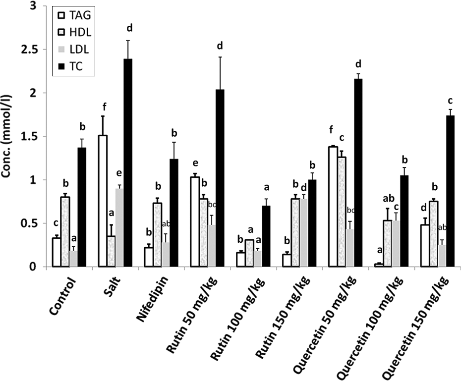
Lipid profile of experimental animals. Results are presented as mean ± SD (n = 6). Values with the same letters are not significantly different (p > 0.05).
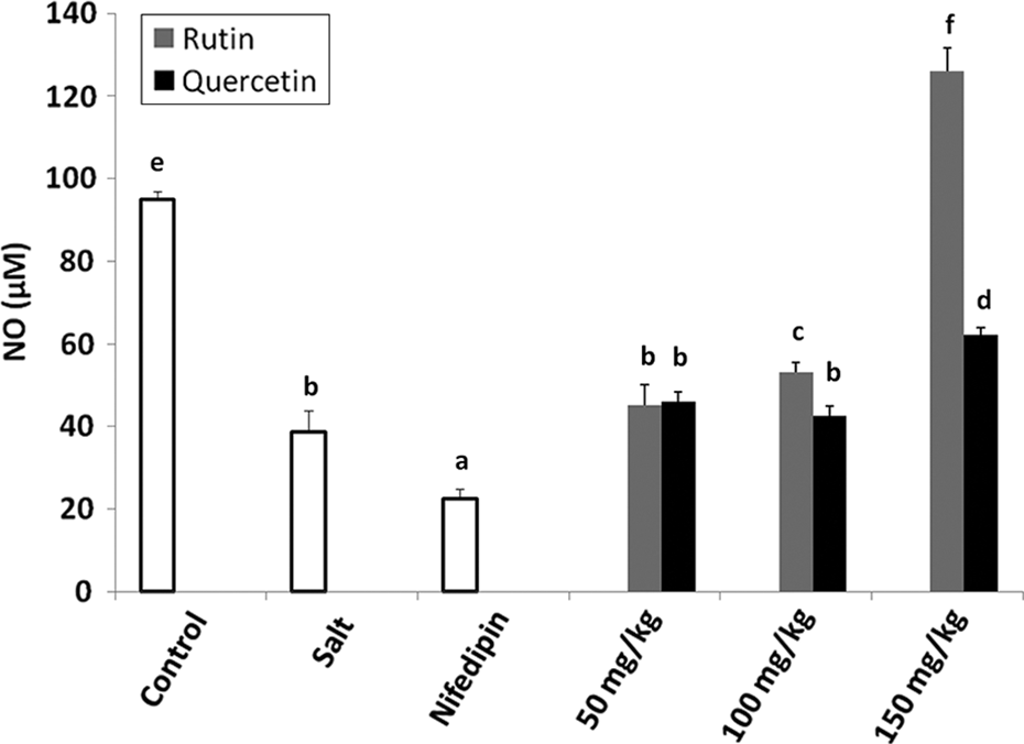
Plasma NO level of experimental animals. Results are presented as mean ± SD (n = 4). Values with the same letters are not significantly different (p > 0.05). NO: nitric oxide.
Footnotes
Conflict of interest
The authors declared no conflicts of interest.
Funding
This research received no specific grant from any funding agency in the public, commercial, or not-for-profit sectors.