
Abstract
This study was carried out to determine the effects of electromagnetic field (EMF) emitted by cellular phones (CPs) on electrocardiograms (ECGs) of guinea pigs. A total of 30 healthy guinea pigs weighing 500–800 g were used. After 1 week of adaptation period, animals were randomly divided into two groups: control group (n = 10) and EMF-exposed group (n = 20). Control guinea pigs were housed in a separate room without exposing them to EMFs of CPs. Animals in second group were exposed to 890–915 MHz EMF (217 Hz of pulse rate, 2 W of maximum peak power and 0.95 wt kg−1 of specific absorption rate) for 12 h day−1 (11 h 45 min stand-by and 15 min speaking mode) for 30 days. ECGs of guinea pigs in both the groups were recorded by a direct writing electrocardiograph at the beginning and 10th, 20th and 30th days of the experiment. All ECGs were standardized at 1 mV = 10 mm and with a chart speed of 50 mm sec−1. Leads I, II, III, lead augmented vector right (aVR), lead augmented vector left (aVL) and lead augmented vector foot (aVF) were recorded. The durations and amplitudes of waves on the trace were measured in lead II. The data were expressed as mean with SEM. It was found that 12 h day−1 EMF exposure for 30 days did not have any significant effects on ECG findings of guinea pigs. However, this issue needed to be further investigated in a variety of perspectives, such as longer duration of exposure to be able to elucidate the effects of mobile phone-induced EMFs on cardiovascular functions.
Introduction
Growing use of mobile phones has recently stimulated discussion about the possible health effects of the electromagnetic fields (EMFs) emitted by these phones. Mobile phones operate on wireless technology, with communication typically occurring via a 900–1800 MHz signal that is pulsed at 217 Hz. Increasing evidences have suggested that the EMFs emitted by cellular phones (CPs) interact with human organism because they represent a potential source of electromagnetic interference. 1 –3 There are several reports that indicate that EMF may elicit a biological effect in target cells or tissues. 4 –6 However, it is not clear whether or not these biological effects lead to an adverse health effects.
The studies of mobile phones and their possible health impact have been focused on changes in electric activity of the brain and reaction times, 7 –9 sleep pattern, 2 subjectively perceived symptoms 10 and malignant tumors. 11 –14 In our previous study, we also demonstrated that EMF emitted from CP produced oxidative stress in brain tissue of guinea pigs. 15 However, none of those studies have given evidence that the use of mobile phones has direct or indirect effects on human bodily functions or carcinogenesis.
Cardiovascular system may also be a potential target for the EMFs emitted by the mobile phones. It has been shown that the signals produced by their operating functions contain components of low frequencies that may interfere with implanted pacemakers. 16 However, whether EMFs emitted by CPs alter cardiac autonomic regulation has not extensively been studied. There are few studies that assessed the influence of EMFs emitted by mobile phone on cardiovascular functions including heart rate, blood pressure and electrocardiogram (ECG) recordings. The obtained results are controversial both in human 17 –19 and in animals. 20,21 Therefore, the present study was designed to evaluate the possible effect of EMFs emitted by mobile phones on ECGs of guinea pigs.
Materials and methods
Treatment of guinea pigs
A total of 30 healthy guinea pigs weighing 500–800 g were used. They were housed in makrolon cages under laboratory conditions (light period of day and night; 21 ± 1°C; chow and tap water available freely) in research laboratory of Physiology Department for 1 week before the experiment. After 1 week of adaptation period, animals were randomly divided into two groups: control group (n = 10) and EMF-exposed group (n = 20). Animals in second group were exposed to 890–915 MHz EMF (217 Hz of pulse rate, 2 W of maximum peak power and 0.95 w kg−1 of specific absorption rate) of a CP for 12 h day−1 (11 h 45 min standby and 15 min speaking mode) for 30 days. The CPs were attached to the cages about 20–30 cm far from the animals. Control guinea pigs were housed in a separate room of the research laboratory with the same conditions as the exposure groups without exposing EMF of a CP. All animals received humane care according to the criteria outlined in the “Guide for the Care and Use of Laboratory Animals” prepared by the National Academy of Sciences and published by the National Institutes of Health. 22
ECG recording
ECGs of guinea pigs in both the groups were recorded by a direct writing electrocardiograph (Cardiofax 6851, Tokyo, Japan) at the beginning and 10th, 20th and 30th days of the experiment. Alligator clip electrodes were attached to the skin at the triceps brachii muscle (caput longum and caput laterale) of the right and left limbs and biceps femoris muscle of the right and left hips. Electrode gel was rubbed onto the skin where the alligator clips were attached to act as a decreasing agent and thereby decrease the resistance of the skin. 23 The guinea pig was placed in a washbowl and waited for 5 min for animal to get calm. They were not anesthetized at any time. All ECGs were standardized at 1 mV = 10 mm, with a chart speed of 50 mm sec−1. Leads I, II, III, lead augmented vector right (aVR), lead augmented vector left (aVL) and lead augmented vector foot (aVF) were recorded. The durations and amplitudes of waves on the trace were measured in lead II recordings regarding their width (second) and height (millivolts), respectively.
Data analysis
Levene and Kolmogorov–Simirnov tests were used to determine the homogeneity and normality of data, respectively. Student’s t test was used to compare the mean of EMF-exposed versus control guinea pigs, with p < 0.05 as the criterion of significance for the statistical comparisons. The data were expressed as mean with SEM.
Results
Heart rates as well as the durations and amplitudes of waves measured in lead II recorded from the control and EMF-exposed groups are shown in Table 1. Representative leads of ECGs of control and EMF-exposed animals are shown in Figures 1 and 2, respectively. It was found that 12 h day−1 EMF exposure for 30 days did not have any significant (p > 0.05) effects on ECG findings of guinea pigs.
Heart rates as well as durations and amplitudes of waves measured in lead II recorded from the control and EMF-exposed groups.
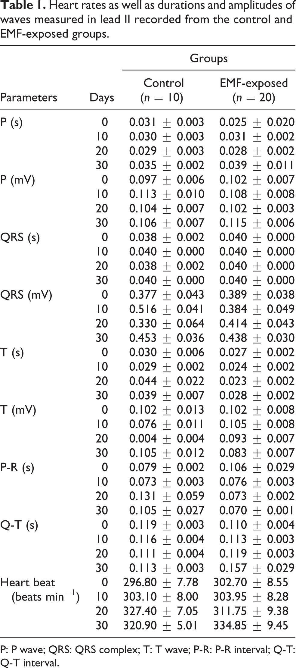
P: P wave; QRS: QRS complex; T: T wave; P-R: P-R interval; Q-T: Q-T interval.

Representative leads of electrocardiograms of control group (standardization, paper speed: 50 mm s−1; 1 mV = 10 mm).

Representative leads of ECGs of EMF-exposed group (standardization: 1 mV = 10 mm; paper speed: 50 mm s−1). ECGs: electrocardiograms; EMF: electromagnetic field.
Discussion
The present study was designed to investigate whether EMFs emitted by mobile phones alters the ECG recordings of guinea pigs. This study is the first one that evaluates the 12h day−1 EMF exposure on ECG recordings of guinea pigs for 30 days. It was found that EMF exposure did not have any significant effects on ECG findings of guinea pigs. These findings are in agreement with previous studies, 24 –26 where no statistically significant changes due to EMF exposure were found on cardiovascular functions.
Despite well-known effects of mobile phones, their effects on autonomic regulation of cardiovascular system have not been extensively investigated. It has been reported that the call with a mobile phone may change the autonomic balance in healthy subjects. 27 They suggested that the changes in heart rate variability during the call with a mobile phone could be affected by EMF but the influence of speaking cannot be excluded. They indicated that the tone of the parasympathetic system measured indirectly by analysis of heart rate variability was increased, while sympathetic tone was lowered during the call with the use of a mobile phone. However, some authors do not confirm connections between the use of mobile telephones and changes in circulatory system. It has been reported that there were no significant changes in arterial blood pressure and heart rate during or after the EMF exposures of 900 or 1800 MHz CPs. 25 It has been suggested that the changes occurred in heart rate and in arterial blood pressure were independent of the EMF exposure with the use of 900 MHz mobile phones. 24 The domination of sympathetic tone in response to EMF exposure was reported, 28 but correlated it with emotional stress related to solving the psychophysiological tests by the participants.
In a previous study, the EMFs emitted by mobile phone enhanced the oxidative stress in the heart tissue and also in normal volunteers and it affected the heart rate and blood pressure. 19 It has been demonstrated that reactive oxygen species (ROS) are directly involved in oxidative damage induced by EMF exposure of CPs on macromolecules (e.g. lipids, proteins and nucleic acids in tissues). ROS can spur myocyte hypertrophy, re-expression of fetal gene programs and apoptosis in cardiac myocytes in culture. In our previous study, we demonstrated that EMF emitted from CP produced oxidative stress in brain tissue of guinea pigs. 15 In the present study, we did not evaluate the cardiac tissue with regard to oxidative stress. Therefore, we do not know whether EMF exposure produces any free radical production on myocardium. However, it did not produce arrhythmia in guinea pigs. This issue needs to be further investigated in a variety of perspectives, such as longer duration of exposure to be able to elucidate the effects of mobile phone-induced EMFs on cardiovascular functions.
Footnotes
Conflict of Interest
The authors declared no conflicts of interest.
Funding
This research received no specific grant from any funding agency in the public, commercial, or not-for-profit sectors.