
Abstract
We examined the effect of exposure to mobile phone 1800 MHz radio frequency radiation (RFR) upon the urinary excretion of 8-oxo-7, 8-dihydro-2′-deoxyguanosine (8-oxodG), one major form of oxidative DNA damage, in adult male Sprague-Dawley rats. Twenty-four rats were used in three independent experiments (RFR exposed and control, 12 rats, each). The animals were exposed to RFR for 2 h from Global System for Mobile Communications (GSM) signal generator with whole-body-specific absorption rate of 1.0 W/kg. Urine samples were collected from the rat while housed in a metabolic cage during the exposure period over a 4-h period at 0.5, 1.0, 2.0 and 4.0 h from the beginning of exposure. In the control group, the signal generator was left in the turn-off position. The creatinine-standardized concentrations of 8-oxodG were measured. With the exception of the urine collected in the last half an hour of exposure, significant elevations were noticed in the levels of 8-oxodG in urine samples from rats exposed to RFR when compared to control animals. Significant differences were seen overall across time points of urine collection with a maximum at 1 h after exposure, suggesting repair of the DNA lesions leading to 8-oxodG formation.
Keywords
Introduction
Mobile phone, as a key device for most people, has a wide range of biological effects, 1 some of which are known while others are not or being investigated. The radio frequency radiation (RFR) has been linked to the development of cancer 2,3 and has been implicated in potential harmful effects on the fertility of male mobile phone users 4,5 and on RFR-exposed rabbits. 6 In his review, Lai 7 reported that RFR could induce genotoxic effects (single- and double-stranded DNA breaks), changes in chromosome conformation and micronuclei formation. In contrast, literature pertaining to the use of mobile phones contains reports of negative association or no significant changes regarding induction of cancer, 8 fertility, 9 and genotoxicity. 7,10 –13 A total of 101 publications on genotoxicity studies (effect on chromosomes, DNA fragmentation and gene mutations) on radio frequency–electromagnetic field (RF-EMF) were exploited, 11 of which 49 reported genotoxic effects, 43 did not and 9 found that RF induces genotoxic events by itself and enhances the genotoxic action of other physical or chemical agents. These studies have been performed with a variety of different test systems—some studies used more than one test system.
The exact mechanism underlying the possible harmful biological effects of exposure to mobile phone radiation is still unknown. One of the proposed mechanisms is the ability of RFR to stimulate oxidative stress (formation of free radicals and DNA oxidative damage). 5,14,15 There is an increasing experimental evidence that the DNA lesion, 8-oxodG, is a critical biomarker for oxidative stress in both clinical 16,17 and occupational 18,19 settings. 8-oxodG is formed in a promutagenic DNA lesion induced by the reaction of hydroxyl radicals with guanine at the C8 site in DNA. 20 It is a miscoding lesion causing G to T transversion. 21 8-oxodG seems to be the most prevalent product of free radical-induced DNA oxidative lesion that can be detected in mammalian genomic DNA. 22 In contrast to the analysis of 8-oxodG in DNA, its analysis in urine is more reproducible because of the lack of artifact formation and the interlaboratory deviation seems to be low. Therefore, it should be easier to assess the effects of lifestyle factors, diet and genotoxic environmental chemicals on cellular oxidative stress by analyzing 8-oxodG in human urine. 23 The nucleoside 8-oxodG is present in substantial amounts in urine. 24 The excretion rate is often assumed to represent the rate of repair of oxidatively generated DNA damage throughout the body and therefore also the rate of input of damage (since these are generally in the equilibrium). Investigators have reported a high concentration of 8-oxodG in urine samples from female patients with carcinoma of genitalia, 25 malignant breast tissues with invasive ductal carcinoma, 26 colorectal tumor tissues 27 and gastric cancer tissues. 28
This study was aimed at assessing the putative effects of exposure of Sprague-Dawley rats to 1800 MHz frequency, usually delivered by mobile phone, by measuring the level of generation of urinary 8-oxodG.
Materials and methods
Animals
Twenty-four male Sprague-Dawley rats (10–12 weeks old, 200–250 g) were randomly divided into 2 groups, 12 animals, each (RFR exposed and control). To reduce the impact of stress, the animals were conditioned by placing them in individual plastic cages for 24 h prior to an experiment in the room in which they would be exposed to mobile phone RFR fields. The animal house was maintained on a 12/12 h light/dark cycle, at an ambient temperature of 22°C and a relative humidity of 65%. The animals involved in this study were maintained and used in accordance with the Animal Welfare Act and the Guide for the Care and Use of Laboratory Animals prepared by Yarmouk University, Animal Ethical Committee.
RFR field exposure system
The exposure system consisted of Global System for Mobile Communications (GSM) signal generator DLW-3000 xp which generates 1800 MHz RFR. The signal generator was located at about 2 cm beside the cage. The antenna was positioned to emit directly toward the inner side of the cage. The Narda model 8616 and detachable probe (Model 8623D) were used extensively to measure the electric field and adjusted to be 15–20 V/m. It is worth noting that the value of 24 V/m is equivalent to specific absorption rate (SAR) value of 1.0 W/kg for 1800 MHz. 29 This simulates the actual exposure levels from the current GSM mobile phones to the direction of the human tissues. The radiation parameters were calculated according to Daniels et al. 30 These parameters were frequently checked throughout the experiments. During exposure, animals were provided with food and drinking water. Animals were individually placed in a metabolic cage (Suzhou Fengshi Laboratory Animal Equipment Co., Ltd, China) with free access to drinking water, but not food, and exposed to the specified field for 2 h between 8:00 and 10:00 a.m. A corresponding number of animals at each collection time served as a control. The control group was maintained in such a way that the signal generator was switched off and placed close to the cage in which the animals were kept. The control cages were treated exactly to match the experimental ones. In the cage, a rat was freely mobile without any restrain. To minimize the effect of metallic objects on the electromagnetic field and thereby on the relative SAR value, the wire led of the cage was removed during exposure and the metal spout of the drinking water bottle was covered with plastic.
Collection of urine samples
Exposed animals were divided into four subgroups (three, each). Urine samples from individual rats were collected for 4 h. From the first subgroup, collection started at the first half an hour of the exposure period. The timing of urine collection for the remaining three subgroups was 1, 2 and 4 h from the beginning of the exposure time (Figure 1). The cage was fitted with a nonmetal funnel attached to the bottom with a ball placed at the hole of the funnel to allow urine but not feces to pass through. Aluminum foil-covered polypropylene, 15 ml, was placed under the funnel to collect the urine without evaporation. Urine specimens were stored in sterile containers at −50°C until later use.
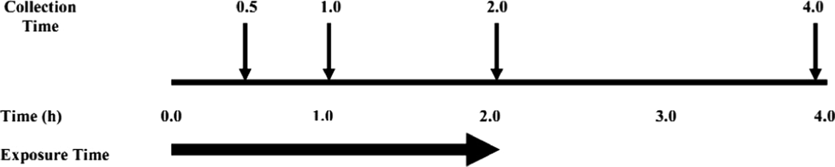
The protocol for exposure of rats to mobile phone radio frequency radiation (RFR) and collection of urine samples.
Determination of urinary 8-oxodG
All reagents and urine samples were brought to room temperature before use. To remove cells and insoluble materials, urine samples were centrifuged at 1500g for 15 min. For determination of 8-oxodG a competitive enzyme-linked immunosorbent assay (ELISA; Northwest Life Science Specialities, USA) was used. Briefly, to each well of the ELISA kit, 50 μl of urine supernatants were added to precoated 8-oxodG protein conjugate microtiter plate, followed by 50 μl of reconstituted primary antibody, anti-8-oxodG monoclonal antibody solution. The plate was covered with adhesive strip, incubated at 37°C and mixed continuously for 1 h. The antibodies bound to the 8-oxodG sample were washed with 0.05% Tween-20/phosphoric acid buffer and an enzyme-labeled secondary antibody was added to the plate, then incubated at 37°C and mixed continuously for 1 h. The unbound enzyme-labeled secondary antibody was washed away and the amount of antibody bound to the plate was determined colorimetrically after the addition of a chromatic substrate (o-phenylenediamine) and read at 490 nm. All standards and samples were typically assayed in a blind manner in triplicate. For each standard 96-well microplate, 6–9 control samples were randomly placed among the unknown samples. Data were corrected by urinary creatinine and urinary 8-oxodG (ng/ml)/creatinine (mg/ml) ratio was abbreviated as urinary 8-oxodG (ng/mg creatinine; Table 1).
Concentration of urinary 8-oxo-7, 8-dihydro-2′-deoxyguanosine (8-oxodG; ng/ml)/creatinine (mg/ml) in rats exposed to mobile phone radiation (1800 MHz) measured by ELISA a and corrected by urinary creatinine

aTriplicate ELISA readings per sample or standard. Three male rats were included per group.
Urine creatinine levels were determined using a creatinine assay kit (Oxford Biomedical Research, Inc. Michigan) based on the method reported before. 31 Briefly, a urine sample or a creatinine standard was mixed with an alkaline picrate solution. The absorbance was measured at 492 nm using a microplate ELISA reader (Stat Fax 3200, Awareness Technologies, USA). The absorbance was read again following the addition of the acid reagent. The difference in absorbance measured at 492 nm before and after acidification is proportional to creatinine concentration. The urinary 8-oxodG concentration was adjusted to the urinary concentration of creatinine to control the variability in urine dilution. The results are expressed as 8-oxodG (ng/ml)/creatinine (mg/ml) ratio.
Statistical methods
The mean and standard error of the means (SEMs) were calculated. For the differences between the exposed and control groups for concentration of 8-oxodG at each time point, an analysis of variance (ANOVA; p < 0.05) was used first to test for significance. Then, unpaired t tests were used. To explore the time effect on the concentration of 8-oxodG ng/mg creatinine within the exposed/control group, ANOVA followed by multiple comparison using Scheffè test. The PASW Statistics 18 Software Package (formerly called SPSS) was used for the ANOVA test.
Results
Using a monoclonal antibody assay to measure 8-oxodG, it was revealed that exposure of rats to RFR led to statistically significantly (p < 0.05 vs. control) increased levels of urinary 8-oxodG with respect to those of controls at all times of urine collection except at 0.5 h where the increase was in favor of the control (Table 1). Also significant differences related to time of urine collection in the concentration of 8-oxod G ng/mg creatinine were noted. The p values were significant at 0.000 for both groups in favor of 1 h time. The RFR exposure led to an increase in 8-oxodG to 1.30 ± 0.03 ng/mg creatinine in the urine collected following the first hour of exposure compared to the corresponding control 0.78 ± 0.16 ng/mg creatinine (Table 1). After these peaks, the levels of 8-oxodG in both groups declined as shown in Figure 2 which presents the adjusted mean (±SEM) of urinary 8-oxodG (ng/mg creatinine) by exposed group at the four time points.

Enzyme-linked immunosorbent assay (ELISA) of urinary 8-oxodG from rats exposed to mobile phone radiation and the controls. The levels were standardized against creatinine concentration.
Discussion
Although more than 20 different modifications of DNA bases have been identified, the major products of DNA oxidatively generated damage are 7, 8-dihydro-8-oxoguanine (8-oxoG) and its nucleoside 7, 8-dihydro-8-oxo-2-deoxyguanosine (8-oxodG), and many researchers have measured 8-oxodG in tissues or urine as a marker of useful oxidative stress. 28 In urine, 8-oxodG appears as a consequence of DNA-repair mechanism in the cell and is thought to reflect the level of oxidatively damaged DNA in the whole body since it is excreted without metabolizing any more 24 and ratio of urinary 8-oxodG to urinary creatinine (8-oxodG/creatinine) as a good biological indicator of DNA oxidation. 32 The urinary 8-oxodG analysis has the following advantages over its measurement in tissues: it is a noninvasive method, results are reproducible, 8-oxodG is not artificially produced during isolation of DNA, 8-oxodG is stable during urine storage, and its concentration is not increased by air oxidation. This may be due to the presence of a high concentration of an antioxidant, uric acid and the low level of the precursor deoxyguanosine in urine. 23 The results reported in the current study appear to disagree with recent trends in the literature on genotoxic effects of RF, showing no significant occurrence of oxidative stress. 7, 10–13 No work regarding the effects of RFR on urinary 8-oxodG is available in the literature. Recently, 33 we have shown that RFR exposure is not associated with the raise in concentrations of tissue and serum 8-oxodG in preliminary experiments on RFR-exposed mice. However, we do not know where and how the effects of this exposure on 8-oxodG formation are induced in vivo. Also, Tomruk et al. 34 reported that whole body exposure of New Zealand white rabbits to 1800 MHz GSM-like RFR did not cause significant formation of radical molecules in hepatocytes. The low levels of 8-oxodG measured in the latter two studies could be explained by the low accumulation of this molecule as a result of an efficient repair system in the investigated tissues.
In the present randomized controlled trial rats that were regularly exposed to 1800 MHz mobile RFR were associated with a statistically significant increase in urinary excretion of 8-OxdG compared with the control. The presented data indicate that RFR could stimulate hydroxyl radical formation, which may imply their potential pro-oxidant action. The collection time of the urine during or following exposure period changed the degree of the observed increase in DNA damage resulting in the formation of 8-oxodG. This may suggest that damaged DNA leading to 8-oxodG is repaired with time. The findings, 35 along with data from a number of literature reports, form an argument against a contribution of these urinary lesions from cell death and diet; and thus in the absence of these confounding factors, urinary measurements may be attributed entirely to the repair of DNA damage and suggest their possible use in studying the associations between DNA repair and exposure to RFR. These conclusions can be further tested using cells in which the base excision repair devoted to 8-oxodG is reduced as in the case of 8-oxoguanine DNA-glcosylase 1 knockout mice. 36
Some limitations of this present study should be noted. Equal exposure to each rat cannot be guaranteed by our methodology. However, we did make sure that RFR was evenly spread throughout the cage by measuring the field strength at different positions and angles. Additionally, since rats in the various groups were passive most of the time, we could arrange/position the cage in such a way that the rats were facing the antenna, anticipating direct exposure to radiation. We cannot explain how the dosage of RFR given to the rats equates to the levels of exposure to human using cell phones. It would have been better to collect the first data at time zero, before the exposure began. Unfortunately, this was not performed. Further, our data regarding the levels of urinary 8-oxodG cannot be compared with baselines in other studies since no such studies are available in the literature. The low levels of urinary 8-oxodG recorded in the present study are perhaps due to the possibility that ELISA is not sensitive enough. For ELISA, other compounds such as oligonucleotides and 8-oxoguanosine may cross-react with antibody to 8-oxodG, although these compounds themselves may be relevant markers of oxidative damage.26,37 Nevertheless, even with the variation in methods, the creatinine-standardized concentrations of 8-oxodG seem broadly similar among different laboratories. 38 Moreover, several studies showed a good correlation between the urinary 8-oxodG values obtained by HPLC-electrochemical detection (ECD) and those obtained by ELISA. 39 Although the measurement of urinary 8-oxodG by HPLC-ECD is reliable, it demands a high technical level and takes a relatively long time. 40,41 In fact, the current gold-standard assessment is by complex chromatographic methods such as HPLC or liquid chromatography–mass spectrometry (LC–MS/MS). Several studies have reported that commercial 8-oxodG ELISA kits correlate sufficiently with chromatographic techniques to be an easier alternative for laboratories without access to gold-standard techniques. 42 One study 35 revealed greater consensus than previously expected, although the concern remains over ELISA. Another point should be addressed which is the possibility of the stress having impact on 8-oxodG in urine. We tried to exclude this by allowing rats to move freely in the cage without restrains. In addition, urine was always collected between 8:00 and 10:00 in the morning to account for possible variation in circadian secretion of the steroids. However, this issue can be further investigated by determining cortisol concentrations in the plasma.
Overall, in view of the good correlation between the 8-oxodG values measured by HPLC-ECD and the ELISA and the ease in performing ELISA, it becomes a reasonable method in molecular epidemiological studies to assess the risk of cancer or other diseases from environmental chemicals. 43,44 Thus, although it is still inconclusive that whether RFR exposure acts as an inducer of reactive oxygen species in vivo, the present results suggest that (a) exposure to RFR may augment oxidative stress; (b) urinary excretion of 8-oxodG may be a good and specific marker of oxidative stress. Further work is also required to understand the origin and validity of excreted urinary 8-oxodG as a marker of oxidative stress. We believe that such studies will benefit from the standardized protocol used in this research trial.
Footnotes
Funding
This work was funded by the Deanship of the Graduate Studies, Yarmouk University (grant number 28/2006).